Table of Contents
Ancylostoma duodenale
Ancylostoma duodenale is a species of the Ancylostoma genus. The hookworm of the Old World is a parasitic nematode worm. It matures and reproduces in the small intestines of humans, cats, and canines. Ancylostoma duodenale and Necator americanus are the two hookworm species commonly cited as the cause of hookworm infection in humans. They have two sexes. Ancylostoma duodenale is widespread across the globe, including in Southern Europe, North Africa, India, China, Southeast Asia, certain regions of the United States, the Caribbean, and South America.

Characteristics of Ancylostoma duodenale
- A. duodenale is a tiny, cylinder-shaped, grayish-white worm. The anterior margin of the buccal capsule features two ventral plates. Each of them has two large fangs with their bases fused together.
- In the depths of the buccal capsule, a pair of small teeth can be discovered. Males are between 8 and 11 mm in length and have a copulatory bursa at the posterior end.
- Females are 10–13 mm in length, with the vulva located at the posterior end; daily egg production ranges from 10,000 to 30,000. One year is the average lifespan of a female A. duodenale.
Historical Retrospect of Ancylostoma Duodenale
- Ancylostoma duodenale, the hookworm of the Old World, is a prevalent nematode parasite of the human small intestine.
- It causes “ancylostomiasis” in humans, and until recently, this hookworm was the most significant helminthic infection of humans. However, in many countries, it has been brought under control.
- In 1838, an Italian physician named A. Dubini discovered the parasite during the autopsy of a woman. Perroncito (1881) described the development of soil-dwelling, free-living larvae.
- In 1898, Looss described the pathogenesis and mode of entry of the larvae into the human intestine.
Geographical Distribution of Ancylostoma Duodenale
- Infection with the parasite has been documented in rural areas of tropical countries, and it may also occur in temperate regions where temperature and humidity are conducive to the development of the soil larvae.
- Europe, Egypt, India, Bangladesh, Sri Lanka, central and northern China, and the Pacific Islands have all reported incidences of hookworm. It is more prevalent in Punjab, Haryana, and Himachal Pradesh, India.
Habitat of Ancylostoma Duodenale
- Adult Ancylostoma duodenale worms are endoparasites that reside in the human intestine, primarily in the jejunum, less frequently in the duodenum, and infrequently in the ileum.
- From the feces-contaminated soil, the infective juveniles enter the human host percutaneously. Hookworms thrive in primordial environments where people walk barefoot, there are no modern sanitary conditions, and human faeces are deposited on the ground.
Structure of Ancylostoma Duodenale
Shape, Size and Colour
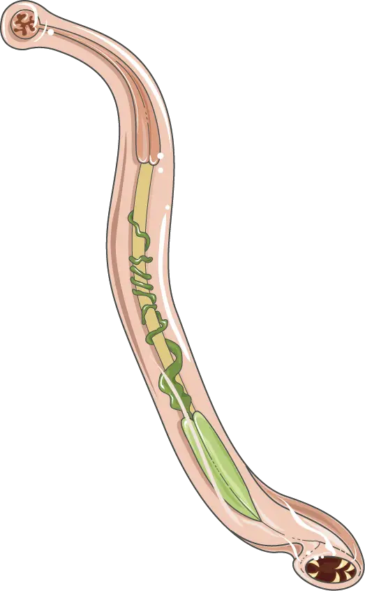
- Ancylostoma duodenale adults are diminutive and cylindrical. Males are approximately 8 mm in length and 0.4 mm in diameter, whereas females are typically 12.5 mm in length and 0.6 mm in diameter. Due to ingested blood in its intestinal tract, freshly expelled faeces are a reddish brown colour.
External and Internal Structures
- Each worm’s proximal end is slightly bent dorsally (hence the name hookworm) and has a large buccal capsule. The large and prominent buccal capsule is lined with a hard substance and has six cutting plates or teeth, four hook-like on the ventral surface and two knob-like (triangular plates) or pointed lancets on the dorsal surface.
- The buccal capsule aids in attachment to the host’s intestinal wall. The female worm’s posterior end tapers abruptly into a brief post-anal tail, whereas the male’s is expanded and umbrella-like. This expanded structure surrounding the cloaca is known as the copulatory bursa.
- The copulatory bursa consists of two lateral lobes with six muscular rays in each and a small median dorsal lobe with one dorsal ray that is only divided at its apex.
- The arrangement of rays is remarkably consistent, and each ray is given a name. The main ray in the dorsal lobe is called a dorsal ray, and in each lateral lobe, starting from the dorsal side, the six rays are named externo-dorsal, postero-lateral, medio-lateral, externo-lateral, and ventro-ventral. The buccal capsule and bursal ray teeth are taxonomically significant.
- Externally, the body of Ancylostoma duodenale is coated with cuticle. It is followed internally by the longitudinally oriented epidermis and musculature. Its body cavity consists of the pseudocoel that surrounds its organ systems.
Morphology of Adult worms
- The mature worms are cylindrical, plump, unyielding, and cream-colored.
- The anterior end is bent dorsally like a hook (hence the name “hookworm”), and the oral aperture is located dorsally.
- On the ventral aspect of the oral aperture are six sharp canines (cutting plates), two on each side, and two on the dorsal surface.
- Large and conspicuous, the buccal capsule is lined with chitin-like substance.
- The cuticle is characterised by thin transverse striations.
- There are tiny, finger-like cervical papillae on each side, a short distance from the anterior extremity.
- The oesophagus is connected to two cephalic glands, a small oesophageal gland, and two pear-shaped cervical glands; the secretion of the oesophageal gland prevents the clotting of ingested blood.
- However, Thorson (1956) reported that the oesophageal gland emerges near the cutting plates (teeth) in the buccal capsule and is involved in extracorporeal digestion. Unknown is the function of cervical glands and cephalic glands.
- Sexual dimorphism is distinguishable. The female is marginally larger and has an end that is straight and pointed. At the caudal end of the male is a bursa copulatrix (an invagination of the body wall around the genital orifice). Thirteen rays sustain the bursa. It has two protruding spicules (1 mm in length) that facilitate the transfer of sperm during copulation.
- Female worms measure approximately 10 to 13 mm x 0.6 mm, while males measure approximately 8 to 10 mm x 0.5 mm.
- The female gonopore is distinct and located at the intersection of the posterior and middle thirds.
- Males have a cloaca at the opening of the ejaculatory duct.
Morphology of Eggs
- The eggs are ovoid, colourless, and roughly 60 m x 40 m in size, with broad, rounded ends.
- Each egg has a thin exterior and a very fine layer of vitelline.
- In a newly laid egg, the segmented ovum is encompassed by a clear space. Typically, it is segmented into 2 to 8 cells.
Life Cycle of Ancylostoma Duodenale
Ancylostoma duodenale completes its life cycle within a single host (man), hence the term monogenetic. In the life cycle of A. duodenale, no intermediate host is recorded.
1. Copulation and Fertilization
During copulation in the intestine of the host, the male’s copulatory bursa is applied to the female’s vulva and sperms are transmitted.
Due to the position of the genital openings, the worms (a male and a female) acquire a Y-shape during copulation. Thus, the sperms are transferred to the seminal receptacles where fertilisation occurs. The fertilised ova are then pushed through the vagina and gonopore into the uterus for implantation.
2. Passage of the eggs from the infected host/Egg Laying
A single female worm produces between 10,000 and 20,000 eggs per day. Fertilised ova are expelled from the host’s body alongside faeces. During its passage through the bowel, the contained ovum undergoes segmentation. The eggs are not infective to humans, but the larvae that emerge from the eggs in soil are infectious.
24 to 48 hours are required for the egg to hatch in moist soil with a temperature of 27 degrees Celsius and sufficient oxygen. Due to the attenuated faeces’ high acidic pH (4.8-5.0), very little development occurs.
Eggs
The eggs are ovoid or elliptical in shape, measure 65 pm by 40 pm, are colourless, and are protected by a transparent hyaline shell membrane. A 4- or 8-celled embryo is contained within an egg that has been released from the host organism. The embryos that were expelled with the faeces are not infectious to humans.
3. Development of larva in soil
- Due to its rhabditiform oesophagus, the newly hatched larva is termed rhabditiform larva or first stage juvenile. It measures approximately 250 m in length and has a convex anterior end and a pointed posterior end.
- It is extremely active and consumes voraciously on the stool and soil’s organic matter. It grows quickly and undergoes two moults per week: the first on the third day and the second on the fifth. At this stage, the larva has reached a length of between 500 and 600 m.
- The oesophageal bulb disappears after the second moult, and the organ becomes simple and muscular. The remaining cuticle serves as a protective sheath. It is now referred to as an infective filariform larva or third-stage juvenile. While still actively motile, the larva ceases feeding and stops growing. In approximately 8 to 10 days, an embryo transforms into a filariform larva.
Biology of the larvae
- The larvae inhabit the top half-inch of soil. In tropical climates, 90% of organisms die within two to three weeks, and the balance within six weeks. Direct sunlight is a potent killer of filariform larvae. They are extremely sensitive to dehydration and temperature extremes. Additionally, they migrate towards the oxygen source.
4. Entrance into a new host
- The filariform larvae obtain access to their host’s (human) body by penetrating the skin. They discarded their sheaths and bore directly through the epidermis or hair follicles.
- It is believed that the filariform larvae are attracted to the warmth of the soles of the feet and palms; therefore, the infection is more prevalent in barefoot individuals. It is also possible to contract an infection from handling feces-soiled apparel that has been wet for four or five days. According to reports, intrauterine infections are also conceivable.
5. Migration after entrance
- The larvae enter lymphatics or microscopic venules several hours after infection. Typically, larvae incapable of reaching the vascular spaces perish or are phagocytosed. On the third day, they are passively transported to the right side of the heart and then to the lungs via the pulmonary artery.
- They then migrate into the alveoli by penetrating the pulmonary capillary wall, where they are stopped due to their size. The larvae are swallowed after ascending the bronchi, trachea, and epiglottis from the lungs to the rear of the pharynx.
- Upon reaching the oesophagus, the third moult occurs with the formation of a temporary buccal capsule that contains four small canines. Between the seventh and tenth day, they reach the jejunum via the stomach and the duodenum. At this stage, the larva has grown swiftly and measures 2 mm x 0.13 mm.
6. Establishment and laying of eggs
- On day 15, the developing larva settles in the jejunum and endures its fourth and final moult. The definitive buccal capsule replaces the temporary buccal capsule with canines during this moult. The worm reaches sexual maturity in three to four weeks, and ova begin to appear in the stool six weeks later.
- Wounds are caused by the sharp fangs of adult worms in the region of attachment to the intestinal wall. Through these incisions, blood is expelled, which is then sucked up by the worms’ suctorial pharynx.
- Blood coagulation is prevented by the production of a secretion from the pharynx that has anticoagulant properties, thereby preventing blood from clotting. A. duodenale has been observed drawing copious amounts of blood at a rate of 0.8 ml per 24 hours.
Pathogenicity of Ancylostoma Duodenale
- Hookworms are the most dangerous parasitic nematodes because their muscular pharynx attaches to the intestinal villi and suckles blood and body fluids from the host. They also cut holes in the intestinal mucosa and leave bleeding lesions. It results in severe anaemia. In children where the prevalence of infection is high, they retard physical and mental development.
- Some toxins secreted by the glands in the head region of worms cause stomachache, food fermentation, diarrhoea, constipation, dyspnea, heart palpitations, eosinophilia, poor health, and eventual collapse of the patient.
- During larval penetration of the epidermis, local irritation and inflammation of the surrounding tissues may result in the formation of tiny sores. In the airways, the migratory larvae can cause haemorrhage and bronchial pneumonitis.
Treatment and Control of Disease Caused by Ancylostoma Duodenale
The treatment and control of disease caused by Ancylostoma duodenale, a parasitic nematode (roundworm) that infects humans, involves several measures. Here are some of the key treatment and control measures:
- Drug treatment: The primary treatment for Ancylostoma duodenale infection involves the use of anthelmintic drugs such as albendazole, mebendazole, or pyrantel pamoate. These drugs work by killing the adult worms and/or the larvae in the host’s body. Treatment is usually given for 1-3 days, depending on the severity of the infection.
- Iron and nutritional supplements: As Ancylostoma duodenale feeds on the host’s blood, it can lead to iron-deficiency anemia and protein malnutrition. Therefore, iron and nutritional supplements may be given to treat these deficiencies.
- Hygiene and sanitation: Proper hygiene and sanitation practices are crucial in preventing the spread of Ancylostoma duodenale. This includes the proper disposal of human waste, the wearing of shoes, and the maintenance of clean water sources.
- Education and awareness: Educating the public about the risks of Ancylostoma duodenale infection and the importance of proper hygiene and sanitation practices can help prevent the spread of the disease.
- Mass drug administration: In areas where Ancylostoma duodenale is endemic, mass drug administration may be used as a control measure. This involves treating the entire population in the affected area with anthelmintic drugs to reduce the overall prevalence of infection.
Overall, a combination of drug treatment, iron and nutritional supplements, hygiene and sanitation, education and awareness, and mass drug administration can help in the treatment and control of disease caused by Ancylostoma duodenale.
FAQ
What is Ancylostoma duodenale?
Ancylostoma duodenale is a parasitic nematode (roundworm) that infects humans and causes a disease known as hookworm disease.
How is Ancylostoma duodenale transmitted?
The larvae of Ancylostoma duodenale can enter the human body through the skin, usually through bare feet, or through ingestion of contaminated food or water.
What are the symptoms of Ancylostoma duodenale infection?
Symptoms of Ancylostoma duodenale infection can include abdominal pain, diarrhea, anemia, and fatigue.
How is Ancylostoma duodenale diagnosed?
Ancylostoma duodenale infection is diagnosed through stool samples that are examined for the presence of hookworm eggs.
What is the treatment for Ancylostoma duodenale infection?
The primary treatment for Ancylostoma duodenale infection involves the use of anthelmintic drugs such as albendazole, mebendazole, or pyrantel pamoate.
Can Ancylostoma duodenale be prevented?
Yes, Ancylostoma duodenale can be prevented through proper hygiene and sanitation practices such as wearing shoes, properly disposing of human waste, and maintaining clean water sources.
Who is at risk for Ancylostoma duodenale infection?
People who live in areas with poor sanitation and hygiene practices are at higher risk for Ancylostoma duodenale infection.
Can Ancylostoma duodenale be fatal?
While Ancylostoma duodenale infection is not usually fatal, it can cause significant health problems, particularly in children and pregnant women.
Is there a vaccine for Ancylostoma duodenale?
Currently, there is no vaccine for Ancylostoma duodenale.
Can pets transmit Ancylostoma duodenale to humans?
While pets can become infected with a different species of hookworm, Ancylostoma caninum, it is rare for humans to become infected with this species.
References
- Aziz MH, Ramphul K. Ancylostoma. [Updated 2022 Jun 14]. In: StatPearls [Internet]. Treasure Island (FL): StatPearls Publishing; 2023 Jan-. Available from: https://www.ncbi.nlm.nih.gov/books/NBK507898/
- https://www.biologydiscussion.com/animals-2/aschelminthes/ancylostoma-duodenale-habitat-morphology-and-life-cycle/32888
- https://www.sciencedirect.com/topics/medicine-and-dentistry/ancylostoma-duodenale
- https://www.sciencedirect.com/topics/agricultural-and-biological-sciences/ancylostoma-duodenale
- https://animaldiversity.org/accounts/Ancylostoma_duodenale/
- https://www.biologydiscussion.com/invertebrate-zoology/phylum-aschelminthes/ancylostoma-duodenale-habitat-structure-and-life-history/28991

Website muhimu sana naomba muendelee kutoa elimu kwa uma