Table of Contents
The Basidiomycetes division together with Ascomycota form the subkingdom Dikarya which is also known as the “higher fungi” within the kingdom Fungi. Basidiomycetes contain the following groups; mushrooms, puffballs, stinkhorns, bracket fungi, other polypores, jelly fungi, boletes, chanterelles, earth stars, smuts, bunts, rusts, mirror yeasts, and also a pathogenic yeast called Cryptococcus.
Basidiomycetes si consider the largest group of fungi because they contain 1100 genera which are consisting of 30,000 species and listed under the sub-division Basidiomycotina.
Basidiomycetes are harmful as well as useful. Their attack foods and ornamental plants, cause many different diseases including seedling diseases, wood rots, root and stem rots, seed diseases (smuts), and rusts, on the other hand, it used as humans foods.
What are Basidiomycetes?
- Basidiomycetes, commonly referred to as club fungi, are a significant division of fungi characterized by their unique reproductive structures and methods. As members of the Basidiomycota division, they stand alongside Ascomycota in the subkingdom Dikarya, often known as the “higher fungi” within the fungal kingdom. This division includes a diverse group of fungi, ranging from well-known mushrooms to less familiar forms like puffballs, jelly fungi, and rusts.
- The defining feature of basidiomycetes is the basidium, a specialized reproductive structure. It’s in the basidium that basidiospores, the primary reproductive spores of these fungi, are produced. The basidium typically forms as a part of a fruiting body known as the basidiocarp, which is commonly observed in mushrooms. These spores play a crucial role in the reproductive cycle of the basidiomycetes, allowing for the spread and propagation of the species.
- In terms of their vegetative structure, basidiomycetes are primarily composed of either primary or secondary mycelium. Mycelium is the vegetative part of a fungus, consisting of a network of fine, thread-like structures called hyphae. This mycelial network is essential for nutrient absorption and growth.
- Reproduction in basidiomycetes can occur both vegetatively and sexually. Vegetative reproduction often occurs through fragmentation, where a part of the mycelium separates and grows independently. Sexual reproduction is a more complex process, involving the fusion of two vegetative or somatic cells, leading to the formation of a basidium where basidiospores are eventually produced.
- This group of fungi is incredibly diverse, including familiar examples such as Agaricus in mushrooms and Puccinia, known for causing rust diseases in plants. Other notable members include puffballs, boletes, chanterelles, and earth stars. Some basidiomycetes, like Cryptococcus, are known to be pathogenic to humans.
- While basidiomycetes predominantly reproduce sexually via basidia, some species are known to reproduce asexually. The identification of these species often relies on certain anatomical features like the clamp connection, cell wall components, and molecular analysis of DNA sequences.
- In summary, basidiomycetes play a crucial role in the fungal kingdom with their unique reproductive structures and methods. Their diverse forms and reproductive strategies make them an important subject of study in mycology and ecology.
Basidiomycetes habitat
Basidiomycetes, a diverse group of fungi, can be found in a variety of habitats across the globe, adapting to different environmental conditions. Here are some common habitats for basidiomycetes:
- Forest Ecosystems:
- Basidiomycetes are frequently found in forests, contributing significantly to the ecosystem. They decompose wood and leaf litter, playing a crucial role in nutrient cycling.
- Soil:
- Many basidiomycetes grow in soil, where they decompose organic matter. Some also form symbiotic relationships with plant roots (mycorrhizae), aiding in nutrient exchange.
- Grasslands and Meadows:
- These open habitats are home to numerous basidiomycetes, especially those that form mycorrhizal associations with grasses and other herbaceous plants.
- Dung and Animal Remains:
- Some basidiomycetes specialize in decomposing animal dung or remains, playing a vital role in recycling nutrients back into the ecosystem.
- Wood and Dead Trees:
- Wood-decaying basidiomycetes are common, breaking down cellulose and lignin in dead trees and fallen logs.
- Living Trees and Plants:
- Certain basidiomycetes are parasitic, living on and deriving nutrients from living trees and plants, sometimes causing diseases.
- Urban and Man-Made Environments:
- Basidiomycetes can also be found in urban areas, growing on wood, mulch, and other organic materials in gardens, parks, and landscaped areas.
- Symbiotic Associations:
- Many basidiomycetes form symbiotic relationships with plants, enhancing plant growth and health by improving nutrient uptake.
- Extreme Environments:
- Some species have adapted to extreme conditions, such as high altitudes or very wet or dry environments.
- Human-Impacted Areas:
- These fungi can also thrive in disturbed habitats, including agricultural fields and areas affected by human activities.
This broad habitat range demonstrates the adaptability and ecological importance of basidiomycetes in both natural and altered environments.
Basidiomycetes Characteristics
- Structure: Basidiomycetes are primarily filamentous fungi composed of hyphae, with the exception of basidiomycota-yeast.
- Sexual Reproduction: They reproduce sexually through the development of club-shaped end cells called basidia, which typically produce external meiospores, usually numbering four.
- Basidiospores: These fungi generate specific spores named basidiospores.
- Asexual Reproduction: A portion of Basidiomycota species are capable of asexual reproduction.
- Ecological Role: Most basidiomycetes are saprophytes, playing a vital role in the decomposition of organic matter like litter, wood, and dung.
- Host Dependency: In nature, they are often confined to living on host plants.
- Mycelium Structure: Their mycelium is well-developed, branched, and septate, differentiating into primary, secondary, or tertiary forms.
- Cell Wall Composition: The cell walls of basidiomycetes consist of chitin and glucans with specific linkages of β-D-glucosyl units.
- Symbiotic Relationships: Some basidiomycetes form symbiotic relationships with trees as mycorrhizae, while others act as harmful parasites to various plants.
- Mycelium Stages: The somatic stage includes a developed, septate, filamentous mycelium, which primarily exists in two stages – primary and secondary (dikaryotic) mycelium.
- Primary Mycelium Features: This stage develops from basidiospores and contains a single haploid nucleus per cell, lacking sex organs, basidia, and basidiospores.
- Secondary Mycelium Role: The secondary or dikaryotic mycelium is the main nutrient-absorbing phase, containing cells with two haploid nuclei, and plays a central role in the life cycle.
- Life Span and Fructifications: In Homobasidiomycetidae, this stage can last for years, producing annual fructifications through hyphal interweaving.
- Teleutospores and Brand Spores: In Heterobasidiomycetidae, the secondary mycelium forms teleutospores or brand spores, which germinate to create basidia with basidiospores.
- Septal Pore Complexity: Except for rusts and smuts, basidiomycetes possess a complex septal pore of the dolipore parenthesome type.
- Absence of Motile Cells: Their life cycle does not feature any motile cells.
- Clamp Connection Formation: Most basidiomycetes form a structure known as a clamp connection.
- Dolipore Septa: A significant number of genera in basidiomycetes contain Dolipore septa.
- Role of Asexual Reproduction: Asexual reproduction, particularly through spore formation, plays a crucial role in their life cycle, varying between Homobasidiomycetidae and Heterobasidiomycetidae.
- Sexual Reproduction Mechanism: They lack traditional sex organs, with sexual reproduction occurring through processes like plasmogamy and karyogamy, where karyogamy is immediately followed by meiosis.
- Basidium Function: The basidium serves as the main reproductive organ, facilitating both karyogamy and meiosis.
- Basidiospore Generation and Germination: Typically, four basidiospores are produced exogenously on the basidium, which then germinate to form primary mycelium.
- Sexual Reproduction Diversity: The dikaryotic cell in sexual reproduction is formed by various processes including spermatization, somatogamy, clamp-connection, or the Buller phenomenon.
Basidiomycetes examples
Examples of basidiomycetes include:
- Mushrooms:
- Mushrooms are the visible, fleshy fruiting bodies of certain fungi, commonly found above ground on soil or a food source.
- They are known for their spore-bearing capacity, contributing to fungal reproduction.
- Puffballs:
- Puffballs are characterized by their unique way of releasing spores.
- When mature, these fungi release clouds of brown, dust-like spores, often triggered by external impact or bursting of the fruit body.
- Smuts:
- These multicellular fungi are notable for producing a large number of teliospores.
- Smuts get their name from the Germanic word for dirt, referencing the dark, thick-walled, dust-like appearance of their teliospores.
- Rusts:
- Rusts are plant diseases caused by fungi in the Pucciniales order.
- They are significant pathogens in horticulture, forestry, and agriculture, known for their damaging effects on various plants.
- Cryptococcus:
- This genus within the Cryptococcaceae family includes both filamentous and yeast forms of fungi.
- Cryptococcus species are notable in both environmental and medical contexts.
- Jelly Fungi:
- Named for their gelatinous, often foliose and irregularly branching fruiting bodies.
- These fungi have a distinctive jelly-like consistency, contributing to their unique appearance and texture.
Classification of Basidiomycetes
Traditional Classification of Basidiomycetes
- Subclasses:
- Basidiomycetes are traditionally divided into two subclasses based on the nature of the basidium’s septation.
- Heterobasidiomycetes (Holobasidiomycetes):
- Characterized by a fragmented basidium.
- Known as obligate parasites, causing diseases in various crops.
- Includes true mushrooms.
- Comprises three main orders:
- Tremellales
- Uredinales
- Ustilaginales
- Homobasidiomycetes:
- Features non-fragmented basidia.
- Common in diverse habitats like grasslands, dung, termite mounds, and wooden logs.
- Includes rust and smut fungi.
- Contains two series:
- Hymenomycetes: With an exposed hymenium, including orders like Exobasidiales, Polyporales, and Agaricales.
- Gastromycetes: With an enclosed hymenium, including orders such as Hymenogastrales, Lycoperdales, Sclerodermatales, Phallales, and Nidulariales.
Recent Classification of Basidiomycetes
- Subclasses and Class-level Taxa:
- The recent classification encompasses three subclasses and two additional class-level taxa.
- Pucciniomycotina:
- Encompasses rust fungi, Septobasidium, some former smut fungi, and diverse, less common fungi.
- Includes eight classes:
- Agaricostilbomycetes
- Atractiellomycetes
- Classiculomycetes
- Cryptomycocolacomycetes
- Crystobasidiomycetes
- Microbotryomycetes
- Mixiomycetes
- Pucciniomycetes
- Ustilaginomycotina:
- Primarily composed of former smut fungi and the Exobasidiales.
- Contains three classes:
- Exobasidiomycetes
- Entorrhizomycetes
- Ustilaginomycetes
- Agaricomycotina (Hymenomycetes):
- Includes most fungi like mushrooms, bracket fungi, and puffballs.
- Comprises three classes:
- Agaricomycetes
- Dacrymycetes
- Tremellomycetes
- Class-level Taxa:
- Wallemiomycetes: Identified as a sister group to Agaricomycotina based on genomic evidence.
- Entorrhizomycetes: Considered a close sister group to the other Basidiomycetes classes.
Economic Importance of Basidiomycetes
- Dual Nature:
- Basidiomycetes play both beneficial and harmful roles, making them of significant economic importance.
- Agricultural Impact:
- Certain basidiomycetes act as pathogens, causing destructive diseases in cereal crops like wheat, oats, and barley.
- Wood Decay:
- Pore fungi, a type of basidiomycete, are known for their ability to rot wood, adversely affecting lumber and timber.
- Culinary Value of Mushrooms:
- Mushrooms, a form of basidiomycetes, are highly valued as food due to their taste and nutritional richness.
- They are widely cultivated for their flavor and various health benefits.
- Edible Puffballs:
- Species like Lycoperdon and Clavatia are notable for their edible young sporophores.
- Clavatia contains a substance called calvacin, which is utilized in cancer treatment.
- Toxicity of Toadstools:
- Some basidiomycetes, such as Amanita, are highly poisonous, potentially causing death, while others may only cause discomfort.
- Mycorrhizal Associations:
- Many basidiomycetes form ectotrophic mycorrhizal connections with forest tree roots.
- These symbiotic relationships benefit both the fungus and the tree, with the fungus transferring essential nutrients to the tree and receiving organic substances in return.
- Decomposition Role:
- Saprophytic basidiomycetes play a crucial role in decomposing dead leaves and other forest litter.
- This decomposition process is vital for recycling nutrients back into the soil ecosystem.
Mycelium of Basidiomycetes
- The mycelium of Basidiomycetes is well-developed, filamentous, and made up of branched, septate hyphae usually developing in a fan-shaped manner.
- The cell wall is chitinous in nature and plasma membrane is present within the cell wall.
- The cytoplasm includes all the organelles which are present in a normal cell, except the chloroplasts.
- The septum, as in the Ascomycetes, begins as an annular outgrowth on the interior of the tubular wall. It develops inwards like a tight shelf decreasing diameter of the pore.
- The hyphae ramify in the substratum and consume food. The mycelium usually is a weft of interlacing and anastomosing hyphae.
- In some genera, the mycelial hyphae move parallel to one another and become bundled together to develop distinct and prominent thick cords of macroscopic size. These are called the rhizomorphs.
- The rhizomorph is surrounded by a sheath (cortex) and acts as a unit.
- The mycelium in various species varies in color and may be white, yellow or orange. It is usually perennial.
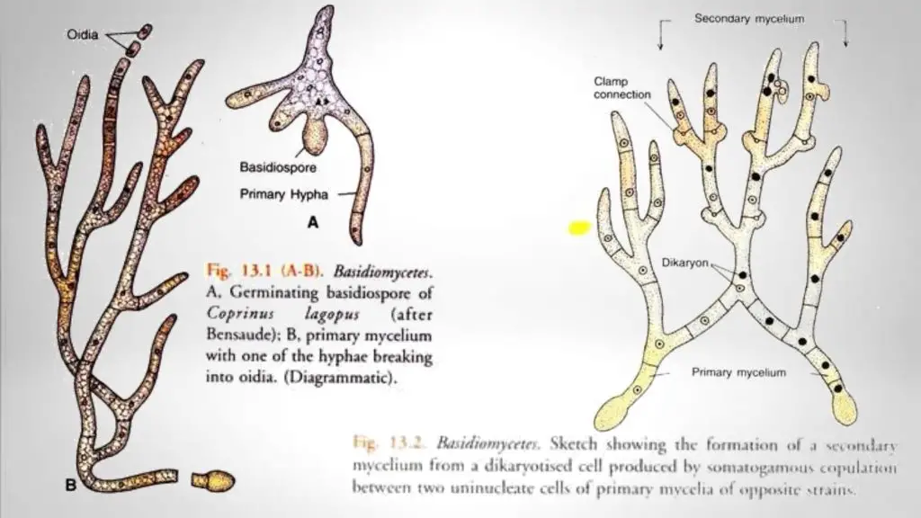
- The mycelium of Basidiomycetes moves through 3 different stages such as the primary, the secondary, and the tertiary stage before the fungus completes its life cycle.
Types of Basidiomycetes mycelium
- Primary mycelium: It develops from the basidiospore. It composed of uninucleate cells and hence they are termed as homokarion, although, the first stage of the primary mycelium may be multinucleate but then septa formation divides it into uninucleate cells.
- Secondary mycelium: It develops from the fusion of two uninucleate cells of primary mycelium; the process of confertion of primary mycelium into secondary mycelium called as dicariotization or dipodization. The basidia formed from binucleate cells of secondary mycelium.
- Tertiary mycelium: The secondary mycelium in some higher basidiomycetes organized into complex tissues to develop sporophores or basidiocarps, this stage known as tertiary mycelium.
Clamp Connections
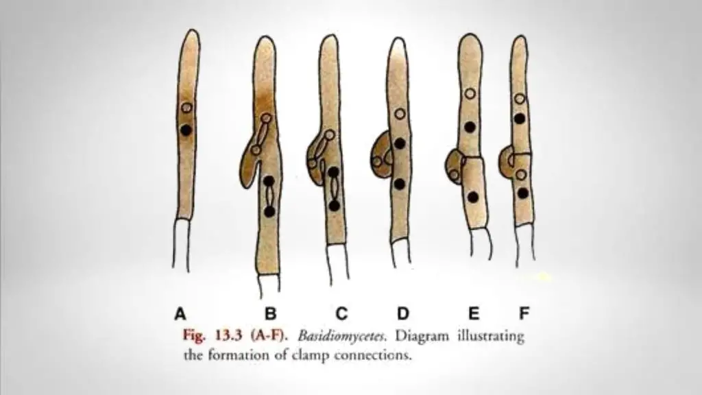
- When the dikaryotic cell (A) is beginning to divide a pouch-like outgrowth occurs from its wall (B), it arises midway between the two nuclei of the dikaryon. The two closely associated nuclei of the cell now divide simultaneously. This is known as conjugate division.
- One of the four daughter nuclei, generally the lower one of the upper pair, passes into the pouch (C). A septum appears at the base of the pouch (D). As a result, the pouch is cut off from the main cell. It may now be called a clamp cell.
- The clamp cell extends and forms a hook-like structure. The tip of the clamp now bends and gets attached to the lateral wall (E) of the parent cell and function as a bridge. This is known as clamp connection.
- An extra septum is attached vertically under the bridge usually at about the level of the upper end of the clamp connection (E). It separates the mother cell into two daughter cells. The terminal daughter cell contains two nuclei.
- All of these are a sister nucleus of the mother dikaryon. The lower or basal daughter cell contain one nucleus. The fourth nucleus lies in the clamp connection. The nucleus of the clamp connection now migrates into the basal daughter cell. The latter also becomes binucleate (F).
- The clamp connection thus simply functions as a bypass. It ensures that the sister nuclei formed by the conjugate division of the dikaryon separate into two newly formed daughter cells. The clamp connections are usually formed on the terminal cells of the hyphae of the secondary mycelium.
- They generally persist on the old dikaryotic hyphae. The presence of hook like clamp connections is a safe criterion for distinguishing a secondary or dikaryotic mycelium from the primary or monokaryotic mycelium.
- The clamp connections by some mycologists are considered homologous to the hooks of the ascogenous hyphae of the Ascomycetes.
- This view is supported by the general occurrence of a clamp connection at the base of a basidium. Moore and Mclear (1962) studied the fine structure of septa of the dikaryotic hyphae of Basidiomycetes.
- They found that the septum which is a cross wall flares sharply and broadly at the centre of the hypha to form a barrel- shaped structure with open ends.
- This is the actual pore surrounded by a swollen rim which is a part of the annular septum. This type of septum is termed a dolipore septum. The septum and the swelling are covered by the cell membrane.
- The opening on either side of the dolipore septum is guarded by a curved or crescent-shaped double membrane pore cap which in section looks like a parenthesis (round bracket).
- The septal pore-cap is thus given the name parenthesome. The parenthesome is similar to the endoplasmic reticulum. The lower parenthesome is seen to be interrupted by gaps in the form of pores. The upper parenthesome may be continuous.
- The dolipore parenthesome septum complex is unique to the Basidiomycetes. It maintains cytoplasmic continuity but prohibits nuclear migration.
- The secondary mycelium in the fruit bodies of the higher Basidiomycetes becomes organised into specialised tissues. It is called the tertiary mycelium. The fructifications are thus formed of the tertiary mycelium.
- The cells of the tertiary mycelium are also binucleate. The distribution of the dolipore parenthe-some septum complex in the Basidiomycetes is widespread.
- It is characteristic of the primary, secondary, and generative tertiary mycelium of the Homo-basidiomycetes as well as of the basidiocarpic mycelium of the Heterobasidiomycetes. The major exceptions are the rusts and smuts.
- The formation of basidia, the dikaryotic mycelium, the dolipore septum with the parenthesomes guarding the pore on both sides and clamp connections are the four diagnostic features of this class.
What is Diploidisation or Dikaryotisatton?
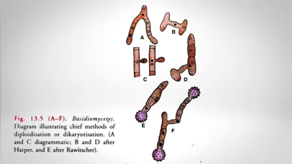
The conversation process of primary mycelium to secondary or dikaryotic mycelium is known as diploidisation or dikaryotisation. In the first step of this process is began with the establishment of a dikaryon in the fusion cell.
The repeated divisions of clamp connections coil the dikaryotised and as a result, a secondary mycelium is formed in which ‘each cell possesses a dikaryon (two neclei).
Methods of Diploidisation
- Hyphal Fusions: In this method, the vegetative cells and two adjacent hyphae of the main mycelia of opposing sexual strains (A) fuse.
- Conjugation of basidiospores (B): In this method, the basidiospore of two opposite strains meet and conjugate. Next, the newly formed binucleate basidiospore germinates to develop a secondary mycelium.
- Diploidisation can take place by the fusion of a germinating oidium of one strain with a cell of the primary mycelium of the opposite strain (C). The binucleate cell formed in this way by elongation and division by clamp connections develops into a secondary mycelium.
- It can take place if the germinating basidiospore and a haploid cell of the basidium (D) fuse. For example, U. violacea
- It can take place if two haploid cells of opposite strains of the basidium are fuse (E). For example, U. carbo.
- It can take place if the basidia produced by the germination of two smut spores (F) are fuse.
Note: Basidia are always produced from the binucleate cells of the secondary mycelium.
Basidiomycetes life cycle or Reproduction
Basidiomycetes reproduced by both sexual and sexual methods.
Asexual Reproduction
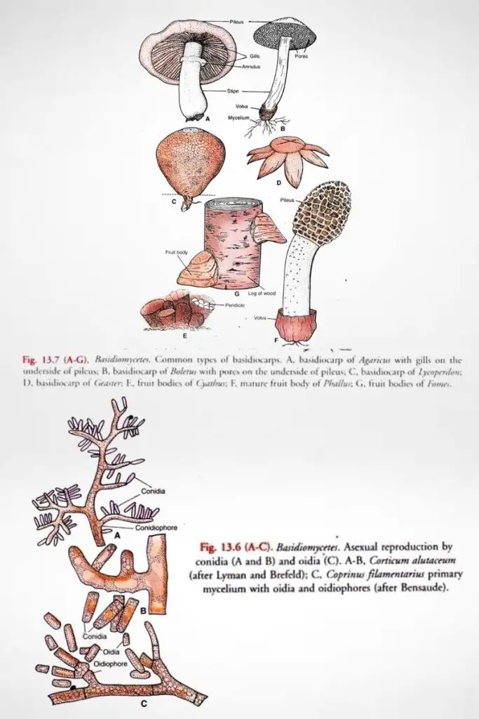
The asexual reproduction of Basidiomycetes is accomplished by either budding or asexual spore formation. During the budding procedure, an outgrowth or bud occurs in the parent cell. Then it gets separated from the parent cell by the formation of transverse septum and give rise to a new daughter cell.
The formation of an asexual spore in Basidiomycetes occurs at the end of a specialized structure known as conidiophores. The septae of concluding cells mature fully defined, dividing a random estimate of nuclei into single cells. The cell walls then harden into a protective coat. The preserved spores break off and are distributed.
Sexual Reproduction
Basidiomycetes reproduce sexually by the formation of basidiospores. Sometimes basidiomycetes are termed as club fungi, because of their microscopic club-shaped basidia (sing., basidium).
Basidia are comparable in function to the asci of ascomycetes. Each basidium is an enlarged hyphal cell that undergoes meiosis to form four basidiospores. Each fungus can produce millions of basidiospores, and each basidiospore develops a new primary mycelium.
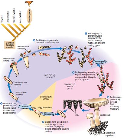
Life cycle
- Initiation of Secondary Mycelium:
- Occurs when a hypha from primary mycelium meets a compatible monokaryotic hypha, typically of a different mating type.
- Fusion of these two hyphae (plasmogamy) leads to the formation of a secondary mycelium.
- The secondary mycelium is dikaryotic, with each cell housing two separate haploid nuclei.
- Growth and Spread:
- The dikaryotic (n + n) hyphae of the secondary mycelium grow rapidly and extensively.
- Formation of Buttons:
- Upon favorable environmental conditions, the hyphae form compact masses known as buttons along the mycelium.
- Development of Fruiting Bodies:
- Each button develops into a fruiting body, commonly recognized as a mushroom or basidiocarp.
- The mushroom consists of a stalk and a cap.
- Structure of Basidiocarp:
- Composed of intertwined, matted hyphae.
- The underside of the cap contains gills, thin plates radiating from the stalk to the cap’s edge.
- Karyogamy in Basidia:
- Takes place on the gills of the mushroom.
- The fusion of haploid nuclei in the dikaryotic cells forms diploid zygote nuclei, the only diploid stage in the basidiomycete’s lifecycle.
- Meiotic Process:
- Meiosis in the diploid cells leads to the formation of four haploid nuclei, each with a distinct genotype.
- Formation of Basidiospores:
- These haploid nuclei move to the edge of the basidium.
- Fingerlike extensions of the basidium develop, housing the nuclei and some cytoplasm, eventually forming basidiospores.
- Discharge of Basidiospores:
- A septum separates each basidiospore from the basidium.
- Basidiospores are forcibly discharged, facilitated by a delicate stalk that breaks during the process.
- Germination of Basidiospores:
- Each basidiospore has the potential to germinate and give rise to a new primary mycelium, continuing the lifecycle of the basidiomycete.
FAQ
- Mushroom-forming fungi and other basidiomycetes spend most of their lives in what life cycle stage?
- Basidiomycetes spend most of their life in the dikaryotic stage, where cells contain two genetically distinct nuclei.
- How did the basidiomycetes get their name?
- The name “basidiomycetes” derives from the structure called a “basidium,” which is the club-shaped, spore-producing structure of these fungi.
- What is the difference between basidiomycetes and ascomycetes?
- Basidiomycetes produce spores on a basidium, while ascomycetes produce spores in an ascus. Basidiomycetes generally have a prolonged dikaryotic stage, unlike ascomycetes.
- Which of the following statements about chytrids and basidiomycetes is incorrect?
- Without specific statements, it’s challenging to determine which is incorrect. Generally, chytrids are unique for their motile spores and primarily aquatic habitat, unlike basidiomycetes.
- How do basidiomycetes disperse spores?
- Basidiomycetes typically disperse spores through a process called ballistic discharge, where spores are forcibly ejected from the basidium.
- What kinds of fungi are classified as basidiomycetes?
- Examples include mushrooms, puffballs, bracket fungi, rusts, smuts, and jelly fungi.
- Where are spores produced in basidiomycetes?
- Spores in basidiomycetes are produced externally on the basidium.
- Four of the following fungi are basidiomycetes. Which one is the exception?
- Without specific examples, it’s not possible to identify the exception.
- How do basidiomycetes move?
- Basidiomycetes are non-motile; they do not move but grow and expand through mycelial growth.
- Why might the term “mushroom” be confusing when referring to basidiomycetes?
- Because not all basidiomycetes form the typical mushroom (basidiocarp) structure; the group also includes rusts, smuts, and other forms without mushroom-like fruiting bodies.
- What are the three stages of sexual reproduction that ascomycetes and basidiomycetes have in common?
- Plasmogamy (fusion of cells), karyogamy (fusion of nuclei), and meiosis (cell division producing spores).
- What are the differences and similarities between ascomycetes and basidiomycetes?
- Both have sexual and asexual reproduction stages, form mycelia, and undergo plasmogamy, karyogamy, and meiosis. Differences lie mainly in their spore-producing structures (ascus for ascomycetes, basidium for basidiomycetes) and life cycle details.
- Where does karyogamy occur in basidiomycetes?
- Karyogamy occurs in the basidium.
- Which structure is typical of the basidiomycetes but not the other two groups?
- The basidium is a structure typical to basidiomycetes.
- Which pair of the following belongs to basidiomycetes?
- Without specific examples, it’s not possible to identify the pair.
- What are five typical basidiomycetes?
- Examples are Agaricus (common mushroom), Ganoderma (bracket fungi), Ustilago (corn smut), Puccinia (rust fungi), and Auricularia (jelly fungi).
- How did basidiomycetes get their name?
- From the basidium, their unique spore-producing structure.
- How do basidiomycetes reproduce?
- Through both sexual and asexual means, with sexual reproduction involving the formation of basidia where karyogamy and meiosis occur to produce spores.
- What is the fruiting structure formed by the secondary mycelium of basidiomycetes?
- The fruiting structure is the basidiocarp, often seen as mushrooms.
- Why are basidiomycetes called club fungi?
- Because of the club-like shape of their spore-producing structure, the basidium.
- What is the common name of basidiomycetes?
- They are commonly known as club fungi.
- What is the most visible portion of the ascomycetes and basidiomycetes?
- The fruiting body or reproductive structure; in basidiomycetes, it’s often the mushroom.
- How do basidiomycetes differ from ascomycetes?
- Primarily in their reproductive structure (basidium vs. ascus) and life cycle stages.
- What is a synapomorphy of basidiomycetes?
- The presence of a basidium is a synapomorphic trait of basidiomycetes.
- What are the characteristics of basidiomycetes?
- Basidiomycetes have a dikaryotic stage, produce spores on basidia, and include forms like mushrooms, rusts, and smuts.
- What structures are heterokaryotic in ascomycetes and basidiomycetes?
- In ascomycetes, it’s the ascogenous hyphae, and in basidiomycetes, it’s the secondary mycelium.
- How do ascomycetes differ from basidiomycetes?
- Ascomycetes form spores in an ascus, while basidiomycetes form spores on a basidium. Their life cycle and mating systems also differ.
- How is the secondary mycelium of basidiomycetes described?
- It is dikaryotic, containing two genetically distinct nuclei in each cell.
- How do basidiomycetes reproduce?
- Through asexual means (like fragmentation) and sexual reproduction involving the formation of basidia and basidiospores.
