Table of Contents
What is Cell membrane or Plasma Membrane?
- The cell membrane, often referred to as the plasma membrane or plasmalemma, is a vital component of both prokaryotic and eukaryotic cells. This biological membrane serves as a protective barrier, meticulously separating the cell’s interior from the external environment. Therefore, it plays a pivotal role in maintaining the integrity of the cell’s internal constituents.
- Composed primarily of a lipid bilayer, the cell membrane features two layers of phospholipids interspersed with cholesterols. These cholesterols are essential lipid components that ensure the membrane retains its fluidity across various temperatures.
- Besides lipids, the membrane is also enriched with proteins, including integral proteins that traverse the membrane and act as transporters. Then, there are peripheral proteins that loosely adhere to the membrane’s exterior, functioning as enzymes and facilitating interactions with the surrounding environment. Additionally, glycolipids embedded in the outer lipid layer play a crucial role in this interaction.
- One of the most significant functions of the cell membrane is its ability to regulate the movement of molecules. Being selectively permeable, it controls the passage of ions and organic molecules, ensuring that only specific substances can enter or exit the cell. This selective permeability is vital for maintaining the cell’s homeostasis and overall health.
- Furthermore, the cell membrane is intricately involved in numerous cellular processes. For instance, it plays a role in cell adhesion, ensuring cells can connect to form tissues. It also aids in ion conductivity and cell signaling.
- The membrane serves as an attachment point for various extracellular structures, such as the cell wall and the carbohydrate-rich layer known as the glycocalyx. Internally, it connects to the cytoskeleton, a network of protein fibers responsible for maintaining the cell’s shape and structure.
- In the realm of synthetic biology, there have been advancements where cell membranes can be artificially reconstructed. However, whether natural or synthetic, the cell membrane’s primary purpose remains consistent: to protect the cell and regulate interactions with its environment.
- Therefore, understanding the cell membrane’s composition and functions is paramount in the field of biology. It not only provides insights into the cell’s workings but also offers a foundation for advancements in medical and synthetic biology research.
Definition of Cell membrane or Plasma Membrane
The cell membrane, also known as the plasma membrane, is a semi-permeable biological barrier that surrounds and protects the cell’s interior from the external environment, regulating the passage of molecules in and out of the cell.
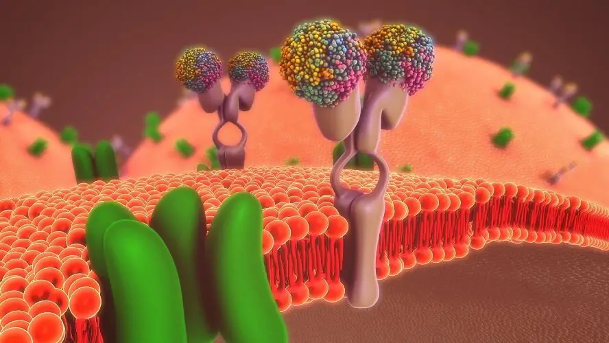
Cell Membrane Composition
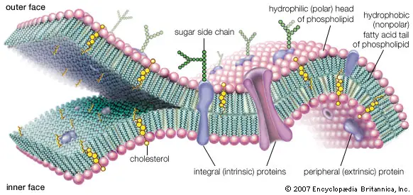
The cell membrane, often referred to as the plasma membrane, is a crucial component of the cell, providing both structural support and functional regulation. Its composition is intricate, consisting of a blend of lipids, proteins, and carbohydrates. Here, we delve into the detailed composition of the cell membrane:
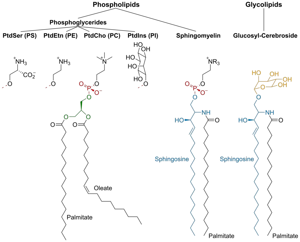
- Lipids:
- Lipids primarily form the structural framework of the cell membrane. The most prominent type of lipid in the cell membrane is the phospholipid. These molecules are arranged in a bilayer, with their hydrophilic (water-attracting) heads facing the aqueous environment both inside and outside the cell, and their hydrophobic (water-repelling) tails oriented inward, away from the water. This bilayer structure is semi-permeable, allowing selective passage of molecules.
- Cholesterol is another vital lipid component found in animal cell membranes. Strategically interspersed between phospholipid molecules, cholesterol prevents the phospholipids from packing too closely together, ensuring the membrane remains fluid and flexible.
- Proteins:
- Proteins are pivotal for the cell membrane’s functionality. They can be broadly categorized into:
- Integral Membrane Proteins: Often referred to as intrinsic proteins, these are permanently embedded within the membrane. Their hydrophobic sections penetrate the phospholipid bilayer, anchoring them firmly to the membrane. These proteins play roles in transport, acting as channels or carriers for molecules to pass through the membrane.
- Peripheral Membrane Proteins: Known as extrinsic proteins, these are temporarily associated with the membrane. Typically hydrophilic, they either attach to integral proteins or bind loosely to the phospholipid heads. They play a pivotal role in cell signaling and are frequently linked with ion channels and transmembrane receptors.
- Proteins are pivotal for the cell membrane’s functionality. They can be broadly categorized into:
- Carbohydrates:
- Though they constitute a smaller fraction of the cell membrane, carbohydrates play significant roles, particularly in cell recognition and signaling. They are typically found on the cell’s exterior surface and can be present in two primary forms:
- Glycoproteins: These are proteins with attached carbohydrate chains. Embedded in the cell membrane, glycoproteins are instrumental in cell-to-cell communication and the transport of substances across the membrane.
- Glycolipids: These are lipids with attached carbohydrate chains. Positioned on the cell membrane’s surface, they extend from the phospholipid bilayer into the extracellular environment. Besides aiding in membrane stability, glycolipids facilitate cellular recognition and communication between cells.
- Though they constitute a smaller fraction of the cell membrane, carbohydrates play significant roles, particularly in cell recognition and signaling. They are typically found on the cell’s exterior surface and can be present in two primary forms:
In conclusion, the cell membrane’s composition is a harmonious blend of lipids, proteins, and carbohydrates. Each component, with its unique structure and function, contributes to the overall integrity and functionality of the membrane, ensuring the cell’s survival and proper functioning.
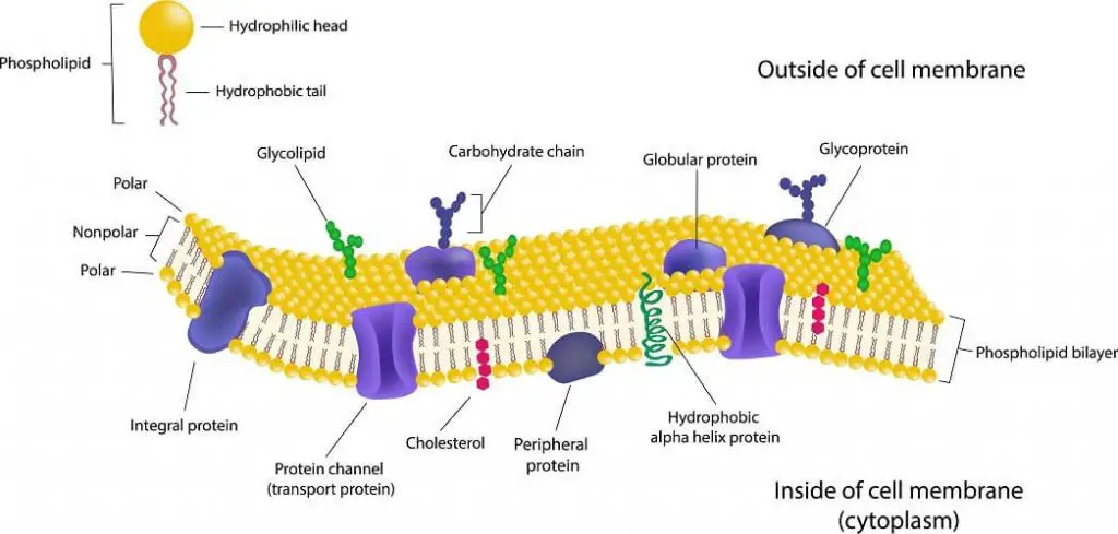
Structure of the cell membrane
The cell membrane, often referred to as the plasma membrane, is a critical component that encloses the cell’s contents and plays a pivotal role in regulating the entry and exit of substances. Over the years, scientists have proposed various models to describe its intricate structure. Two of the most notable models are the Sandwich model and the Fluid Mosaic model.
1. Sandwich Model (Lamellar Model)
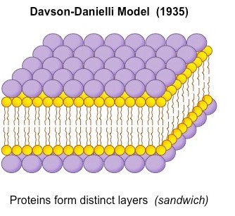
Proposed in 1935 by James Danielli and Hugh Davson, the Sandwich model presented the idea that the plasma membrane is a solid and stable structure. According to this model, the membrane comprises four molecular layers: two layers of phospholipids sandwiched between two layers of proteins, forming a P-L-L-P pattern (P: Protein, L: Lipid). Each phospholipid molecule in this model has a hydrophilic (water-attracting) head and hydrophobic (water-repelling) tails. The hydrophilic heads face the peripheral proteins, while the hydrophobic tails orient towards the center. Despite its detailed description, the Sandwich model faced several limitations. It couldn’t explain the dynamic nature of the plasma membrane, the variability in different biomembranes, or the transport mechanisms across the membrane. Due to these shortcomings, this model was eventually rejected.
Limitations of the Sandwich model
- Nature of the Plasma Membrane: The Sandwich model portrays the plasma membrane as a solid and stable structure. However, in reality, the plasma membrane is dynamic and exhibits a semi-solid or quasifluid nature. This means that the components of the membrane, such as lipids and proteins, can move laterally, allowing for flexibility and adaptability.
- Variability in Biomembranes: The model fails to account for the variability observed in different biomembranes. Biomembranes can vary in their form, composition, and thickness, depending on the cell type and its specific functions. The Sandwich model’s rigid structure cannot accommodate these variations.
- Transport Mechanisms: One of the primary functions of the cell membrane is to regulate the transport of solutes and solvents across it. The Sandwich model does not provide a mechanism to explain how substances move through the biological membrane, especially given its depiction of the membrane as a solid structure.
- Active and Bulk Transports: The model is also inadequate in explaining active and bulk transport mechanisms. Active transport refers to the movement of molecules against their concentration gradient, often requiring energy. Bulk transport involves the movement of large molecules or particles across the membrane through vesicle formation. The Sandwich model lacks the structural components and flexibility to explain these processes.
- Lipid-Protein Ratio: The composition of the cell membrane, specifically the ratio of lipids to proteins, is crucial for its function. The Sandwich model’s depiction does not align with observed lipid-protein ratios in actual cell membranes. This discrepancy further undermines the model’s accuracy.
2. Fluid Mosaic Model
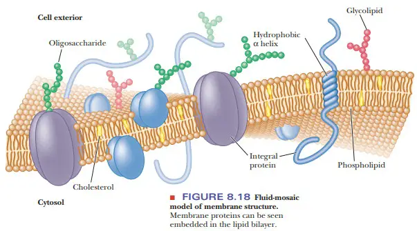
Introduced in 1972 by Singer and Nicolson, the Fluid Mosaic Model offers a more dynamic and fluid depiction of the cell membrane. This model describes the membrane as a sea of phospholipids with protein “icebergs” floating within. Similar to the Sandwich model, the phospholipids have hydrophilic heads and hydrophobic tails. However, in this model, proteins are present in both floating and suspended forms. These proteins can be categorized as:
- Extrinsic Proteins: Also known as peripheral proteins, they are present on the membrane’s surfaces in a floating form.
- Intrinsic Proteins: These are integral or tunnel proteins that can be either fully or partially suspended within the phospholipid bilayer.
The Fluid Mosaic Model also identifies five primary functions of membrane proteins:
- Structural Proteins: Provide stability to the membrane.
- Channel Proteins: Facilitate the transport of water and dissolved substances.
- Carrier Proteins: Involved in active transport mechanisms.
- Enzymes: Participate in various metabolic activities.
- Receptor Proteins: Play roles in hormone transport and nerve impulse conduction.
Additionally, on the membrane’s outer surface, lipids and extrinsic proteins can combine with oligosaccharides to form glycolipids and glycoproteins, respectively.
The Fluid Mosaic Model’s advantages are numerous. It explains the dynamic nature of the cell membrane, its semi-solid state, and the variability in different biomembranes. It also provides insights into the transport mechanisms across the membrane, both passive and active.
Advantages of the Fluid Mosaic Model
- Dynamic Nature: One of the significant advantages of the Fluid Mosaic Model is its depiction of the cell membrane as a dynamic structure. This means that the components of the membrane, such as lipids and proteins, can move laterally, allowing the membrane to be flexible and adaptable. Therefore, if the membrane incurs minor damage, it can repair itself quickly, ensuring the cell’s continued protection and functionality.
- Semi-Solid Structure: The model portrays the plasma membrane as a semi-solid or quasifluid structure. This characteristic is essential as it allows the membrane to be both stable and flexible, accommodating various cellular processes, including vesicle formation and fusion.
- Variability in Biomembranes: The Fluid Mosaic Model accounts for the variability observed in different biomembranes. It recognizes that biomembranes can differ in their form, composition, and thickness, depending on the cell type and its specific functions. This adaptability ensures that the model is applicable to a wide range of cells.
- Transport Mechanisms: A crucial function of the cell membrane is to regulate the transport of solutes and solvents across it. The Fluid Mosaic Model provides mechanisms to explain how substances move through the biological membrane, both passively and actively. It can elucidate processes like diffusion, osmosis, and active transport.
- Active and Bulk Transports: The model adeptly explains active and bulk transport mechanisms. It details how molecules move against their concentration gradient in active transport and how large molecules or particles are transported across the membrane through vesicle formation in bulk transport.
- Lipid-Protein Ratio: The composition of the cell membrane, specifically the ratio of lipids to proteins, plays a pivotal role in its function. The Fluid Mosaic Model’s depiction aligns with observed lipid-protein ratios in actual cell membranes, further bolstering its accuracy and relevance.
In conclusion, while the Sandwich model provided initial insights into the cell membrane’s structure, it is the Fluid Mosaic Model that offers a more comprehensive and widely accepted depiction of this vital cellular component. This model not only elucidates the membrane’s composition but also emphasizes the functions of its various components, enhancing our understanding of cellular interactions and processes.
Difference between the Sandwich and the Fluid Mosaic Model
The cell membrane is a critical component of cells, providing a barrier and regulating the transport of substances in and out of the cell. Over the years, various models have been proposed to describe its structure and function. Two of the most notable models are the Sandwich model and the Fluid Mosaic Model. Here, we will delve into the differences between these two models based on the provided content:
- Cell Membrane Structure:
- Sandwich Model: This model portrays the cell membrane as a rigid and stable structure. It suggests a fixed arrangement where proteins are sandwiched between two layers of lipids.
- Fluid Mosaic Model: Contrary to the Sandwich model, the Fluid Mosaic Model describes the cell membrane as dynamic and less rigid. It emphasizes the fluidity of the membrane, where proteins and lipids can move laterally, allowing for flexibility and adaptability.
- Variability of the Cell Membrane:
- Sandwich Model: This model does not account for the variability observed in different biomembranes. It presents a one-size-fits-all approach, which does not align with observed variations in actual cell membranes.
- Fluid Mosaic Model: This model recognizes that biomembranes can differ in their form, composition, and thickness, depending on the cell type and its specific functions. Therefore, it offers a more accurate representation of the diverse nature of cell membranes.
- Phospholipid Bilayer:
- Sandwich Model: According to this model, the phospholipid bilayer is solid, suggesting a more static nature.
- Fluid Mosaic Model: In contrast, this model describes the phospholipid bilayer as a semi-solid or quasifluid structure. This depiction aligns with the observed fluid nature of cell membranes, where molecules can move laterally within the lipid bilayer.
- Transport:
- Sandwich Model: This model falls short in explaining the transport mechanisms of the cell membrane. It does not provide mechanisms for the active and bulk transport of materials or the passage of electrolytes and non-electrolytes through the membrane.
- Fluid Mosaic Model: This model adeptly explains various transport mechanisms, including active and bulk transport. It details how molecules move across the membrane, both passively and actively, and provides mechanisms for the transport of both electrolytes and non-electrolytes.
In conclusion, while the Sandwich model provided initial insights into the structure of the cell membrane, it had several limitations. The Fluid Mosaic Model, on the other hand, offers a more comprehensive and accurate representation of the cell membrane’s structure and functions, making it the widely accepted model in modern cell biology.
Intracellular membranes
Intracellular membranes play a pivotal role in the organization and function of eukaryotic cells. These membranes compartmentalize the cell, ensuring that specific biochemical processes occur in designated locations. Let’s delve into the intricacies of these membranes and their associated organelles:
- Mitochondria and Chloroplasts:
- Both mitochondria and chloroplasts are believed to have originated from bacteria, a concept known as the endosymbiotic theory. This theory posits that during evolution, a eukaryotic cell engulfed certain bacteria, leading to the formation of mitochondria and chloroplasts within eukaryotic cells. The evidence supporting this theory includes the presence of their own DNA within these organelles.
- The dual-membrane system of these organelles is a result of this engulfment. The outer membrane is derived from the host’s plasma membrane, while the inner membrane originates from the engulfed bacterium’s plasma membrane.
- Nuclear Membrane:
- The nuclear membrane, a defining feature of eukaryotic cells, encloses the nucleus, segregating it from the cytoplasm. This membrane is composed of two layers: an inner and an outer membrane.
- Nuclear pores punctuate the nuclear membrane, facilitating the exchange of materials between the nucleus and the cytosol. The number of pores correlates with the transcriptional activity of the nucleus.
- The protein composition within the nuclear membrane can differ significantly from the cytosol, primarily because many proteins cannot diffuse through the pores. Notably, only the outer membrane is continuous with the endoplasmic reticulum (ER) membrane and is adorned with ribosomes, which synthesize and transport proteins into the space between the two nuclear membranes.
- During mitosis, the nuclear membrane disassembles in the early stages and reassembles in the later stages, ensuring proper chromosome segregation.
- Endoplasmic Reticulum (ER):
- The ER, a major component of the endomembrane system, constitutes a significant portion of the cell’s total membrane content. This intricate network of tubules and sacs is involved in protein synthesis and lipid metabolism.
- The ER is categorized into two types: rough ER, which is studded with ribosomes and is involved in protein synthesis, and smooth ER, which plays roles in detoxification processes and calcium regulation within the cell.
- Golgi Apparatus:
- The Golgi apparatus is characterized by its interconnected round Golgi cisternae. These compartments form multiple tubular-reticular networks that are crucial for organization, stack connection, and cargo transport. This structure also showcases a continuous string of vesicles, resembling a grape-like formation.
- The primary function of the Golgi apparatus is to modify, sort, and package proteins and lipids for transport to targeted destinations.
Transport across the Cell Membrane
The cell membrane, often referred to as the plasma membrane, serves as a barrier that separates the internal environment of the cell from the external environment. One of its primary functions is to regulate the transport of substances in and out of the cell. This transport is essential for maintaining the cell’s internal environment and ensuring its proper functioning.
- Selective Permeability:
- The cell membrane exhibits selective permeability, meaning it allows certain substances to pass through while excluding others. This selectivity is primarily due to the lipid bilayer structure of the membrane. The hydrophobic interior of the lipid bilayer allows only small, nonpolar substances, such as oxygen, carbon dioxide, and lipids, to move through it by simple diffusion.
- Polar or charged substances, such as glucose, amino acids, and ions, cannot easily cross the lipid bilayer due to their incompatibility with the hydrophobic core. Therefore, these substances require specialized transport mechanisms to enter or exit the cell.
- Passive Transport:
- Passive transport is a method by which substances move across the cell membrane without the use of cellular energy. This movement is driven by concentration gradients, where molecules move from an area of higher concentration to an area of lower concentration.
- Diffusion: At the heart of passive transport is the process of diffusion. It is the natural movement of molecules from a region of higher concentration to one of lower concentration. For instance, perfume sprayed in a room or sugar in tea will eventually spread out uniformly, illustrating diffusion.
- Osmosis: Specifically for water molecules, osmosis is the process by which water diffuses across a semipermeable membrane. The direction of water movement is determined by the concentration of solutes on either side of the membrane. Cells must maintain an isotonic environment, where the solute concentration is balanced inside and outside the cell, to prevent cell shrinking (in a hypertonic solution) or swelling (in a hypotonic solution).
- Facilitated Diffusion: For substances that cannot freely cross the lipid bilayer, facilitated diffusion comes into play. This process involves protein channels or carriers that assist in transporting specific molecules across the membrane. For example, glucose enters cells through a specialized carrier protein, utilizing facilitated diffusion.
- Active Transport:
- Unlike passive transport, active transport requires energy, typically derived from ATP, to move substances against their concentration gradients.
- Pumps: Active transport often involves protein pumps that move ions or molecules across the membrane. A classic example is the sodium-potassium pump, which maintains the electrical gradient in nerve cells by pumping sodium out and potassium into the cell.
- Endocytosis and Exocytosis: These are bulk transport mechanisms. Endocytosis allows the cell to engulf external substances or fluids, while exocytosis enables the cell to release substances outside. Within endocytosis, there are specific types like phagocytosis (ingestion of large particles) and pinocytosis (ingestion of liquids).
- Receptor-mediated Endocytosis: This is a selective form of endocytosis where specific molecules bind to receptors on the cell membrane, leading to their ingestion.
- Secondary Active Transport:
- This involves the use of energy from primary active transport to drive the transport of other substances. Symporters and antiporters are proteins involved in this process. Symporters move two substances in the same direction, while antiporters move them in opposite directions.
Origin of Plasma Membrane
- There is hardly a structure within a cell that is more vital to its immediate vitality than the plasma membrane.
- If the cell is damaged or harmed, it loses its ability to maintain gradients, transport nutrients selectively, and contain the pool of enzymes and organelles necessary for homeostasis.
- Thus, new membranes can be introduced to existing membranes without affecting their barrier and selective transporter capabilities.
- In order to preserve the characteristic asymmetry of the membrane, the membrane must also be built with the exact proper molecular topography.
- Consequently, all cellular membranes originate from pre-existing membranes that serve as templates for the insertion of new precursors.
- Daughter cells receive a full complement of membrane systems, which grow until the next division and are passed on to successive offspring.
- Meanwhile, the membrane’s molecules undergo continual replenishment. Plasma membrane protein molecules are produced on both connected and unbound ribosomes.
- Following their completion and release from the ribosomes, proteins produced by free ribosomes may be introduced into the plasma membrane.
- Plasma membrane proteins generated on attached ribosomes of rough ER are first inserted into the membrane of RER, then moved to the Golgi apparatus, processed there (e.g., glycosylation), and finally delivered to the plasma membrane via secretory vesicles.
- Similarly, phospholipid molecules of the plasma membrane are synthesised by the smooth ER (SER). Like newly generated proteins, newly synthesised lipids are inserted into SER membranes, transferred to the Golgi apparatus for processing, and finally sent to the plasma membrane via small secretory vesicles.
- In addition, the cytosol contains phospholipid transport proteins that move phospholipid molecules from one cellular membrane to another (e.g., from ER membranes to plasma membranes).
- In actuality, glycosylation (or glycosidation, i.e. the addition of oligosaccharides containing sugars such as galactose, fucose, and/or sialic acid to the molecules of plasma membrane proteins and phospholipids) is completed at the Golgi apparatus level. However, certain sugars are added to the RER lumen’s proteins.
Functions of the cell membrane
The cell membrane, often referred to as the plasma membrane, plays a pivotal role in maintaining the integrity of the cell and regulating the movement of substances in and out of the cell. This membrane is not just a passive barrier; it is an active and dynamic structure with multifaceted functions. Here, we will explore the various functions of the cell membrane based on the provided content:
- Physical Barrier:
- The cell membrane acts as a protective shield around the cell, separating the cell’s internal components from its external environment. This ensures that the intracellular components, such as organelles and cytoplasm, are kept distinct from the extracellular matrix.
- Besides providing physical integrity, the cell membrane also anchors the cytoskeleton, which gives shape to the cell. Furthermore, it assists in attaching cells to the extracellular matrix or other cells, facilitating the formation of tissues.
- Selective Permeability:
- One of the primary functions of the cell membrane is its selective permeability. This means that the membrane can regulate what enters and exits the cell.
- The selective nature of the membrane is facilitated by various transport mechanisms, including passive osmosis, diffusion, and active transport through transmembrane protein channels and transporters. This ensures that essential nutrients can enter the cell, while waste products are efficiently expelled.
- Transport Mechanisms:
- Passive Transport: Small molecules like oxygen (O2) and carbon dioxide (CO2) can move across the plasma membrane by diffusion, a process that doesn’t require energy. Similarly, osmosis allows for the movement of solvent molecules across the membrane.
- Transmembrane Protein Channels and Transporters: These proteins span the lipid bilayer and aid in the transport of specific molecules. They can be selective, allowing only certain molecules to pass through. For instance, aquaporins facilitate the movement of water molecules.
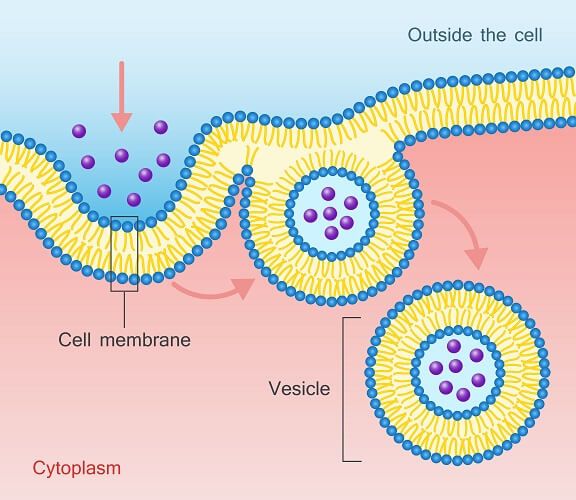
- Endocytosis: This process allows the cell to take in larger molecules by engulfing them. The cell membrane forms an invagination, capturing the substance, which then pinches off to form a vesicle inside the cell.
- Exocytosis: Conversely, exocytosis enables the cell to expel substances. Vesicles containing waste or other substances fuse with the plasma membrane, releasing their contents outside the cell.
- Signal Reception and Transmission:
- The cell membrane contains receptor sites for specific biochemicals, such as hormones and neurotransmitters. These receptors allow the cell to detect and respond to signals from its external environment, facilitating communication between cells.
- Cell Recognition and Adhesion:
- Glycolipids and glycoproteins present on the cell membrane play a role in cell recognition. This is crucial for processes like immune responses and the acceptance or rejection of transplants.
- The cell membrane also aids in the formation of structures like microvilli, which assist in digestion, and pseudopodia, which aid in movement.
- Maintaining Electrochemical Gradients:
- The cell membrane is instrumental in maintaining cell potential, which is essential for various cellular processes, including the transmission of nerve impulses.
Different cell membrane models
Different models have been proposed over the years to explain the structure and function of the cell membrane. These models have evolved based on scientific discoveries and technological advancements. Here, we delve into the various cell membrane models that have been postulated:
- Overton Model (1910):
- This was the pioneering model to elucidate the structure of the plasma membrane.
- Overton posited that the plasma membrane is enveloped with lipid-soluble material.
- He further proposed that non-polar solutes can readily traverse the membrane, whereas polar solutes are unable to do so.
- Irving Langmuir Model (1925):
- Langmuir was the first to suggest that the cell membrane is constituted of monolayers.
- These monolayers are formed by a single layer of amphipathic phospholipid molecules. Here, the hydrophobic tails are oriented away from water, while the hydrophilic heads face the water.
- Gorter and Grendel Model (1924):
- This model introduced the idea that the cell membrane components comprise various types of lipids arranged in a double layer.
- Davson and Danielli Model (1935):
- Also known as the sandwich model, this was the first to incorporate proteins into the plasma membrane structure.
- The model depicted the membrane as a protein-lipid-protein sandwich, emphasizing the trilaminar nature of the structure.
- Robertson Model (1950s):
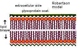
- Termed the “Unit Membrane Model,” Robertson’s model emphasized the uniformity of the membrane structure.
- The model proposed that there’s no space between the phospholipid bilayers and that the membrane’s thickness is quantifiable.
- After conducting experiments on various cell types, Robertson concluded that the fundamental structure of the cell membrane remains consistent across different cells.
- Singer and Nicholson Model (1972):
- Currently the most widely accepted model, the Fluid Mosaic Model offers a comprehensive understanding of the membrane structure.
- The model defines the plasma membrane as a mosaic of proteins embedded in a fluid-like lipid bilayer. This suggests that proteins are not uniformly distributed but are interspersed in a discontinuous manner within the lipid matrix.
- The analogy used is that of proteins being akin to icebergs floating in a sea, representing the fluid lipid component of the membrane.
In conclusion, the understanding of the cell membrane’s structure and function has evolved over time, with each model building upon the discoveries of its predecessors. The Fluid Mosaic Model, being the most recent and comprehensive, provides a detailed and dynamic representation of the cell membrane, accounting for both its lipid and protein components.
Quiz
FAQ
What is the cell membrane?
The cell membrane, also known as the plasma membrane, is a thin, flexible layer that surrounds the cytoplasm of all cells. It is composed of a double layer of phospholipid molecules with embedded proteins and other lipids.
What is the function of the cell membrane?
The cell membrane has several important functions, including regulating the transport of substances in and out of the cell, providing structural support, and facilitating communication between cells.
What is the structure of the cell membrane?
The cell membrane is composed of a lipid bilayer, with hydrophobic tails facing each other and hydrophilic heads facing outward. Embedded in the lipid bilayer are proteins, which may serve as channels, pumps, or receptors.
What are the types of proteins in the cell membrane?
There are two main types of proteins in the cell membrane: integral proteins, which are embedded within the lipid bilayer, and peripheral proteins, which are attached to the surface of the membrane.
How does the cell membrane regulate transport?
The cell membrane uses various mechanisms, such as diffusion, facilitated diffusion, and active transport, to regulate the movement of substances in and out of the cell.
What is the role of cholesterol in the cell membrane?
Cholesterol is a lipid molecule that is embedded within the cell membrane and helps to maintain its fluidity and stability.
Can the cell membrane repair itself?
Yes, the cell membrane has the ability to repair itself through a process called membrane remodeling, which involves the rearrangement of lipids and the removal of damaged proteins.
What is the glycocalyx?
The glycocalyx is a layer of carbohydrates that coats the outer surface of the cell membrane and helps to protect the cell from damage and infection.
Can the cell membrane communicate with other cells?
Yes, the cell membrane contains receptors and other signaling molecules that allow it to communicate with other cells and regulate various physiological processes.
How does the cell membrane contribute to cell identity?
The composition and organization of the cell membrane can vary between different cell types and can contribute to their unique identity and function.
