Table of Contents
What is a cell?
Your body is currently doing a lot of things simultaneously. Your body is sending electrical impulses, pumping blood and filtering urine. It also makes protein, digests food, and stores fat. And that’s not all! All of this is possible because cells are tiny units of life, which are full of machinery and machinery that help you do your job. Every living thing is made up of cells, including blue whales and the archaebacteria found in volcanoes. Cells can take on many shapes and sizes, just like the organisms that they make up. Giant squids’ nerve cells can grow to as long as 12m (39 feet) in length. Human eggs, which are the largest cells in the body, measure only 0.1mm in width. Plant cells have protective walls made out of cellulose, which also makes celery stringy and difficult to eat. Fungal cell walls are made out of the same material as lobster shells. All these factories share the same basic machinery, despite their vast differences in size, function, and shape.
There are two types of cells: prokaryotic or eukaryotic. Eukaryotes have membrane-bound nuclei. Prokaryotes don’t. We will be focusing on eukaryotes for the rest of this discussion. Consider what a factory requires to be able to function properly. A factory must have a building, a product and a method to make it. Every cell has membranes (the building), DNA and ribosomes. They can also make proteins (the product, let’s say toys). Since eukaryotes are the type of cell that has organelles, this article will be focused on them.
What’s found inside a cell
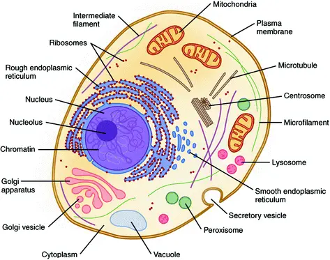
An organelle, also known as an internal organ of a cell, is a membrane-bound structure within a cell. These mini-organs, just like the cells that have membranes to keep everything in, are also bound in double layers of phospholipids to protect their small compartments in larger cells.
Organelles can be thought of as small rooms in a factory with special conditions that allow them to perform their task. This could include a break area with snacks or a research space with cool gadgets and an air filter. The cytoplasm is the liquid that carries the organelles within cells. It is a viscous liquid inside the cell membrane. This table shows the organelles that are found in the basic human cells. It will be our guide for the discussion.
| Organelle | Function | Factory part |
|---|---|---|
| Nucleus | DNA Storage | Room where the blueprints are kept |
| Mitochondrion | Energy production | Powerplant |
| Smooth Endoplasmic Reticulum (SER) | Lipid production; Detoxification | Accessory production – makes decorations for the toy, etc. |
| Rough Endoplasmic Reticulum (RER) | Protein production; in particular for export out of the cell | Primary production line – makes the toys |
| Golgi apparatus | Protein modification and export | Shipping department |
| Peroxisome | Lipid Destruction; contains oxidative enzymes | Security and waste removal |
| Lysosome | Protein destruction | Recycling and security |
What are Cell Organelles?
Cell organelles are the cellular components. These cell organelles can be found in both membrane-bound organelles and non-membrane organelles. They are different in their functions and structures. They are essential for cell function and coordination. Some of them provide shape and support while others are involved in the movement and reproduction of cells.
Organelle is derived from the notion that organelles are part of cells. The suffix -elle refers to a diminutive. Organelles can be either enclosed in their own lipid bilayers (also known as membrane-bound organelles), or they are functionally distinct units that are not bound by a surrounding bilayer (nonmembrane bound organelles). While most organelles function within cells, there are some functional units outside cells that are called organelles. These include the flagellum, archaellum and cilia.
Microscopy can identify organelles and can be purified using cell fractionation. Organelles come in many forms, especially in eukaryotic cells. These structures include the internal endomembrane systems (such as nuclear envelope, endoplasmic retina, and Golgi apparatus) and other structures like mitochondria and plastids.
Although prokaryotes don’t have eukaryotic organelles but some prokaryotes contain protein-shelled bacteria microcompartments that are thought to act like primitive prokaryotic organismelles. There is also evidence for other membrane-bounded structures. Organelles are also often referred to as the prokaryotic flagellum, which protrudes from the cell, and its motor.
Examples of Cell Organelles
Major eukaryotic organelles
| Organelle | Main function | Organisms |
|---|---|---|
| cell membrane | separates the interior of all cells from the outside environment (the extracellular space) which protects the cell from its environment. | all eukaryotes |
| cell wall | The cell wall is a rigid structure composed of cellulose that provides shape to the cell, helps keep the organelles inside the cell, and does not let the cell burst from osmotic pressure. | plants, protists, rare kleptoplastic organisms |
| chloroplast (plastid) | photosynthesis, traps energy from sunlight | plants, protists, rare kleptoplastic organisms |
| endoplasmic reticulum | translation and folding of new proteins (rough endoplasmic reticulum), expression of lipids (smooth endoplasmic reticulum) | all eukaryotes |
| flagellum | locomotion, sensory | some eukaryotes |
| Golgi apparatus | sorting, packaging, processing and modification of proteins | all eukaryotes |
| mitochondrion | energy production from the oxidation of glucose substances and the release of adenosine triphosphate | most eukaryotes |
| nucleus | DNA maintenance, controls all activities of the cell, RNA transcription | all eukaryotes |
| vacuole | storage, transportation, helps maintain homeostasis | eukaryotes |
Minor eukaryotic organelles and cell components
| Organelle/Macromolecule | Main function | Organisms |
|---|---|---|
| acrosome | helps spermatozoa fuse with ovum | most animals |
| autophagosome | vesicle that sequesters cytoplasmic material and organelles for degradation | all eukaryotes |
| centriole | anchor for cytoskeleton, organizes cell division by forming spindle fibers | animals |
| cilium | movement in or of external medium; “critical developmental signaling pathway”. | animals, protists, few plants |
| cnidocyst | stinging | cnidarians |
| eyespot apparatus | detects light, allowing phototaxis to take place | green algae and other unicellular photosynthetic organisms such as euglenids |
| glycosome | carries out glycolysis | Some protozoa, such as Trypanosomes. |
| glyoxysome | conversion of fat into sugars | plants |
| hydrogenosome | energy & hydrogen production | a few unicellular eukaryotes |
| lysosome | breakdown of large molecules (e.g., proteins + polysaccharides) | animals |
| melanosome | pigment storage | animals |
| mitosome | probably plays a role in Iron-sulfur cluster (Fe-S) assembly | a few unicellular eukaryotes that lack mitochondria |
| myofibril | myocyte contraction | animals |
| nucleolus | pre-ribosome production | most eukaryotes |
| ocelloid | detects light and possibly shapes, allowing phototaxis to take place | members of the family Warnowiaceae |
| parenthesome | not characterized | fungi |
| peroxisome | breakdown of metabolic hydrogen peroxide | all eukaryotes |
| porosome | secretory portal | all eukaryotes |
| proteasome | degradation of unneeded or damaged proteins by proteolysis | all eukaryotes, all archaea, and some bacteria |
| ribosome (80S) | translation of RNA into proteins | all eukaryotes |
| stress granule | mRNA storage | most eukaryotes |
| TIGER domain | mRNA encoding proteins | most organisms |
| vesicle | material transport | all eukaryotes |
Prokaryotic organelles and cell components
| Organelle/macromolecule | Main function | Organisms |
|---|---|---|
| anammoxosome | anaerobic ammonium oxidation | “Candidatus” bacteria within Planctomycetes |
| carboxysome | carbon fixation | some bacteria |
| chlorosome | photosynthesis | green sulfur bacteria |
| flagellum | movement in external medium | some prokaryotes |
| magnetosome | magnetic orientation | magnetotactic bacteria |
| nucleoid | DNA maintenance, transcription to RNA | prokaryotes |
| pilus | Adhesion to other cells for conjugation or to a solid substrate to create motile forces. | prokaryotic cells |
| plasmid | DNA exchange | some bacteria |
| ribosome (70S) | translation of RNA into proteins | bacteria and archaea |
| thylakoid membranes | photosynthesis | mostly cyanobacteria |
Types of Cell Organelles
Classification of Cell Organelles based on presence or absence of membrane
There are many organelles within the cell. They are divided into three groups based on whether or not there is membrane.
- Organelles without membrane: Cell wall, Ribosomes and Cytoskeleton all are organelles that are not bound by membranes. They can be found in both prokaryotic cells and eukaryotic cells.
- Single membrane-bound organelles: Single membrane-bound organelles are Vacuole and Lysosome, Golgi Apparatus and Endoplasmic Reticulum. These organelles can only be found in a eukaryotic cells.
- Double membrane-bound organelles: Nucleus, mitochondria and chloroplast are double membrane-bound organelles present only in a eukaryotic cell.
Classification of Cell Organelles based on location
Cell organelles are classified into these following three groups;
- General cell organelles: General cell organelles are found in all cells, including animal and plant. They include cell membrane, cytosol and cytoplasm, nucleus and mitochondrion.
- Temporal cell organelles: Temporal cell organelles are found only at certain stages of a cell’s life cycle: chromosomes, autophagosomes, chromosomes, and endosomes.
- Cell type specific cell organelles: Cell type-specific cell organelles are only found in plant cells: chloroplast, central vacuumole and cell wall.
Many unique cell organelles/structures only exist in specific cell types. The food vacuoles of amoeba or the paramecia trichocysts are examples of unique cell organelles/structures that can only be found in specific cell types. However, there are some organelles in human cells that cannot be found elsewhere, such as the Weibel-Palade body in blood vessel cells.
The cell membrane (Plasma membrane/ Plasmalemma)
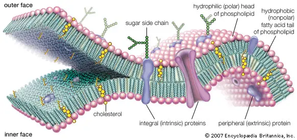
The plasma membrane is made up of proteins and lipids. It can fluctuate depending on fluidity and external environment.
Structure of Cell membrane
The structure of the cell membrane is composed of various components that work together to fulfill its functions. Here is an explanation of the structure of the cell membrane:
- Phospholipid Bilayer: The main structural component of the cell membrane is a phospholipid bilayer. Phospholipids are molecules that have a polar, hydrophilic (water-loving) head group and two hydrophobic (water-repelling) fatty acid tails. The phospholipids arrange themselves in a double layer, with the hydrophilic heads facing outward towards the watery environments both inside and outside the cell, and the hydrophobic tails pointing inward, forming a hydrophobic core.
- Embedded Proteins: Embedded proteins are proteins that are embedded within the phospholipid bilayer. These proteins span the entire width of the membrane and act as channels or transporters, facilitating the movement of particles, such as ions and molecules, across the cell membrane. Some embedded proteins also function as receptors, allowing the cell to recognize and bind to specific molecules or signals in the environment.
- Peripheral Proteins: Peripheral proteins are proteins that are loosely attached to the surface of the cell membrane, either on the inner or outer side. These proteins do not penetrate the phospholipid bilayer but associate with the membrane through weak non-covalent interactions. Peripheral proteins play various roles, including providing mechanical support to the cell membrane and contributing to its fluidity. They can also participate in cell signaling and act as enzymes or structural elements.
The combination of the phospholipid bilayer and the embedded and peripheral proteins forms the basic structure of the cell membrane. This arrangement allows the membrane to provide a protective barrier, regulate the passage of molecules, facilitate cell signaling, and maintain the integrity and shape of the cell. The phospholipid bilayer provides a hydrophobic barrier, while the embedded proteins and peripheral proteins contribute to the membrane’s functionality and dynamic nature.
Functions of Cell membrane
- Mechanical Support: The cell membrane provides structural integrity to the cell, maintaining its shape and preventing it from collapsing.
- Cell Enclosure: It forms a boundary that separates the cell from its surroundings, encapsulating all the cellular contents within.
- Semi-Permeability: The cell membrane acts as a selectively permeable barrier, regulating the movement of substances into and out of the cell. It allows the passage of essential compounds while preventing the entry of harmful substances.
- Exchange of Essential Compounds: Channels and transport proteins present in the cell membrane facilitate the exchange of essential molecules, such as nutrients and ions, required for the survival and functioning of the cell.
- Signal Generation and Distribution: The cell membrane is involved in the generation and distribution of signals both within and outside the cell. It contains receptors and proteins that can detect external signals and transmit them to the cell’s interior, triggering specific cellular responses.
- Inter-Cellular Communication: The cell membrane enables interactions between neighboring cells, which is essential for processes like tissue formation and cell fusion. Cell adhesion molecules on the membrane surface facilitate cell-cell interactions and play a role in various physiological processes.
These functions collectively highlight the importance of the cell membrane in maintaining cell integrity, regulating the transport of substances, transmitting signals, and facilitating cellular interactions.
Cell Wall
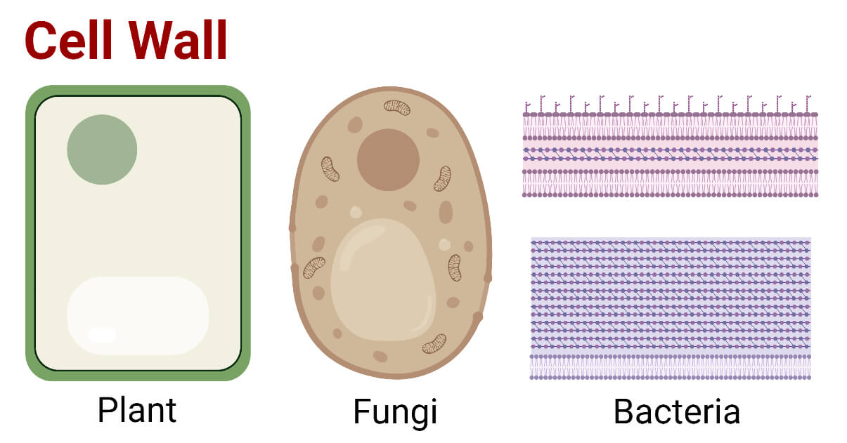
The cell wall is an additional layer that exists outside of the cell membrane. It provides structure, protection, and filtering mechanisms to the cells.
Structure of Cell Wall
The structure of the cell wall varies between different organisms, but it serves as an important component providing support and protection. Here is an explanation of the structure of the cell wall:
- Composition in Plant Cells: In plant cells, the cell wall is primarily composed of cellulose, a complex carbohydrate, along with other polysaccharides such as hemicellulose and pectin. Proteins are also present in the cell wall.
- Composition in Fungal Cells: In fungal cells, the cell wall is predominantly composed of a tough polysaccharide called chitin. Chitin is a nitrogen-containing polysaccharide that provides strength and rigidity to the fungal cell wall.
- Multilayered Structure: The cell wall has a multilayered structure, consisting of three main layers in plant cells:
- Middle Lamina: The middle lamina is the outermost layer of the cell wall. It contains various polysaccharides, including pectin, which provides adhesion between neighboring cells and allows binding of cells to one another.
- Primary Cell Wall: The primary cell wall lies beneath the middle lamina. It is primarily composed of cellulose microfibrils embedded in a matrix of hemicellulose and other polysaccharides. The primary cell wall provides strength, flexibility, and elasticity to the plant cell.
- Secondary Cell Wall: In some plant cells, a secondary cell wall may form beneath the primary cell wall. The secondary cell wall is a thicker layer and provides additional support and protection. It is composed of cellulose, hemicellulose, and lignin, a complex polymer that further strengthens the cell wall.
It’s important to note that not all plant cells have a secondary cell wall, and its presence varies depending on the specific cell type and function.
Overall, the cell wall serves as a protective layer that maintains the shape, integrity, and rigidity of plant and fungal cells. It provides mechanical strength, protection against physical stresses, and allows for the exchange of nutrients and water between cells. The specific composition and structure of the cell wall contribute to the unique properties and functions of different organisms.
Functions of Cell Wall
The cell wall plays essential roles in maintaining cell integrity, providing support, and regulating molecular exchange. Here are the functions of the cell wall based on the provided information:
- Protection and Shape Maintenance: One of the primary functions of the cell wall is to protect the cell and maintain its shape. The cell wall provides a rigid structure that acts as a physical barrier, shielding the cell from external stresses and preventing it from collapsing under its own internal pressure. It helps to withstand the turgor pressure exerted by the cell’s contents, maintaining the shape of the cell.
- Signal Initiation for Cell Division: The cell wall is involved in initiating cell division by providing signals to the cell. During the cell cycle, the cell wall sends specific signals that trigger the division process, ensuring proper growth and development of tissues and organisms.
- Regulation of Molecular Passage: The cell wall acts as a selective barrier, allowing the passage of certain molecules while blocking others. It controls the entry and exit of substances into and out of the cell, helping to maintain the internal environment necessary for cellular processes. The composition and structure of the cell wall determine its permeability, which influences the exchange of nutrients, water, and other essential molecules required for the cell’s survival and functioning.
By performing these functions, the cell wall contributes to the overall stability, growth, and survival of the cell. It provides protection, maintains cell shape, facilitates cellular communication, and regulates molecular exchange, ensuring the proper functioning of the cell and supporting the development of tissues and organisms.
Centriole
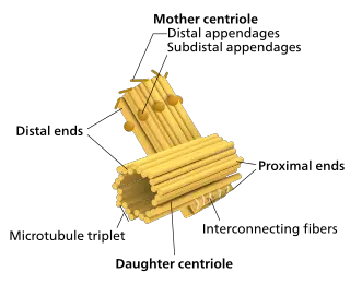
Centrioles, which are tubular structures found mostly in eukaryotic cells, are mainly composed of the protein tubulin.
Structure of Centriole
The structure of a centriole is composed of specific components that contribute to its function in cell division and organization of the cytoskeleton. Here is an explanation of the structure of the centriole based on the provided information:
- Cylindrical Structure: A centriole has a cylindrical shape and is made up of nine triplets of microtubules arranged in a circular pattern around the periphery. Each triplet consists of three microtubules that are connected to each other, forming a tubular structure.
- Y-shaped Linker and Barrel-like Structure: At the center of the centriole, there is a Y-shaped linker. This linker connects the triplets of microtubules, providing stability and structural integrity to the centriole. Additionally, there is a barrel-like structure that surrounds the linker, further contributing to the stability of the centriole.
- Cartwheel Structure: Within the centriole, there is another structure called the cartwheel. The cartwheel is made up of a central hub with nine spokes or filaments radiating from it. The filaments extend outward and connect to the triplets of microtubules through pinheads, forming a connection between the cartwheel and the microtubules.
The arrangement of the triplets of microtubules, the Y-shaped linker, the barrel-like structure, and the cartwheel collectively form the structure of the centriole. This structure provides the centriole with stability and plays a crucial role in its function as a microtubule-organizing center during cell division. Centrioles are involved in the formation of the mitotic spindle, which helps separate chromosomes during cell division, and they also contribute to the organization of the cytoskeleton, influencing cell shape and movement.
Functions of Centriole
Centrioles play important roles in various cellular processes, primarily in cell division and the formation of specialized cellular structures. Here are the functions of centrioles based on the provided information:
- Formation of Spindle Fibers: One of the critical functions of centrioles is their involvement in cell division. During cell division (both mitosis and meiosis), centrioles play a crucial role in the formation of the spindle apparatus. The centrioles migrate to opposite poles of the cell, and from there, they organize and anchor microtubules to form spindle fibers. These spindle fibers help in the movement and alignment of chromatids (sister chromatids or homologous chromosomes) during chromosome segregation, ensuring accurate distribution of genetic material to daughter cells.
- Formation of Cilia and Flagella: Centrioles are also involved in the formation of cilia and flagella, which are specialized cellular structures involved in cell motility and sensory functions. In cells that possess cilia or flagella, centrioles give rise to basal bodies. Basal bodies serve as the template for the assembly of microtubules that form the axoneme, the structural core of cilia and flagella. The coordinated movement of microtubules in the axoneme enables the waving or rotation of cilia and flagella, allowing cells to move or generate fluid flow.
These functions highlight the crucial roles of centrioles in cell division and the formation of specialized structures involved in cellular movement and sensory functions. Centrioles ensure the accurate distribution of genetic material during cell division and contribute to cell motility and sensory perception through the formation of spindle fibers, cilia, and flagella.
Cilia and Flagella
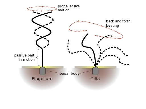
Flagella and Cilia are microtubule-based projections of the cell that look like hairs. They are covered by the plasma membrane.
Structure of Cilia and Flagella
The structure of cilia and flagella varies between different organisms and cell types. Here is an explanation of the structure of cilia and flagella based on the provided information:
- Cilia Structure: Cilia are hair-like projections found on the surface of certain cells. They consist of a complex structure composed of microtubules. The primary structure of cilia includes:
- Microtubule Arrangement: Cilia have a 9+2 arrangement of microtubules. This means that there are nine pairs of microtubule doublets forming a ring around the periphery, surrounding two singlet microtubules in the center. The nine doublets are connected by radial spokes, giving cilia a characteristic appearance.
- Basal Body: Cilia are anchored to the cell by a basal body. The basal body is derived from a centriole and serves as a template for microtubule assembly in cilia formation. It provides a stable base for the attachment of cilia to the cell membrane.
- Flagella Structure: Flagella are elongated whip-like structures used for cell movement. The structure of flagella differs between prokaryotes and eukaryotes:
- Prokaryotic Flagella: In prokaryotes, flagella are composed of a protein called flagellin. Flagellin molecules self-assemble in a helical manner to form the flagellar filament. This results in a hollow, filamentous structure that extends throughout the length of the flagellum. Prokaryotic flagella rotate, propelling the cell through its environment.
- Eukaryotic Flagella: In eukaryotes, the structure of flagella is different. The protein flagellin is absent, and instead, eukaryotic flagella are made up of microtubules. The microtubules are organized in a 9+2 arrangement similar to cilia. The central pair of microtubules is surrounded by nine outer doublets, and these microtubules provide the structural integrity and movement of eukaryotic flagella.
The structure of cilia and flagella reflects their function in cellular movement. Cilia and flagella play crucial roles in the movement of cells, fluid flow, and sensory perception in various organisms. The arrangement of microtubules or flagellin molecules contributes to their unique structures and functions in different cell types and organisms.
Functions
Cilia and flagella serve important functions in various organisms, primarily related to movement and sensory perception. Here are the functions of cilia and flagella based on the provided information:
- Movement: The primary function of both cilia and flagella is to facilitate movement. They enable cells or organisms to move through their environment. Cilia and flagella can generate coordinated beating or waving motions, propelling cells or organisms through liquid or air. For example, in single-celled organisms like Paramecium, the coordinated beating of cilia allows them to move and navigate in their aquatic habitats. In multicellular organisms, cilia and flagella play roles in the movement of sperm cells, the clearance of mucus in the respiratory tract, and the movement of fluids in various tissues.
- Sensory Functions: In some specialized cases, cilia have sensory functions. Certain cilia found in specific organs can act as sensory antennae, detecting and responding to external signals. For instance, cilia present in the olfactory epithelium of the nose are involved in sensing and detecting odor molecules, playing a role in our sense of smell. Additionally, some cilia found in the lining of the respiratory tract have sensory functions and help in detecting and responding to environmental stimuli, such as irritants or changes in airflow.
- Flow Control: In certain organs, cilia have a specific function in regulating fluid flow. For example, cilia in the lining of the respiratory tract and the oviducts (fallopian tubes) beat in a coordinated manner, creating a directional flow of mucus or eggs, respectively. Similarly, in the blood vessels, cilia can be found that help control blood flow by sensing mechanical forces and transmitting signals to regulate the diameter of blood vessels.
These functions highlight the importance of cilia and flagella in various biological processes. They enable movement at the cellular and organismal level, contribute to sensory perception, and have specialized roles in fluid flow regulation. The specific functions of cilia and flagella vary depending on their location, organization, and the organisms they are found in.
Chloroplast
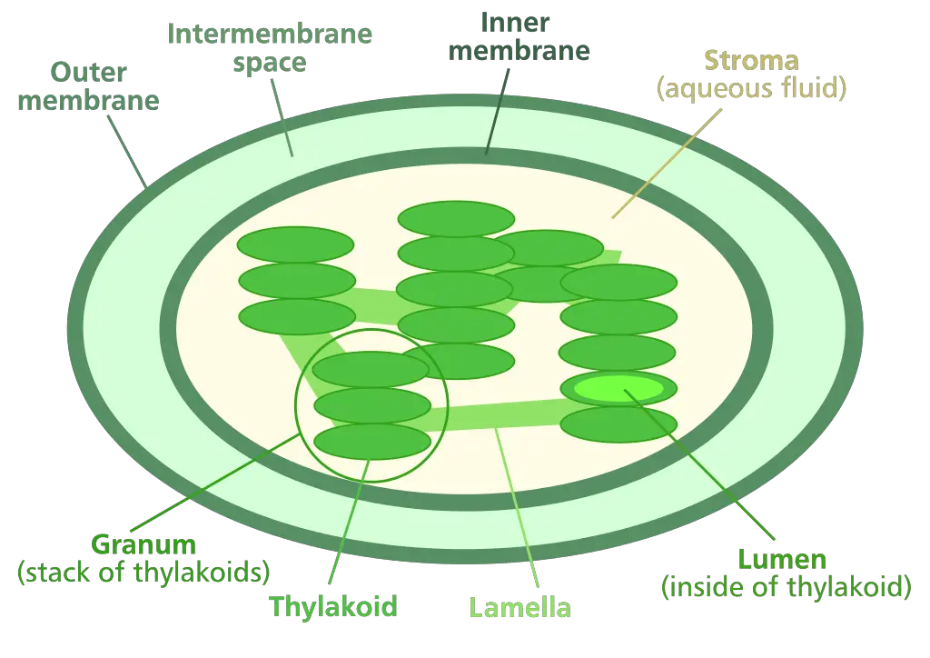
A chloroplast, a type of plastic, is involved in photosynthesis in plants or algae. Chloroplast has a vital pigment called chlorophyll that traps sunlight to produce glucose.
Structure of Chloroplast
The structure of a chloroplast is complex and highly specialized for its function in photosynthesis. Here is an explanation of the structure of the chloroplast based on the provided information:
- Double-Membraned Structure: Chloroplasts are bounded by a double membrane. The outer membrane serves as a protective barrier, while the inner membrane controls the passage of molecules in and out of the chloroplast. This double-membraned structure provides compartmentalization and maintains the integrity of the chloroplast.
- Thylakoid Membrane System: Within the chloroplast, there is a system of internal membranes called the thylakoid membrane. The thylakoid membrane is arranged in stacks called grana (singular: granum) and forms flattened, disk-like structures known as thylakoids. The thylakoid membrane contains various pigments, including chlorophyll, which are crucial for capturing light energy during photosynthesis.
- Stroma: The space between the inner membrane and the thylakoid membrane is called the stroma. The stroma is a gel-like matrix that fills the chloroplast. It contains a mixture of enzymes, DNA, chloroplast ribosomes, proteins, and starch granules. The stroma plays a vital role in the synthesis of organic molecules through the Calvin cycle, which is the second stage of photosynthesis.
The outer and inner membranes of the chloroplast are porous, allowing the transport of materials such as small molecules and ions. The stroma, on the other hand, houses the DNA and other components necessary for the chloroplast’s metabolic activities. This includes the production of proteins through the translation of chloroplast DNA into proteins using chloroplast ribosomes, as well as the storage of starch granules for energy reserves.
The complex structure of the chloroplast, including its double-membraned structure, thylakoid membrane system, and stroma, enables it to carry out the intricate process of photosynthesis. The chloroplast captures light energy, converts it into chemical energy, and utilizes that energy to produce organic molecules, such as glucose, which are essential for the plant’s growth and metabolism.
Functions of Chloroplast
The chloroplast plays crucial roles in the process of photosynthesis, encompassing both light-dependent and light-independent reactions. Here are the functions of the chloroplast based on the provided information:
- Light-Dependent Reactions: The chloroplast is the primary site where light-dependent reactions of photosynthesis occur. Within the thylakoid membrane of the chloroplast, chlorophyll and other pigments absorb light energy. This energy is then used to power the conversion of light energy into chemical energy in the form of ATP (adenosine triphosphate) and NADPH (nicotinamide adenine dinucleotide phosphate).
- Light-Independent Reactions (Calvin Cycle): The chloroplast is also responsible for carrying out the light-independent reactions, commonly known as the Calvin cycle or C3 cycle. These reactions occur in the stroma of the chloroplast. In the Calvin cycle, carbon dioxide is converted into glucose and other organic molecules, utilizing the ATP and NADPH generated during the light-dependent reactions. This process plays a critical role in the synthesis of carbohydrates and other organic compounds necessary for plant growth and metabolism.
- Regulation of Photorespiration: Photorespiration is a process that occurs in plants under certain environmental conditions, where oxygen competes with carbon dioxide in the active site of the enzyme involved in the Calvin cycle. Chloroplasts have various proteins, including specific proteins associated with chlorophyll, that are involved in the regulation and control of photorespiration. These proteins help minimize the detrimental effects of photorespiration and optimize the efficiency of carbon fixation in the Calvin cycle.
The functions of the chloroplast in light-dependent and light-independent reactions allow for the conversion of light energy into chemical energy and the subsequent synthesis of organic compounds. The chloroplast’s ability to capture and utilize light energy is essential for the growth, development, and metabolic processes of plants. Additionally, chloroplast proteins associated with chlorophyll contribute to the regulation of photorespiration, ensuring the efficient utilization of resources during photosynthesis.
Cytoplasm
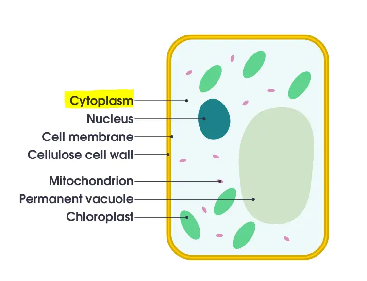
The cytoplasm is everything inside a cell, except for the nucleus. The cytoplasm is found in animal and plant cells. They are jelly-like compounds, located between the cell membrane and the nucleus. They’re mostly made up of organic and inorganic substances. The cytoplasm is among the fundamental cells in which all organelles within the cell reside. Cell organelles are home to enzymes that are responsible for controlling the metabolic activity that takes in the cell. They are the location for the majority reaction reactions that occur within cells.
Structure of Cytoplasm
The structure of the cytoplasm, the fluid-filled region of the cell, can be described based on the provided information:
- Cytosol: The cytoplasm consists of a gel-like substance called cytosol. Cytosol is a semi-transparent, colorless fluid that makes up the majority of the cytoplasm. It is primarily composed of water, comprising approximately 80% of its volume. In addition to water, the cytosol contains various dissolved nutrients, ions, enzymes, and other molecules necessary for cellular metabolism.
- Cell Organelles: Within the cytoplasm, there are various membrane-bound organelles. These organelles include the mitochondria, endoplasmic reticulum, Golgi apparatus, lysosomes, peroxisomes, and more. Each organelle has a specific structure and function that contributes to the overall cellular processes.
- Cytoplasmic Inclusions: The cytoplasm may also contain cytoplasmic inclusions, which are insoluble molecules or particles that are not enclosed by any membrane. These inclusions can vary in composition and function. Examples of cytoplasmic inclusions include lipid droplets, glycogen granules, pigment granules, and crystal-like structures. These inclusions often store energy reserves or play specific roles within the cell.
The cytoplasm exhibits properties of both viscous and elastic matter. It can flow and change shape to accommodate various cellular processes. One notable phenomenon facilitated by the cytoplasm’s elasticity is cytoplasmic streaming, also known as cyclosis. Cytoplasmic streaming is the movement of cytoplasm within the cell, enabling the transport of organelles, nutrients, and other substances. This dynamic movement is essential for maintaining cellular homeostasis and supporting cellular functions.
Overall, the cytoplasm’s structure, comprising cytosol, organelles, and cytoplasmic inclusions, provides a supportive environment for cellular activities, housing various components necessary for cellular metabolism, and allowing for the movement of materials within the cell.
Functions of Cytoplasm
The cytoplasm, with its various components and dynamic properties, performs several essential functions within the cell. Here are the functions of the cytoplasm based on the provided information:
- Cellular Reactions: The cytoplasm serves as the site for numerous vital cellular reactions. It is involved in key metabolic processes such as cellular respiration, where energy is generated by breaking down molecules like glucose to produce ATP (adenosine triphosphate). Additionally, the cytoplasm is where the translation of mRNA into proteins occurs, a process essential for the synthesis of cellular components and the functioning of enzymes.
- Protective Role: The cytoplasm acts as a protective buffer within the cell. It serves as a barrier, shielding the genetic material (such as DNA and RNA) and other organelles from potential damage caused by collisions or fluctuations in the pH of the cytosol. This protection ensures the integrity and stability of cellular structures and genetic information.
- Cytoplasmic Streaming: Cytoplasmic streaming, also known as cyclosis, is a process facilitated by the cytoplasm. It involves the movement of the cytoplasm within the cell, resulting in the distribution of various nutrients and the movement of cell organelles. This streaming aids in the efficient transport of essential substances and molecules throughout the cell, ensuring proper cellular functioning and the distribution of resources.
Overall, the cytoplasm plays a vital role in supporting cellular activities. It provides a suitable environment for important metabolic reactions, protects cellular components from potential damage, and facilitates the distribution of nutrients and the movement of organelles. The functions of the cytoplasm are essential for cellular metabolism, maintenance of cellular homeostasis, and the overall functioning and survival of the cell.
Cytoskeleton
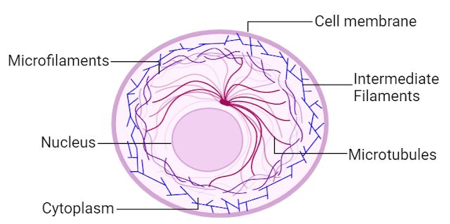
The cytosol contains a number of fibrous structures that help give shape to cells and support cellular transport. It is a continuous system of filamentous proteinsaceous structures that extend throughout the cytoplasm starting from the nucleus and ending at the plasma membrane. It is present in all living cells, and is particularly present in eukaryotes.
The cytoskeleton matrix comprises of a variety of proteins that are able to divide quickly or break down depending on the needs of cells. The main function of the cytoskeleton matrix is providing the form and resistance of cells against deformation. The contractile nature of the filaments aids in cytokinesis as well as in motility.
Structure of Cytoskeleton
The cytoskeleton is a dynamic and complex network of protein fibers that provides structural support and regulates various cellular processes. Its structure can be categorized into three main classes of fibers: microtubules, microfilaments, and intermediate filaments. Here’s a breakdown of the structure of the cytoskeleton based on the provided information:
- Microtubules: Microtubules are hollow, tubular structures made up of the protein called tubulin. They are the largest components of the cytoskeleton, with a diameter of about 25 nanometers. Microtubules are composed of alpha-tubulin and beta-tubulin subunits that arrange themselves in a cylindrical shape. These tubules provide structural support, serve as tracks for intracellular transport, and play a role in cell division, including the formation of the mitotic spindle.
- Microfilaments: Microfilaments, also known as actin filaments, are thin, solid fibers composed of the protein actin. They have a diameter of about 7 nanometers. Microfilaments form a dense network throughout the cytoplasm and are involved in various cellular functions, including cell movement, cell division, and maintenance of cell shape. They also play a crucial role in muscle contraction.
- Intermediate Filaments: Intermediate filaments are the most diverse and stable components of the cytoskeleton. They are formed by a variety of proteins, including keratins, vimentin, and neurofilaments, depending on the cell type. Intermediate filaments have a diameter of about 10 nanometers and provide mechanical strength and structural stability to cells. They are particularly important for maintaining the integrity of tissues and resisting mechanical stress.
These three classes of fibers, microtubules, microfilaments, and intermediate filaments, collectively contribute to the overall structure and function of the cytoskeleton. They interact with each other and with other cellular components to maintain cell shape, support intracellular transport, facilitate cell division, and participate in various cellular processes.
In summary, the cytoskeleton is composed of microtubules, microfilaments, and intermediate filaments, each made up of specific proteins. These fibers form an intricate network within the cell and play essential roles in maintaining cell structure, supporting cellular functions, and regulating various cellular processes.
Functions of Cytoskeleton
The cytoskeleton, with its diverse network of protein fibers, performs several important functions within the cell. Here are the functions of the cytoskeleton based on the provided information:
- Shape and Mechanical Support: One of the primary functions of the cytoskeleton is to provide shape and mechanical support to the cell. The interconnected network of microtubules, microfilaments, and intermediate filaments helps maintain the structural integrity of the cell and prevents it from undergoing deformation or collapsing under external forces. The cytoskeleton gives the cell its characteristic shape and rigidity, allowing it to withstand mechanical stresses.
- Cell Movement: The cytoskeleton plays a crucial role in cell movement. The dynamic properties of microfilaments and microtubules allow the cell to undergo expansion and contraction, enabling various forms of cellular movement. For example, microfilaments are involved in the formation of actin-based structures, such as lamellipodia and filopodia, which aid in cell crawling and migration. Microtubules, on the other hand, play a key role in organizing the cellular machinery involved in cell division and intracellular transport.
- Intracellular and Extracellular Transport: The cytoskeleton serves as a transportation system within the cell, facilitating the movement of various organelles, vesicles, and other cellular components. Microtubules act as tracks for intracellular transport, allowing motor proteins to transport cargo along their length. This transport system ensures the proper distribution of materials within the cell. Additionally, the cytoskeleton is involved in the movement of cilia and flagella, which are hair-like structures that enable cell movement and the transport of substances across the cell surface.
Overall, the cytoskeleton performs critical functions in maintaining cell shape, providing mechanical support, facilitating cell movement, and enabling intracellular and extracellular transport. It is a dynamic and versatile network that contributes to the overall organization and functionality of the cell.
Endoplasmic Reticulum (ER)
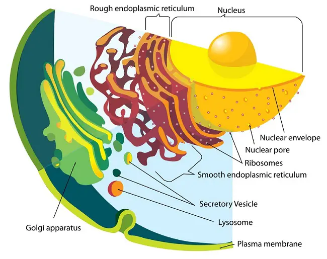
Endoplasmic means inside (endo) the cytoplasm(plasm). The Latin word for the net is reticulum. An endoplasmic reticulum is a plasma membrane that forms inside a cell. It folds inwards to create an inner space called the lumen. The lumen is actually continuous and is connected to the perinuclear area.
The Endoplasmic Reticulum, a network membranous canals that are filled with fluid, is the largest of these. They act as the transport system for the cell and transport materials within the cell.
- Rough Endoplasmic Reticulum – These are made up of cisternae and tubules and vesicles that are present throughout the cell and involved in protein production.
- Smooth Endoplasmic Reticulum – These are the storage organelles, which are associated with the production and use of steroids, lipids, and detoxification.
Rough Endoplasmic Reticulum
Because its surface is covered with ribosomes (molecules responsible for protein production), the rough endoplasmic retina is named so. A ribosome may find a particular RNA segment and tell it to go to the rough endoplasmic retina to embed itself. The protein that is created from this segment will be found inside the lumen in the rough endoplasmic retina. There it will fold and be tagged with a (usually carbohydrate-based) molecule. This marks the protein for transport into the Golgi apparatus. The rough endoplasmic retina is continuous with the nuclear membrane and appears like a series canals close to the nucleus. The rough endoplasmic retina is destined for proteins to be either part of a cell membrane or secreted out of the cell. It would be much more difficult to tell the difference between proteins that should be left and those that should stay in the cell without a rough endoplasmic retina. The rough endoplasmic retina allows cells to specialize, and the organism to be more complex.
Smooth Endoplasmic Reticulum
Instead of being involved with protein synthesis, the smooth endoplasmic retina makes lipids. These fat-based molecules are vital in energy storage, membrane structure and communication. (Steroids can also act as hormones). The cell’s detoxification is also done by the smooth endoplasmic retina. It is tubularer than the rough endoplasmic retinal, and it is not always continuous with the nuclear membrane. Each cell has a smooth, endoplasmic retinalum. However, the amount of this reticulum will vary depending on how it is used. The liver, which is responsible primarily for detoxification in the body, has a greater amount of smooth endoplasmic retinaum.
Structure of Endoplasmic Reticulum (ER)
The Endoplasmic Reticulum (ER) is a complex membranous structure found in eukaryotic cells. It consists of three distinct forms: cisternae, vesicles, and tubules. Here is a breakdown of the structure of the ER based on the provided information:
- Cisternae: Cisternae are sac-like structures that are flattened and unbranched. They are arranged in stacks, with one cisterna stacked on top of another. These stacked cisternae are often referred to as “rough” or “smooth” ER, depending on the presence or absence of ribosomes on their surfaces. The rough ER is involved in protein synthesis and modification, while the smooth ER plays a role in lipid synthesis and detoxification.
- Vesicles: Vesicles are small, spherical structures that bud off from the ER membrane. They act as transport carriers, carrying proteins and other molecules synthesized in the ER to their target destinations within the cell. Vesicles are involved in intracellular trafficking, delivering proteins to the Golgi apparatus, lysosomes, plasma membrane, or other organelles.
- Tubules: Tubules are tubular branched structures that connect the cisternae and vesicles within the ER. They form a network of interconnected tubes throughout the cytoplasm of the cell. The tubules play a crucial role in maintaining the continuity of the ER membrane and facilitating the exchange of materials between different parts of the ER. They also provide a pathway for the movement of molecules within the ER and contribute to the dynamic nature of the ER structure.
Overall, the structure of the Endoplasmic Reticulum consists of cisternae, vesicles, and tubules. The cisternae are stacked, flattened sacs that can be rough or smooth. Vesicles are small spherical structures involved in intracellular transport, while tubules form a branched network connecting the cisternae and vesicles. This intricate structure of the ER allows it to carry out its diverse functions, including protein synthesis, lipid metabolism, and intracellular transport.
Functions of Endoplasmic Reticulum (ER)
The Endoplasmic Reticulum (ER) is involved in various vital functions within the cell. Here are the functions of the ER based on the provided information:
- Metabolic Processes: The ER contains numerous enzymes required for various metabolic processes. These enzymes play essential roles in the synthesis, modification, and breakdown of molecules such as lipids, carbohydrates, and proteins. The ER’s surface is crucial for processes like diffusion, osmosis, and active transport, allowing for the movement of molecules in and out of the ER.
- Lipid Synthesis: The ER is responsible for synthesizing lipids, including cholesterol and steroids. It plays a crucial role in lipid metabolism and is involved in the production of lipid components that are essential for cell membranes, hormone production, and energy storage.
- Protein Modification: The rough ER, with its ribosomes attached to the surface, is involved in protein synthesis. It allows for the modification of polypeptides as they emerge from the ribosomes. The rough ER assists in the folding and shaping of proteins, preparing them for their functional structure. This process includes the formation of secondary and tertiary protein structures.
- Synthesis of Membrane Proteins: The ER is involved in synthesizing various membrane proteins that are essential for the structure and function of cell membranes. These proteins play crucial roles in cell signaling, transport of molecules across the membrane, and maintaining membrane integrity.
- Nuclear Envelope Formation: After cell division, the ER plays a crucial role in preparing the nuclear envelope. The ER membranes contribute to the formation of the nuclear membrane, providing structural support and facilitating the organization of the genetic material within the nucleus.
In summary, the Endoplasmic Reticulum is involved in a wide range of functions within the cell. It participates in metabolic processes, lipid synthesis, protein modification, synthesis of membrane proteins, and the formation of the nuclear envelope. These functions are essential for the proper functioning, structure, and organization of the cell.
Endosomes
Endosomes are membrane-bound cells within cells that are derived from the Golgi network
Structure of Endosomes
Endosomes are membrane-bound organelles involved in the process of endocytosis, which allows the cell to internalize and transport materials from the cell surface. Here is the structure of endosomes based on the provided information:
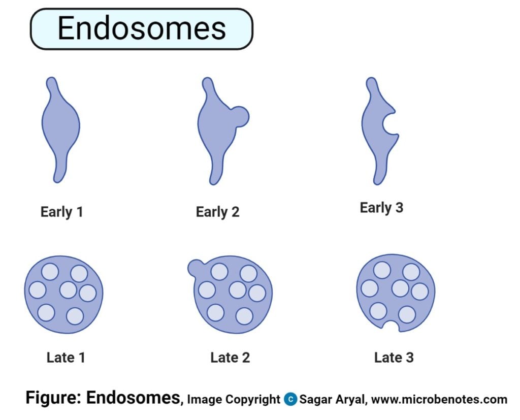
- Early Endosomes: Early endosomes have a tubular-vesicular network structure. They are typically located closer to the plasma membrane and receive newly internalized materials through endocytosis. Early endosomes act as sorting stations, sorting and directing molecules to different cellular destinations. They receive cargo from the plasma membrane and sort them for recycling back to the cell surface, transport to the trans-Golgi network, or further processing in late endosomes.
- Late Endosomes: Late endosomes lack tubules but instead contain numerous close-packed intraluminal vesicles. They are located deeper within the cell compared to early endosomes. Late endosomes are involved in the degradation and recycling of cellular components. They receive cargo from early endosomes and are responsible for delivering materials to lysosomes for degradation or recycling. The intraluminal vesicles within late endosomes are formed through a process called invagination, where the endosomal membrane buds inward, creating vesicles within the endosome.
- Recycling Endosomes: Recycling endosomes are specialized endosomes involved in the recycling of molecules back to the cell surface. They are associated with microtubules and are mainly composed of tubular structures. Recycling endosomes receive cargo from early endosomes and sort specific molecules for recycling back to the plasma membrane. This allows the cell to retrieve and reuse certain proteins and lipids, maintaining cellular homeostasis and membrane composition.
In summary, endosomes exhibit different structures based on their function and stage of the endocytic pathway. Early endosomes have a tubular-vesicular network structure, late endosomes contain many intraluminal vesicles, and recycling endosomes primarily consist of tubular structures. These structural differences reflect the diverse roles of endosomes in sorting, directing, recycling, and degrading cellular components.
Functions of Endosomes
Endosomes play vital roles in the intracellular transport and sorting of materials. Here are the functions of endosomes based on the provided information:
- Sorting of Internalized Materials: Endosomes are responsible for receiving and sorting internalized materials that have been taken up from the cell surface through the process of endocytosis. They act as intermediate compartments between the plasma membrane and other cellular destinations. Endosomes receive cargo such as proteins, lipids, and receptors and determine their subsequent fate.
- Delivery to the Golgi Apparatus: Some materials internalized by endocytosis are destined for further processing in the Golgi apparatus. Endosomes play a crucial role in transporting these materials from the cell surface to the Golgi. They serve as an intermediate station for sorting and packaging cargo into transport vesicles that carry them to the Golgi for further modification, sorting, and distribution within the cell.
- Transport to Lysosomes: Endosomes also function in delivering materials to lysosomes for degradation. Late endosomes, in particular, mature into lysosomes or fuse with existing lysosomes. The intraluminal vesicles present in late endosomes, which are formed through invagination of the endosomal membrane, contain cargo that is targeted for degradation by lysosomal enzymes. This process helps maintain cellular homeostasis by removing damaged or unwanted molecules.
- Recycling of Internalized Materials: Recycling endosomes specialize in the recycling of specific molecules back to the cell surface. These endosomes sort and direct molecules, such as receptors and membrane proteins, for recycling, allowing the cell to retrieve and reuse them. This recycling process helps maintain the integrity of the cell membrane and regulate cellular processes.
In summary, endosomes play critical roles in sorting, directing, and transporting internalized materials within the cell. They facilitate the delivery of cargo to the Golgi apparatus for further processing and to lysosomes for degradation. Additionally, recycling endosomes enable the recycling of specific molecules back to the cell surface, contributing to the maintenance of cellular homeostasis and membrane composition.
Golgi Apparatus/ Golgi Complex/ Golgi Body
The Golgi Apparatus is the main organelle in cells of eukaryotrd that helps in the packing of macromolecules into vesicles that can be transported out to the site of action.
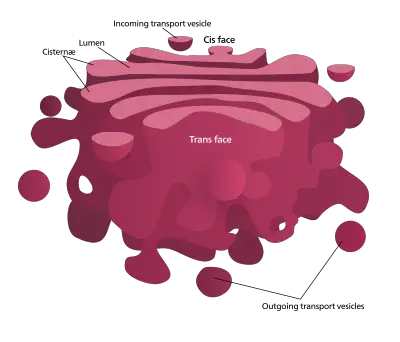
Golgi Apparatus is also known by its name Golgi Complex. This is an elastomeric organelle made up of a set of pouches flattened, stacked and known as Cisternae. The cell organelle is principally involved in transporting, changing and packaging proteins and lipids to specific locations. Golgi Apparatus is located within the cell’s cytoplasm and is present in animal and plant cells.
Structure of Golgi Apparatus/ Golgi Complex/ Golgi Body
The Golgi apparatus, also known as the Golgi complex, is a cellular organelle involved in the processing, sorting, and packaging of proteins and lipids. Here is a description of its structure based on the provided information:
- Cisternae: The Golgi apparatus is composed of stacks of flattened, membrane-bound sacs called cisternae. These cisternae are the smallest unit of the Golgi complex and are arranged in parallel bundles. The cisternae serve as the main structural component of the Golgi apparatus and are involved in the modification and sorting of molecules.
- Tubules: Tubules are tubular and branched structures that radiate from the cisternae. These tubules extend throughout the Golgi apparatus and provide a network for transport and communication within the organelle. The tubules are fenestrated at the periphery, allowing the exchange of materials between the cisternae and other cellular compartments.
- Vesicles: The Golgi apparatus contains various types of vesicles involved in different functions:
- Transitional Vesicles: Transitional vesicles are small vesicles that bud off from the endoplasmic reticulum (ER) and transport newly synthesized proteins and lipids to the Golgi apparatus. They serve as a connection between the ER and the Golgi, facilitating the transport of cargo for further processing.
- Secretory Vesicles: Secretory vesicles are spherical bodies that form in the Golgi apparatus. They contain processed and modified molecules, such as proteins or lipids, destined for secretion outside the cell. These vesicles transport their cargo to the plasma membrane, where they fuse and release their contents through exocytosis.
- Clathrin-Coated Vesicles: Clathrin-coated vesicles are involved in receptor-mediated endocytosis, a process in which specific molecules from the cell surface are internalized. These vesicles are coated with a protein called clathrin, which aids in the formation and targeting of vesicles for internalization.
In summary, the Golgi apparatus consists of cisternae, tubules, and vesicles. The cisternae form the stacked structure of the Golgi and are involved in the modification and sorting of molecules. Tubules provide a network for transport within the Golgi, while vesicles mediate the transport of molecules to and from the Golgi apparatus. Together, these structural components allow the Golgi apparatus to carry out its essential functions in processing, sorting, and packaging various cellular components.
Functions of Golgi Apparatus/ Golgi Complex/ Golgi Body
The Golgi apparatus, also known as the Golgi complex or Golgi body, plays vital roles in the cell’s protein and lipid processing, sorting, and transport. Here are the functions of the Golgi apparatus based on the provided information:
- Protein and Lipid Sorting: The Golgi apparatus acts as a central hub for directing proteins and lipids to their specific destinations within the cell. It receives newly synthesized proteins and lipids from the endoplasmic reticulum (ER) and modifies them, ensuring they are correctly sorted and prepared for transport to their target locations. This sorting process involves adding specific molecular tags, such as carbohydrates or phosphate groups, to the molecules, which act as signals for their subsequent transport.
- Exocytosis: The Golgi apparatus is involved in the process of exocytosis, where secretory vesicles derived from the Golgi fuse with the cell membrane, releasing their contents outside the cell. This process allows the Golgi to export various products and proteins, such as zymogen (inactive enzyme precursors), mucus, lactoprotein, and components of the thyroid hormone, to be used in other parts of the body or to carry out specific functions.
- Organelle Synthesis: The Golgi apparatus is responsible for synthesizing and assembling certain cellular organelles. It plays a role in the formation and maintenance of the cell membrane by synthesizing lipids and incorporating them into the membrane structure. Additionally, the Golgi apparatus is involved in the production of lysosomes, which are organelles responsible for intracellular digestion and waste disposal.
- Sulfation: The Golgi apparatus is involved in the sulfation process, where sulfate groups are added to molecules. This modification plays a crucial role in the functional properties of molecules, such as proteins and carbohydrates. Sulfation can affect protein stability, activity, and interactions with other molecules. The Golgi apparatus facilitates this sulfation process, ensuring the proper modification of molecules in the cell.
In summary, the Golgi apparatus serves as the “traffic police” of the cell, directing proteins and lipids to their intended destinations. It is involved in exocytosis, organelle synthesis, and sulfation. Through these functions, the Golgi apparatus contributes to the proper functioning and organization of cellular processes.
Intermediate filaments
The third filament class that makes up the cytoskeleton is the intermediate filaments. They are classified as intermediate filaments due to the diameter intermediate of the filaments compared to myosin and microfilaments.
Structure of Intermediate filaments
The structure of intermediate filaments is characterized by the arrangement of a family of related proteins into coiled-coil structures. Here are some key points regarding the structure of intermediate filaments based on the provided information:
- Family of Related Proteins: Intermediate filaments are composed of a diverse family of related proteins. These proteins include keratins, vimentin, desmin, lamin, and neurofilaments, among others. Different types of intermediate filaments are expressed in various cell types and tissues, contributing to their structural and functional diversity.
- Coiled-Coil Structure: The individual filaments within intermediate filaments are organized in a unique helical arrangement called a coiled-coil structure. The coiled-coil structure is formed by two or more alpha helices wrapping around each other in a rope-like manner. This coiling provides stability and strength to the intermediate filaments.
- Heteropolymeric Assembly: Intermediate filaments are assembled through the process of heteropolymeric assembly. This means that multiple protein subunits, typically of different types, come together to form the intermediate filament structure. The specific combination of protein subunits varies depending on the cell type and tissue, contributing to the diverse functions of intermediate filaments.
- Cross-Linking Proteins: Intermediate filaments are cross-linked by specific proteins that help stabilize their structure and regulate their function. These cross-linking proteins, such as plectin and desmoplakin, bind to intermediate filaments and link them to other cellular structures, including other intermediate filaments, cell junctions, and the cell membrane. This cross-linking enhances the mechanical strength and integrity of the cytoskeleton.
- Tissue-Specific Expression: Different types of intermediate filaments exhibit tissue-specific expression patterns. For example, keratins are predominantly found in epithelial cells, while vimentin is commonly expressed in mesenchymal cells. This tissue-specific expression of intermediate filaments contributes to the structural characteristics and functions of different cell types.
Overall, the structure of intermediate filaments is characterized by the coiled-coil arrangement of a family of related proteins. This structure provides stability and strength to the filaments, allowing them to support the cytoskeleton, maintain cell shape, and fulfill their specific functions in different cell types and tissues.

Functions of Intermediate filaments
Intermediate filaments have several important functions in the cell, primarily related to providing structural support and maintaining tissue integrity. Here are some key functions of intermediate filaments based on the provided information:
- Structural Support: Intermediate filaments contribute to the overall structural integrity of the cell. They form a supportive framework within the cytoplasm, providing mechanical strength and resistance to external forces. Intermediate filaments help to maintain cell shape and integrity, preventing the cell from easily deforming or collapsing under stress.
- Tissue Integrity: Intermediate filaments play a crucial role in holding tissues together in various organs. They are particularly important in tissues that experience mechanical stress, such as the skin, muscles, and epithelial linings. Intermediate filaments anchor cells to each other and to the extracellular matrix, providing stability and cohesion within tissues. This function is essential for maintaining tissue architecture and integrity.
- Mechanical Strength: Intermediate filaments contribute to the mechanical strength of cells and tissues. By forming a network of interconnected filaments, they provide resistance to stretching and compression forces. Intermediate filaments help cells withstand mechanical stress and maintain their structural integrity, especially in tissues subjected to frequent mechanical strain.
- Cell Junctions: Intermediate filaments are involved in the formation and stabilization of various cell junctions. They interact with proteins located at cell-cell junctions, such as desmosomes and hemidesmosomes, helping to anchor cells together and provide structural support. These junctions play a critical role in tissue organization and stability.
- Cell Signaling and Regulation: Recent studies have also revealed additional functions of intermediate filaments in cell signaling and regulation. Intermediate filaments have been implicated in modulating cellular processes such as cell migration, cell division, and intracellular transport. They interact with signaling molecules and participate in signaling pathways, influencing cellular responses and behaviors.
Overall, the functions of intermediate filaments include providing structural support, maintaining tissue integrity, contributing to mechanical strength, and participating in cell signaling. These versatile filaments are essential components of the cytoskeleton and play a vital role in the overall organization and function of cells and tissues throughout the body.
Lysozyme
Lysozymes, also known as lysosomes, are membrane-bound organelles found in the cytoplasm of animal cells. They play a crucial role in intracellular digestion and the breakdown of various macromolecules. Lysozymes contain a variety of hydrolytic enzymes, including lipases, amylases, proteases, and nucleases, which are involved in the degradation of lipids, carbohydrates, proteins, and nucleic acids, respectively.
Lysozymes are spherical vesicles enclosed by a single membrane called the lysosomal membrane. This membrane helps maintain the acidic pH inside the lysosome, which is necessary for the optimal activity of the hydrolytic enzymes. The lysosomal membrane also acts as a barrier, preventing the enzymes from leaking into the cytoplasm and ensuring their confined action within the lysosome.
Within the cell, there are no distinct types of lysozymes referred to as primary and secondary lysozymes. However, it is important to note that lysosomes can fuse with other vesicles or organelles, allowing the enzymes contained within the lysosome to degrade the engulfed materials. This fusion process helps in the breakdown and recycling of various cellular components, including damaged organelles and engulfed molecules or organelles.
The primary function of lysozymes is intracellular digestion and the recycling of cellular waste. They are involved in the degradation of worn-out organelles, the turnover of cellular components, and the clearance of foreign materials, such as bacteria or cellular debris. Lysozymes also play a role in various cellular processes, including autophagy, where they participate in the degradation of unwanted cellular components.
In summary, lysozymes are membrane-bound organelles found in animal cells. They contain hydrolytic enzymes involved in the degradation of macromolecules. Lysozymes function in intracellular digestion, the recycling of cellular waste, and the clearance of foreign materials.
- Primary lysosome, which contains hydrolytic enzymes such as lipases, proteases, amylases, and nucleases.
- Secondary lysozyme created by the combination of primary lysozymes that contain organelles or molecules that have been engulfed.
Structure of Lysozyme
- Shape: Irregular or pleomorphic, but often found in spherical or granular structures.
- Enclosed by a lysosomal membrane.
- Lysosomal membrane protects the cytosol and the rest of the cell from the harmful actions of the enzymes.
- Irregular shape provides a larger surface area for enzyme-substrate interactions.
- Lysosomal membrane acts as a selective barrier, allowing the passage of small molecules and ions while restricting the movement of larger enzymes.
- Lysosomal membrane contains specific transport proteins for uptake of substrates into the lysosome and export of degradation products out of the lysosome.
- Maintains the integrity of the lysosome and ensures confinement of enzymes to their designated compartments.
- Enables efficient digestion and recycling processes within the lysosome.
Functions of Lysozyme
- Intracellular Digestion: Lysozyme aids in the hydrolysis of glycosidic bonds in peptidoglycan, contributing to the degradation of bacterial cells and the immune defense against bacterial infections.
- Autolysis: Lysozyme participates in the self-destruction of unwanted or damaged organelles within the cytoplasm, maintaining cellular homeostasis and recycling cellular components.
- Secretion: Lysozyme is involved in the packaging and transport of cellular products, including hormones, enzymes, and antibodies, ensuring their timely release.
- Plasma Membrane Repair: In case of membrane damage, lysosomes fuse with the plasma membrane and release lysozyme to initiate the repair process, removing damaged components and restoring membrane integrity.
- Cell Signaling: Lysozyme and lysosomes regulate the release and degradation of signaling molecules, influencing cell growth, differentiation, and apoptosis.
- Energy Metabolism: Lysozyme aids in the breakdown of complex molecules such as glycogen and glycosaminoglycans, releasing energy for cellular activities and supporting cellular respiration.
Microfilaments
Microfilaments form part of the cytoskeleton in cells, made of actin proteins as parallel polymers. They are the smallest filaments in the cytoskeleton that have the highest rigidity and flexibility giving strength and flexibility to the cells.
Structure of Microfilaments
- Microfilaments, also known as actin filaments, are a crucial component of the cytoskeleton in eukaryotic cells. They play a vital role in providing structural support, facilitating cell movement, and enabling various cellular processes. The structure of microfilaments involves the arrangement of protein chains in a helical fashion, with distinct polarity at each end.
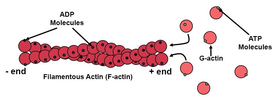
- Microfilaments are composed of globular protein subunits called actin monomers. These actin monomers polymerize to form long, thin filaments. The filaments can exist in two different forms: cross-linked networks or bundled arrangements. In cross-linked networks, the filaments form a mesh-like structure, providing strength and stability to the cell’s cytoskeleton. In bundled arrangements, the filaments come together to form thicker, parallel bundles, which can contribute to cellular processes such as cell contraction.
- The structure of microfilaments is characterized by a helical arrangement of the actin monomers. The chains of protein wrap around each other, creating a twisted structure. One end of the filament, known as the barbed end, is positively charged. It is called the barbed end because it appears as if actin monomers are pointing outward like the bristles of a barbed wire. The barbed end is involved in rapid actin polymerization and is crucial for processes such as cell protrusion and movement.
- On the other hand, the opposite end of the microfilament is referred to as the pointed end. It is negatively charged and has a pointed appearance. The pointed end is involved in actin depolymerization and plays a role in regulating the length and dynamics of microfilaments. It is important for processes such as cellular remodeling and cell division.
- The polarity of microfilaments, with the barbed end and the pointed end having opposite charges, enables the directed assembly and disassembly of actin filaments. This polarized structure allows for the formation of actin networks and provides a framework for the coordination of cellular activities such as cell migration, cytokinesis, and intracellular transport.
- In summary, the structure of microfilaments consists of protein chains twisted around each other in a helical arrangement. One end, the barbed end, is positively charged and barbed, while the other end, the pointed end, is negatively charged and pointed. This polarized structure is essential for the various functions of microfilaments within the cell.
Functions of Microfilaments
- Generation of Cell Structure and Movement: Microfilaments, in association with the motor protein myosin, contribute to the generation of strength and the maintenance of cell structure. They form the basis of the contractile machinery responsible for muscle contraction. In non-muscle cells, microfilaments and myosin enable cell movement by facilitating the formation of cellular protrusions, such as lamellipodia and filopodia, allowing the cell to migrate and change shape.
- Cell Division: Microfilaments play a crucial role in cell division. During cytokinesis, microfilaments form a contractile ring called the cleavage furrow. The contractile ring contracts, leading to the physical separation of the dividing cell into two daughter cells. Microfilaments provide the necessary force for the division process and ensure the accurate distribution of cellular components.
- Cell Surface Projections: Microfilaments are involved in the production of various cell surface projections. They contribute to the formation of microvilli, which are tiny finger-like projections on the cell surface. Microvilli increase the cell’s surface area, allowing for efficient nutrient absorption and sensory functions. Additionally, microfilaments are essential for the formation of other cell surface projections, such as stereocilia, which are specialized microvilli involved in mechanosensation, and filopodia, which have roles in cell adhesion, signaling, and environmental exploration.
Microtubules
Microtubules also form a part of the cytoskeleton, which differs from microfilaments because of that they contain tubulin.
Structure of Microtubules
- Tubular Structure: Microtubules are long, hollow structures with a tubular shape, approximately 24nm in diameter. They form rigid cylinders that provide structural support within the cell.
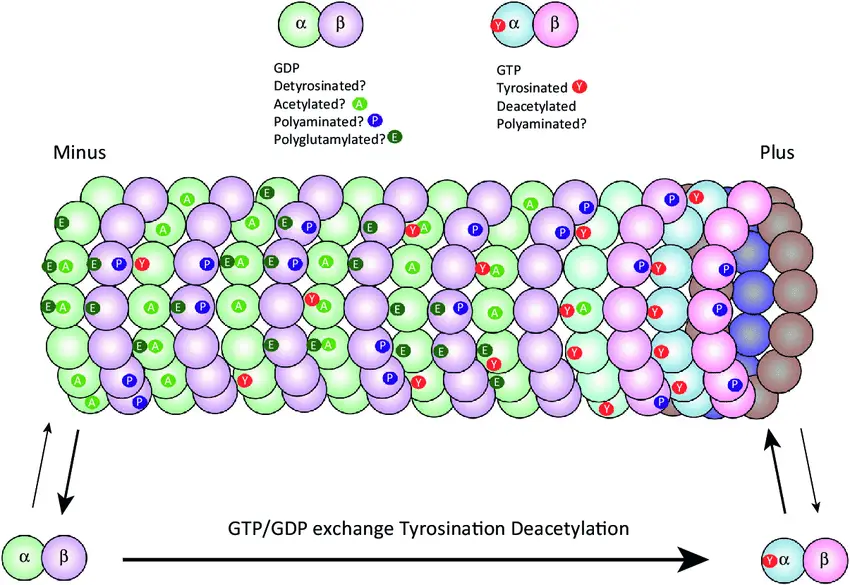
- α and β Tubulin Subunits: The wall of the microtubule consists of globular subunits known as α and β tubulin. These subunits arrange in a helical array, forming protofilaments.
- Protofilaments: Protofilaments are linear chains of α and β tubulin subunits that align side by side to create the cylindrical structure of the microtubule. Typically, microtubules consist of 13 protofilaments.
- Polarity: Microtubules exhibit polarity, with distinct plus and minus ends. The plus end is often located towards the outer periphery of the cell and has a positive charge. The minus end is typically anchored near the microtubule organizing center (MTOC), such as the centrosome, and has a negative charge.
- Dynamic Instability: Microtubules display dynamic instability, characterized by phases of growth (polymerization) and shrinkage (depolymerization). This dynamic behavior is regulated by the addition and removal of tubulin subunits at the ends of the microtubule.
- Cellular Functions: Microtubules play a crucial role in various cellular processes. They are involved in cell division, where they form the mitotic spindle to separate chromosomes. Microtubules also serve as tracks for intracellular transport, facilitating the movement of organelles, vesicles, and other cellular components. Additionally, microtubules contribute to the formation of cellular structures like cilia and flagella.
Functions of Microtubules
- Cell Shape and Support: Microtubules are integral components of the cytoskeleton and play a crucial role in providing shape and support to the cell. They form a structural framework, along with other cytoskeletal elements, that helps maintain the overall shape and integrity of the cell.
- Cell Movement: Microtubules are involved in various forms of cell movement. They form the core structure of cilia and flagella, which are specialized appendages on the cell surface. Microtubules within cilia and flagella undergo coordinated sliding, generating movement that allows cells or extracellular fluids to be propelled. This movement is important for processes such as the movement of sperm cells or the clearance of mucus in the respiratory tract.
- Intracellular Transport: Microtubules serve as tracks for intracellular transport within the cell. Specialized motor proteins, such as dynein and kinesin, bind to microtubules and transport various cell organelles, vesicles, and molecules to their specific destinations within the cell. Microtubules provide the infrastructure for this movement, facilitating the precise delivery and distribution of cellular components.
- Cell Division: Microtubules play a critical role in cell division. During mitosis, microtubules form the mitotic spindle, a complex structure that helps separate replicated chromosomes into two daughter cells. The spindle fibers, composed of microtubules, attach to the chromosomes and exert forces that ensure accurate chromosome segregation.
- Cell Signaling and Regulation: Recent studies have revealed additional functions of microtubules in cell signaling and regulation. Microtubules interact with signaling molecules and participate in signaling pathways, influencing cellular responses and behaviors. They also play a role in the organization and positioning of various cellular structures, including the nucleus and cell organelles.
In summary, microtubules serve multiple important functions in the cell. They provide shape and support, contribute to cell movement, facilitate intracellular transport, and play a crucial role in cell division. Additionally, microtubules have implications in cell signaling and regulation, impacting various cellular processes.
Microvilli
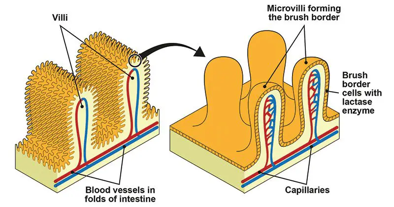
Microvilli are tiny structures resembling fingers that extend from or onto cells. They can be found on their alone or when they are in combination with villi.
Structure of Microvilli
- Surface Bundles: Microvilli are small, finger-like protrusions that are loosely arranged on the surface of cells. They extend outward from the cell membrane, increasing the cell’s surface area for various functions.
- Lack of Cellular Organelles: Microvilli generally do not contain significant cellular organelles. They consist primarily of cytoplasmic extensions supported by a network of microfilaments.
- Plasma Membrane Enclosure: Each microvillus is surrounded by a plasma membrane, forming a distinct boundary between the microvillus and the external environment. The plasma membrane of microvilli contains various proteins and receptors that contribute to their specific functions.
- Cytoplasmic Core: Inside each microvillus, there is a core of cytoplasm. This cytoplasm contains essential components, including microfilaments and associated proteins.
- Actin Filament Bundles: The microvillus structure is primarily composed of bundles of actin filaments. These actin filaments provide the rigidity and structural support to the microvillus. They form a dense network within the cytoplasmic core of the microvillus.
- Binding Proteins: The actin filaments in microvilli are bound and cross-linked by specific proteins. Fimbrin, villin, and epsin are among the proteins involved in bundling and stabilizing the actin filaments within the microvillus structure.
In summary, microvilli are cellular protrusions that extend from the cell surface. They consist of bundles of actin filaments held together by binding proteins such as fimbrin, villin, and epsin. Microvilli lack cellular organelles and are enclosed by a plasma membrane, with a core of cytoplasm containing the necessary components for their function.
Functions of Microvilli
- Increased Surface Area: One of the primary functions of microvilli is to increase the surface area of the cell. By extending from the cell surface in a multitude of small finger-like projections, microvilli greatly expand the cell’s surface area. This increased surface area is crucial for enhancing absorption and secretion processes.
- Enhanced Absorption and Secretion: Microvilli play a vital role in absorption and secretion functions. The presence of microvilli on the cell membrane allows for a larger surface area available for nutrient absorption and the secretion of molecules. The densely packed microvilli on the cell surface maximize the contact between the cell and its surrounding environment, enabling efficient absorption and secretion processes.
- Enzymatic Breakdown: The membrane of microvilli is packed with enzymes that aid in the breakdown of larger molecules into smaller ones. These enzymes facilitate the digestion and absorption of nutrients, such as proteins, carbohydrates, and lipids. By breaking down these complex molecules into smaller components, microvilli contribute to more effective absorption and utilization of nutrients by the cell.
- Anchoring Agent: Microvilli also serve as anchoring agents in certain cell types. For example, in white blood cells (leukocytes), microvilli help anchor the cell to the endothelial lining of blood vessels, facilitating their migration through tissues during immune responses. In sperm cells, microvilli play a role in anchoring the sperm to the surface of the egg during fertilization, aiding in the process of fertilization.
In summary, microvilli serve important functions related to increasing the surface area of the cell. They enhance absorption and secretion processes by providing a larger contact area with the environment. The presence of enzymes in microvilli aids in the breakdown of molecules for more effective absorption. Additionally, microvilli act as anchoring agents in certain cells, facilitating cell migration and fertilization processes.
Mitochondria
Mitochondria are cells with two membranes that are responsible for the production as well as storage of fuel to the cell. The oxidation process of different substances in cells and the release of energy as a form ATP (Adenosine Triphosphate) is the main purpose of mitochondria.
Structure of Mitochondria
- Double Membrane: Mitochondria have a unique structure consisting of two membranes. The outer membrane is smooth and surrounds the entire organelle, acting as a protective barrier. The inner membrane is highly folded and contains numerous finger-like structures called cristae, which greatly increase its surface area.
- Inner Mitochondrial Membrane: The inner mitochondrial membrane is the site of various crucial processes. It contains enzymes, coenzymes, and components of multiple metabolic cycles, such as the citric acid cycle (Krebs cycle). This membrane is also equipped with transport pores that allow the movement of substrates, ATP (adenosine triphosphate), and phosphate molecules.
- Matrix: Within the inner membrane is the matrix, a gel-like substance that fills the space inside the mitochondria. The matrix contains numerous enzymes involved in metabolic processes, including the citric acid cycle. It also houses the mitochondrial DNA (mtDNA), which is responsible for producing a portion of the proteins found within the mitochondria.
- mtDNA: Mitochondria have their own unique DNA, known as mitochondrial DNA (mtDNA). This DNA is present in the form of single or double-stranded molecules within the mitochondria. mtDNA is capable of encoding and producing approximately 10% of the proteins found in mitochondria. It plays a critical role in the synthesis of proteins necessary for the proper functioning of the mitochondria.
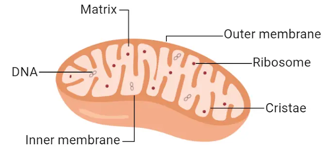
Functions of Mitochondria
- ATP Synthesis: The primary function of mitochondria is the synthesis of adenosine triphosphate (ATP), which serves as the main energy currency of the cell. Through a process known as oxidative phosphorylation, mitochondria generate ATP by utilizing the energy obtained from the breakdown of nutrients, such as glucose and fatty acids. This ATP synthesis provides energy for various cellular processes and is essential for the proper functioning of all cell organelles.
- Calcium Ion Regulation: Mitochondria play a role in maintaining the balance of calcium ions (Ca^2+) within the cell. They have the ability to take up and release calcium ions, thus contributing to the regulation of intracellular calcium levels. This regulation is crucial for various cellular processes, including muscle contraction, neurotransmitter release, and cellular signaling.
- Apoptosis Regulation: Mitochondria are involved in the regulation of programmed cell death, known as apoptosis. They release specific proteins, such as cytochrome c, which activate a cascade of events leading to apoptosis. This process plays a vital role in removing damaged or unwanted cells and maintaining tissue homeostasis.
- Hormone Synthesis: Within mitochondria, certain segments of hormones and components of the blood are synthesized. For example, the synthesis of steroid hormones, such as cortisol, aldosterone, and sex hormones, occurs in specialized cells within the adrenal cortex and gonads, where mitochondria play a crucial role.
- Ammonia Detoxification: Mitochondria in the liver possess the ability to detoxify ammonia, a toxic byproduct of amino acid metabolism. Through a series of enzymatic reactions, ammonia is converted into urea, which can be safely eliminated from the body. This process, known as the urea cycle, occurs primarily in the mitochondria of liver cells and is essential for maintaining nitrogen balance in the body.
In summary, mitochondria serve multiple important functions within cells. They are primarily responsible for ATP synthesis, providing energy for cellular processes. Mitochondria also contribute to calcium ion regulation, apoptosis regulation, hormone synthesis, and ammonia detoxification. These diverse functions highlight the essential role of mitochondria in cellular energy metabolism and overall cellular homeostasis.
Nucleus
Nucleus is a dual membrane-bound structure that is responsible for controlling every cell’s activity and is also a central point for genetic materials and also for transferring. It is among the cell organelles that are large, taking up 10% of the total cell space. It is commonly referred to as”the “brain of the cell” because it is the one that gives commands to the functioning of other organelles within the cell. The term “nucleus” is defined when it comes to cells that are eukaryotic, but it is not present in prokaryotic cells that have the genetic material that is distributed within the cytoplasm.
Structure of Nucleus
- Nuclear Envelope: The nucleus is enclosed by a nuclear envelope, which is structurally and compositionally similar to the cell membrane. The nuclear envelope consists of two phospholipid bilayers with an intervening space. It has pores, known as nuclear pores, that allow the movement of proteins and RNA molecules in and out of the nucleus. The nuclear envelope enables communication and interaction between the nucleus and other cell organelles while maintaining the integrity of the nucleoplasm and chromatin within the nucleus.
- Chromatin: The nucleus contains chromatin, which is a complex of DNA, RNA, and proteins. Chromatin is the genetic material responsible for carrying the hereditary information from one generation to another. It is present in a dispersed and less condensed form during interphase, allowing access to the genes for transcription. However, under powerful magnification, chromatin can condense and become visible as distinct structures called chromosomes during cell division.
- Nucleolus: The nucleolus is a distinct region within the nucleus. It is a membrane-less organelle responsible for the synthesis of ribosomal RNA (rRNA) and the assembly of ribosomes, which are essential for protein synthesis. The nucleolus contains specific regions where rRNA genes are transcribed and processed, resulting in the production of rRNA molecules. These rRNA molecules combine with proteins to form ribosomal subunits that are then exported from the nucleus for further assembly into functional ribosomes in the cytoplasm.
In summary, the nucleus has a distinct structure consisting of a nuclear envelope, chromatin, and a nucleolus. The nuclear envelope encloses the nucleus and contains nuclear pores for the movement of molecules. Chromatin, composed of DNA, RNA, and proteins, carries the genetic information. The nucleolus is involved in the synthesis of rRNA and the assembly of ribosomes. Together, these components contribute to the nucleus’s functions, including gene expression, DNA replication, and the synthesis of RNA and ribosomes.
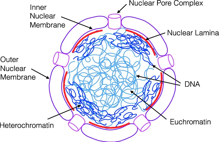
Functions of Nucleus
- Storage and Transfer of Genetic Material: The nucleus is responsible for the storage and transfer of genetic material in the form of DNA (deoxyribonucleic acid) and RNA (ribonucleic acid). DNA, located within the chromatin, contains the instructions for protein synthesis and carries the hereditary information from one generation to another. RNA molecules, such as messenger RNA (mRNA), are transcribed from DNA and play a crucial role in protein synthesis.
- Transcription: The nucleus plays a key role in the process of transcription, which is the synthesis of mRNA molecules. Transcription involves the copying of genetic information from DNA into mRNA, which serves as a template for protein synthesis. The nucleus houses the machinery and enzymes required for transcription, allowing the synthesis of mRNA molecules from specific regions of DNA.
- Control of Organelle Activity: The nucleus serves as the control center of the cell, regulating the activity of all other organelles. It contains the genetic instructions that determine the characteristics and functions of the cell. Through gene expression and the production of specific proteins, the nucleus influences and directs cellular processes, including metabolism, growth, and differentiation.
- Cell Growth and Cell Division: The nucleus plays a critical role in cell growth and cell division. It coordinates and regulates the processes involved in cell growth, such as DNA replication, RNA synthesis, and protein production. During cell division, the nucleus ensures the accurate distribution of genetic material to daughter cells. It participates in mitosis and meiosis, the two processes that lead to the formation of new cells, enabling growth, tissue repair, and reproduction.
- Protein Synthesis: The nucleus is involved in the synthesis of proteins, which are essential for various cellular functions. Through the process of transcription, mRNA molecules are produced in the nucleus. These mRNA molecules then carry the genetic code from the nucleus to the cytoplasm, where they are translated into proteins on ribosomes. The nucleus controls the regulation and expression of genes, determining which proteins are synthesized and when.
In summary, the nucleus serves vital functions in the cell. It stores and transfers genetic material, aids in transcription, controls organelle activity, facilitates cell growth and division, and participates in protein synthesis. These functions highlight the nucleus’s crucial role in maintaining cellular integrity, genetic information flow, and overall cellular processes.
Peroxisomes
Peroxisomes are membrane-bound organelles that undergo oxidation that are found in the cell cytoplasm of all Eukaryotes. Their name is attributed to their hydrogen peroxide-generating and eliminating activities.
Structure of Peroxisomes
- Single Membrane: Peroxisomes are membrane-bound organelles that consist of a single membrane surrounding their contents. This membrane separates the peroxisomal matrix from the cytoplasm of the cell. The membrane is selectively permeable, allowing the movement of specific molecules in and out of the peroxisome.
- Granular Matrix: Within the peroxisome, there is a granular matrix that is scattered throughout the organelle. This matrix contains various enzymes and other molecules necessary for the peroxisome’s metabolic functions. The granular matrix is involved in carrying out specific metabolic reactions and serves as the site for various enzymatic activities.
- Interconnected Tubules or Individual Peroxisomes: Peroxisomes can exist in the form of interconnected tubules or as individual organelles in the cytoplasm. The specific structure and distribution of peroxisomes can vary depending on the cell type and metabolic requirements. In some cells, peroxisomes may form a network of interconnected tubules, facilitating communication and exchange of metabolites between different peroxisomes. In other cases, individual peroxisomes may be scattered throughout the cytoplasm.
- Compartments for Different Metabolic Activities: Peroxisomes contain compartments within their structure that allow the creation of optimized conditions for different metabolic activities. These compartments help separate specific enzymatic reactions and prevent the interference of different metabolic processes within the peroxisome. The organization of these compartments enables efficient functioning of peroxisomal metabolic pathways.
- Enzymes: Peroxisomes contain various types of enzymes, with the major groups including urate oxidase, D-amino acid oxidase, and catalase. These enzymes are involved in different metabolic pathways occurring within the peroxisome. Urate oxidase is responsible for the breakdown of uric acid, D-amino acid oxidase is involved in the metabolism of certain amino acids, and catalase is responsible for the degradation of hydrogen peroxide, a byproduct of peroxisomal reactions.
In summary, peroxisomes consist of a single membrane and a granular matrix scattered in the cytoplasm. They can exist as interconnected tubules or individual organelles. Peroxisomes contain compartments for different metabolic activities and various enzymes that facilitate specific reactions. The unique structure of peroxisomes allows them to carry out important metabolic functions within the cell.
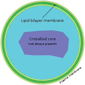
Functions of Peroxisomes
- Detoxification of Hydrogen Peroxide: One of the key functions of peroxisomes is the production and elimination of hydrogen peroxide (H2O2). Peroxisomes contain the enzyme catalase, which breaks down hydrogen peroxide into water and oxygen, preventing the accumulation of this potentially harmful molecule. By detoxifying hydrogen peroxide, peroxisomes help maintain cellular homeostasis and protect cells from oxidative damage.
- Fatty Acid Oxidation: Peroxisomes play a crucial role in the oxidation of fatty acids. Within peroxisomes, fatty acids are broken down into smaller molecules through a series of enzymatic reactions. This process, known as beta-oxidation, generates energy and produces acetyl-CoA, which can be further utilized in various cellular metabolic pathways. Peroxisomes are particularly important in tissues with high energy demands, such as the liver and muscles.
- Synthesis of Lipids: Peroxisomes are involved in the synthesis of specific lipids, including cholesterol and plasmalogens. Cholesterol, an essential component of cell membranes and a precursor for various molecules like hormones, is synthesized within peroxisomes. Plasmalogens, which are important membrane lipids with antioxidant properties, are also synthesized within these organelles. The synthesis of these lipids within peroxisomes contributes to cellular membrane integrity and function.
- Detoxification of Xenobiotics: Peroxisomes are involved in the detoxification of various xenobiotics, which are foreign or harmful substances that enter the body. Certain enzymes present in peroxisomes, such as urate oxidase and D-amino acid oxidase, are responsible for the breakdown and elimination of specific xenobiotics. This detoxification process helps protect cells from the damaging effects of these substances.
- Metabolism of Reactive Oxygen Species: In addition to detoxifying hydrogen peroxide, peroxisomes are also involved in the metabolism of other reactive oxygen species (ROS). ROS are highly reactive molecules that can cause oxidative stress and damage cellular components. Peroxisomes contain enzymes involved in the breakdown of ROS, contributing to the overall cellular antioxidant defense system.
In summary, peroxisomes play vital roles in cellular metabolism and homeostasis. They are involved in the detoxification of hydrogen peroxide, oxidation of fatty acids, synthesis of lipids, detoxification of xenobiotics, and metabolism of reactive oxygen species. These functions highlight the importance of peroxisomes in maintaining cellular health and overall organismal well-being.
Plasmodesmata
Plasmodesmata are tiny channels or passages which allow for the transfer of materials and communications between cells.
Structure of Plasmodesmata
Plasmodesmata are specialized channels that connect two adjacent plant cells, allowing direct communication and transport of materials between them. They have a specific structure consisting of three main components:
- Plasma Membrane: Plasmodesmata have a plasma membrane that is continuous with the plasma membrane of the connected cells. The plasma membrane surrounding the plasmodesma consists of a phospholipid bilayer, similar to the plasma membrane of the cells. This continuous membrane connection helps maintain the integrity and fluidity of the cell boundaries.
- Cytoplasmic Sleeve: The cytoplasmic sleeve is a central region within the plasmodesma that connects the cytosol of the two adjacent cells. It forms a narrow channel through which various molecules and substances can move between the cells. This cytoplasmic sleeve allows for the exchange of ions, small molecules, and even larger macromolecules, such as proteins and RNA, facilitating intercellular communication and transport.
- Desmotubule: The desmotubule is a specialized structure found at the center of the plasmodesma. It is derived from the endoplasmic reticulum (ER) and provides a network of tubules or strands between the two connected cells. The desmotubule plays a role in maintaining the stability and structure of the plasmodesma. It also serves as a conduit for the transport of specific molecules, such as proteins and other macromolecules, between the cells.
The overall structure of plasmodesmata, including the plasma membrane, cytoplasmic sleeve, and desmotubule, enables direct communication and exchange of materials between adjacent plant cells. These channels play a crucial role in coordinating cellular activities, facilitating nutrient and signaling molecule transport, and allowing for the spread of signals and substances throughout the plant organism.
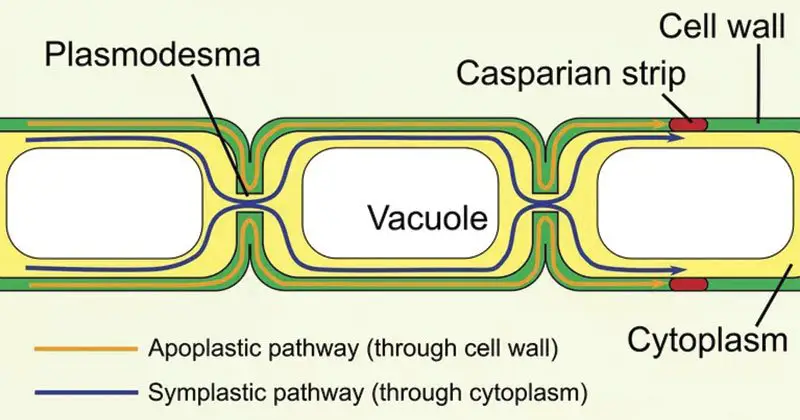
Functions of Plasmodesmata
- Intercellular Communication: Plasmodesmata serve as the primary site for direct communication between adjacent plant cells. They create channels that enable the exchange of various molecules, including proteins, RNA, and viral genomes, between cells. This intercellular communication allows for coordinated responses, signaling, and the spread of essential molecules throughout the plant.
- Transport of Small Molecules: Plasmodesmata facilitate the transport of small molecules, such as ions, sugars, and metabolites, between neighboring cells. These channels provide a direct pathway for the movement of these molecules, allowing for the efficient exchange of nutrients, signaling molecules, and waste products between cells.
- Movement of Macromolecules: Plasmodesmata also play a crucial role in the transport of larger macromolecules, including proteins and RNA. These channels allow for the passage of specific proteins involved in developmental processes, defense responses, and signaling between cells. Additionally, plasmodesmata facilitate the movement of RNA molecules, including messenger RNA (mRNA), small interfering RNA (siRNA), and microRNA (miRNA), which are important for gene regulation and cellular functions.
- Cell-to-Cell Signaling: Plasmodesmata contribute to cell-to-cell signaling in plants. They provide a direct route for the transmission of signaling molecules, such as hormones, secondary messengers, and defense-related compounds, between cells. This allows for the rapid and coordinated response of cells to environmental stimuli, developmental signals, and biotic or abiotic stresses.
- Viral Movement: Plasmodesmata also play a role in the spread of viral infections within plants. Viruses can exploit these channels to move from one cell to another, allowing for the systemic infection of the entire plant. Viral genomes can pass through plasmodesmata, facilitating the dissemination of viral particles and infection throughout the plant tissue.
In summary, plasmodesmata serve as crucial conduits for intercellular communication and transport in plants. They facilitate the exchange of molecules, including proteins, RNA, and viral genomes, between adjacent cells. Plasmodesmata play a significant role in coordinating cellular activities, transporting nutrients and signaling molecules, and contributing to the spread of viral infections within plants.
Plastids
Plastids are membrane-bound double structures found in plants as well as other eukaryotes involved with the production and storage of nutrients. Based on the kind of pigments the plastids come in three kinds:
- Chloroplasts – Chloroplasts are organelles with two membranes that typically vary in their shape – ranging from disc-like shapes to discoid-spherical, spherical oval, and ribbon. They are found in mesophyll cells found in leaves, where they contain chloroplasts as well as the other pigments of carotenoid. They help in capturing sunlight energy to enable photosynthesis. The membrane’s interior is enclosed by the stroma, a space. Flattened disc-like chlorophyll-containing structures known as thylakoids are arranged in a stacked manner like a pile of coins. Each pile is known as the granum (plural: the word grana) as well as the thylakoids from various grana are linked by membranous tubules that are flat, referred to as lamella stromal. Similar to mitochondrial matrix, the chloroplast’s stroma also has the double-stranded circular DNA 70S ribosomes and enzymes that are require the synthesis of proteins and carbohydrates.
- Chromoplasts – The chromoplasts contain carotenoid pigments, fat-soluble like carotene, xanthophylls, etc. that provide plants with their unique color, namely red, yellow, orange, etc.
- Leucoplasts – Leucoplasts are plastids that have no color and store nutrients. Amyloplasts are able to store carbohydrate (like the starch found in potatoes) and aleuroplasts store proteins and elaioplasts store fats and oils.
Structure of Plastids
Plastids are membrane-bound organelles found in the cells of plants and algae. They exhibit a distinctive structure that includes the following components:
- Outer and Inner Membrane: Plastids possess an outer membrane and an inner membrane, which are separated by the intermembrane space. The outer membrane forms the outer boundary of the plastid, while the inner membrane encloses the internal components. These membranes regulate the passage of molecules into and out of the plastid, providing a selective barrier.
- Intermembrane Space: The intermembrane space is the region between the outer and inner membranes of plastids. It acts as a buffer zone, allowing the exchange of certain molecules between the cytoplasm and the plastid.
- Stroma: The inner membrane encloses a matrix called the stroma. The stroma is a gel-like substance that contains various enzymes, proteins, and metabolic pathways necessary for plastid function. It is involved in processes such as carbon fixation, lipid synthesis, and the synthesis of various metabolites.
- Grana and Thylakoids: Within the stroma, plastids contain structures known as grana. Each granum consists of several stacked, disc-like structures called thylakoids. Thylakoids are membrane-bound compartments where photosynthetic pigments, such as chlorophyll, are located. They are involved in capturing light energy and conducting photosynthesis. The thylakoids within a granum are interconnected by stroma lamellae, which are thin regions of stroma between adjacent thylakoids.
- Plastid DNA and RNA: Plastids have their own DNA and RNA molecules, allowing them to synthesize necessary proteins and perform specific functions independently. Plastid DNA (plastome) encodes essential genes involved in plastid-specific processes, such as photosynthesis and pigment synthesis. Plastid RNA molecules are involved in gene expression and protein synthesis within the plastid.
The distinct structure of plastids, with their outer and inner membranes, intermembrane space, stroma, grana, and thylakoids, provides a specialized environment for various plastid functions. Plastids are involved in processes such as photosynthesis, the synthesis and storage of pigments, lipid metabolism, starch synthesis, and the production of essential metabolites in plants and algae.
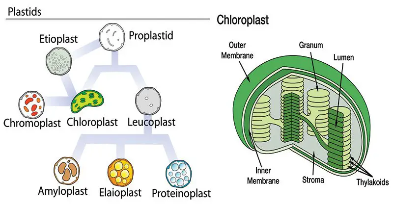
Functions of Plastids
- Photosynthesis: Chloroplasts, a type of plastid, are the primary sites of photosynthesis in plant cells. They contain specialized pigments, such as chlorophyll, which capture light energy. Within the chloroplasts, photosynthesis occurs, converting light energy into chemical energy in the form of glucose. Enzymes and other components present in the chloroplasts facilitate the biochemical reactions of photosynthesis, including the light-dependent and light-independent reactions.
- Starch Synthesis and Storage: Plastids, particularly amyloplasts, are responsible for the synthesis and storage of food reserves, primarily starch. Starch granules are formed within the plastids, and these granules serve as a long-term storage form of glucose in plants. Amyloplasts are abundant in storage tissues, such as seeds, tubers, and roots, where they accumulate large quantities of starch.
- Pigment Synthesis and Storage: Plastids are involved in the synthesis and storage of various pigments that contribute to the coloration of plants. Chloroplasts contain chlorophyll pigments essential for photosynthesis. Other plastids, such as chromoplasts, produce and store pigments like carotenoids and anthocyanins, which provide colors ranging from red, orange, and yellow to blue and purple in flowers, fruits, and other plant parts. These pigments play important roles in attracting pollinators, seed dispersal, and protecting against photodamage.
- Lipid Metabolism: Plastids, specifically proplastids and lipid-containing plastids called oleosomes, are involved in lipid metabolism. They synthesize and store lipids, including triglycerides and phospholipids. Plastids contribute to the production of lipids used for energy storage, membrane synthesis, and the formation of lipid-based signaling molecules in plants.
- Biosynthesis of Secondary Metabolites: Plastids are involved in the biosynthesis of various secondary metabolites in plants. These metabolites include pigments, essential oils, alkaloids, and other specialized compounds. Plastids, particularly chromoplasts, produce and accumulate these metabolites, which serve roles in defense against herbivores, attraction of pollinators, and other ecological functions.
In summary, plastids, particularly chloroplasts, are primarily responsible for photosynthesis and the synthesis of glucose, the primary energy source for plants. Plastids also play roles in starch synthesis and storage, pigment synthesis and storage, lipid metabolism, and the biosynthesis of secondary metabolites. These diverse functions of plastids contribute to plant growth, development, energy storage, and various physiological and ecological processes.
Ribosomes
Ribosomes are ribonucleoproteins that contain equal amounts of proteins and RNA together with a variety of other components essential to synthesize proteins. In prokaryotes they can be found freely and in eukaryotes they are either found in the form of on their own or in the endoplasmic retina.
Structure of Ribosomes
Ribosomes are cellular structures involved in protein synthesis. They have a distinct structure composed of the following components:
- Two Subunits: Ribosomes are composed of two subunits, each with a specific size. In prokaryotic cells, ribosomes are classified as 70S ribosomes. The larger subunit is known as the 50S subunit, while the smaller subunit is called the 30S subunit. In eukaryotic cells, ribosomes are classified as 80S ribosomes. The larger subunit is the 60S subunit, and the smaller subunit is the 40S subunit. The subunits come together during protein synthesis and dissociate afterward.
- Ribonucleoprotein Composition: Ribosomes are composed of ribonucleic acid (RNA) molecules and proteins. The RNA molecules are known as ribosomal RNA (rRNA) and play a crucial role in the ribosome’s structure and function. The proteins associated with the rRNA form a complex called ribonucleoprotein. The specific composition of rRNA and proteins varies between prokaryotic and eukaryotic ribosomes.
- Structural Differences: Prokaryotic and eukaryotic ribosomes differ in size and composition. Prokaryotic ribosomes are smaller and consist of fewer proteins compared to eukaryotic ribosomes. This size difference is reflected in the classification of 70S and 80S ribosomes for prokaryotes and eukaryotes, respectively. The distinct subunit sizes allow ribosomes to interact with different cellular components and participate in protein synthesis processes specific to each cell type.
- Dynamic Nature: Ribosomes are dynamic structures that can disassemble and reassemble as needed. After protein synthesis, the ribosomal subunits separate and can either be reused for subsequent rounds of protein synthesis or remain dissociated until they are needed again. This dynamic nature allows ribosomes to adapt to the changing demands of protein production within the cell.
In summary, ribosomes consist of two subunits, with the specific sizes and composition varying between prokaryotes and eukaryotes. These ribonucleoprotein structures play a crucial role in protein synthesis within cells. The dynamic nature of ribosomes allows for their disassembly and reassembly, ensuring their availability for future protein synthesis processes.
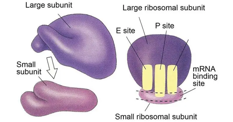
Functions of Ribosomes
- Protein Synthesis: Ribosomes are responsible for the synthesis of proteins in all living organisms. They serve as the site where the genetic information encoded in messenger RNA (mRNA) is translated into the specific sequence of amino acids that make up a protein. Ribosomes function by arranging amino acids in the order indicated by transfer RNA (tRNA) molecules, which recognize and bind to specific codons on the mRNA. Through a process called translation, ribosomes catalyze the formation of peptide bonds between amino acids, ultimately leading to the production of a polypeptide chain.
- mRNA Decoding: Ribosomes play a crucial role in decoding the information contained in mRNA molecules. They interact with the mRNA strand and read its sequence of codons, which are triplets of nucleotides that specify particular amino acids or signal the termination of protein synthesis. By recognizing and pairing the codons with the corresponding tRNA molecules carrying the appropriate amino acids, ribosomes ensure the accurate translation of mRNA into the correct sequence of amino acids in a protein.
- Protein Folding: Ribosomes can also assist in the folding of newly synthesized proteins. As the polypeptide chain emerges from the ribosome, it undergoes folding into its three-dimensional structure, which is critical for its proper function. Ribosomes can provide an environment conducive to protein folding and facilitate the initial stages of this process. However, further protein folding and maturation often require the involvement of additional cellular factors and chaperone proteins.
- Regulation of Protein Synthesis: Ribosomes are involved in the regulation of protein synthesis in response to cellular needs. The rate of protein synthesis can be modulated by altering the availability and activity of ribosomes. This regulation allows cells to control the production of specific proteins, responding to changing environmental conditions, developmental stages, and metabolic demands.
In summary, ribosomes are the primary sites of protein synthesis in all living organisms. They decode the information contained in mRNA molecules, arrange amino acids in the correct order, and catalyze the formation of peptide bonds to synthesize proteins. Ribosomes also play a role in protein folding and participate in the regulation of protein synthesis to ensure the proper production of proteins based on cellular needs.
Storage granules
Storage granules, which are membrane-bound organelles also known as zymogen granules that store cells’ energy reserve as well as other compounds.
Structure of Storage granules
Storage granules are specialized compartments found within cells that store various substances for future use. The structure of storage granules can vary depending on their specific composition and function. However, some general characteristics can be described:
- Lipid Bilayer: Storage granules are surrounded by a lipid bilayer membrane, similar to other cellular organelles. This membrane provides a barrier that separates the contents of the granule from the surrounding cytoplasm, allowing for controlled storage and release of substances.
- Composition: The contents of storage granules can vary depending on their location within the body and their specific function. In general, these granules contain high concentrations of phosphorus and oxygen, indicating the presence of phosphate-containing compounds. These compounds may include glycogen, lipids, proteins, or other molecules that serve as a reserve for energy, nutrients, or specialized substances.
- Location-Specific Components: Different types of storage granules may have specific components based on their location and function within the body. For example, in certain cells involved in digestion, storage granules may contain degradative enzymes that are yet to participate in digestive activities. These enzymes are stored in an inactive form until they are needed for specific physiological processes.
Overall, the structure of storage granules includes a lipid bilayer membrane surrounding the granule and the specific components stored within it. These granules serve as reservoirs for various substances and play an important role in maintaining cellular homeostasis by storing and releasing these substances as needed.
Functions of Storage granules
- Nutrient Storage: Storage granules serve as reservoirs for storing nutrients and reserves in both prokaryotic and eukaryotic cells. These granules allow cells to store excess nutrients, such as carbohydrates, lipids, proteins, or other molecules, for later use. The stored nutrients can be utilized during times of nutrient scarcity or when energy demands increase, providing a readily available source of energy and building blocks for cellular processes.
- Energy Reserve: Storage granules play a crucial role in storing energy within cells. By storing molecules rich in energy, such as glycogen or lipids, cells can efficiently maintain energy balance. During periods of high energy demand or limited nutrient availability, the stored molecules can be broken down and metabolized to generate ATP, the primary energy currency of cells. This ensures a constant supply of energy for cellular activities and metabolic processes.
- Metabolic Adaptation: Storage granules enable cells to adapt to changing environmental conditions or fluctuations in nutrient availability. When nutrients are abundant, cells can accumulate reserves within storage granules to prepare for times of scarcity. Conversely, during periods of nutrient abundance, cells can use excess nutrients to synthesize and store reserves for future use. This adaptive strategy allows cells to maintain metabolic homeostasis and survive under varying conditions.
- Specialized Energy Sources: Some storage granules, such as sulfur granules found in certain prokaryotes, serve as specific energy sources for particular organisms. Prokaryotes that utilize hydrogen sulfide as a source of energy can store sulfur granules within their cells. These granules contain elemental sulfur, which can be oxidized or reduced to generate energy through specific metabolic pathways. The presence of sulfur granules allows these organisms to adapt to their unique ecological niches and utilize alternative energy sources.
In summary, storage granules function as nutrient reserves and energy sources within cells. They provide a means for cells to store excess nutrients and energy for future use, allowing for metabolic adaptation and maintaining cellular energy balance. In some cases, storage granules can also serve as specialized energy sources, enabling organisms to utilize unique energy substrates.
Vacuole
Vacuoles are membrane-bound cells that vary in size within cells of various species of organisms.
Structure of Vacuole
Vacuoles are membrane-bound organelles found in the cells of many organisms, including plants, fungi, and some protists. They play diverse roles depending on the cell type and organism. The structure of vacuoles can be described as follows:
- Membrane: Vacuoles are surrounded by a single membrane known as the tonoplast. The tonoplast is a lipid bilayer that separates the contents of the vacuole from the cytoplasm of the cell. It controls the movement of substances into and out of the vacuole, maintaining its internal environment.
- Fluid-Filled Space: The vacuole contains a fluid-filled space called the vacuolar lumen. This fluid, known as cell sap, is composed of a mixture of water, inorganic ions, organic molecules, nutrients, pigments, enzymes, and other dissolved substances. The composition of the vacuolar fluid can vary depending on the cell type and its specific functions.
- Vesicle Fusion: Vacuoles are formed by the fusion of various smaller vesicles derived from the Golgi apparatus and endoplasmic reticulum. These vesicles fuse together and eventually form a larger central vacuole in plant cells. In some other cell types, vacuoles may be smaller and more numerous.
- Vacuolar Membrane Proteins: The tonoplast of vacuoles contains specific proteins that are responsible for the transport of substances into and out of the vacuolar lumen. These proteins regulate the movement of water, ions, and other molecules, maintaining the osmotic balance and contributing to the overall function of the vacuole.
Overall, the structure of vacuoles consists of a single tonoplast membrane surrounding a fluid-filled space. They are formed through the fusion of smaller vesicles and share similarities in structure with vesicles. The vacuolar lumen contains a variety of substances that contribute to the diverse functions of vacuoles in different cell types and organisms.
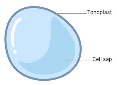
Functions of Vacuole
Vacuoles are organelles with diverse functions that vary depending on the cell type and organism. They play important roles in maintaining cellular homeostasis and carrying out various metabolic processes. The functions of vacuoles can be described as follows:
- Storage of Nutrients and Waste Materials: Vacuoles act as storage compartments for a wide range of substances, including nutrients, ions, sugars, proteins, pigments, and waste materials. They help regulate the concentration of these substances within the cell, ensuring their availability when needed and protecting the cell from the toxic effects of accumulated waste materials.
- Osmotic Balance and Homeostasis: Vacuoles play a crucial role in maintaining osmotic balance and cellular homeostasis. By controlling the movement of ions, particularly protons (H+ ions), into and out of the vacuole, they help regulate the pH and ion concentration within the cytoplasm. This balance is important for the overall functioning of the cell and its various metabolic processes.
- Regulation of Cell Volume and Turgor Pressure: Vacuoles contribute to the regulation of cell volume and turgor pressure in plant cells. By storing water and ions, they can control the osmotic pressure within the cell. When the vacuole absorbs water, it increases in size, exerting pressure against the cell wall and providing support to the plant structure. This turgor pressure is essential for maintaining plant rigidity and shape.
- Detoxification and Defense: Vacuoles can sequester and detoxify harmful substances, protecting the cell from their toxic effects. They store and isolate waste materials, such as metabolic byproducts and toxic compounds, preventing their interference with essential cellular processes. Additionally, vacuoles can also store defense compounds, such as alkaloids and secondary metabolites, which play a role in plant defense against pathogens and herbivores.
- Metabolic Functions: Vacuoles contain various enzymes that participate in different metabolic processes. These enzymes can be involved in the breakdown of macromolecules, the synthesis of secondary metabolites, and the recycling of cellular components. Vacuoles also participate in the degradation and turnover of organelles through autophagy.
In summary, vacuoles have multiple functions in cells. They serve as storage compartments, contribute to osmotic balance and homeostasis, regulate cell volume and turgor pressure, aid in detoxification and defense, and participate in various metabolic processes. The diverse functions of vacuoles are essential for the overall health and functioning of cells in both plant and animal organisms.
Vesicles
Vesicles are the structures in cells. They can be formed naturally through processes such as exocytosis, endocytosis or transport of material within the cell, or they may be created artificially, they are referred to as liposomes. There are different kinds of vesicles such as vacuoles, transport and secretory vesicles, based on their functions.
Structure of Vesicles
Vesicles are small membrane-bound structures found within cells. They play important roles in intracellular transport, storage, and communication. The structure of vesicles can be described as follows:
- Lipid Bilayer: Vesicles are surrounded by a lipid bilayer, similar to the structure of the plasma membrane. The lipid bilayer consists of phospholipids arranged with their hydrophobic tails facing inward and their hydrophilic heads facing outward. This arrangement creates a barrier that separates the contents of the vesicle from the surrounding cytoplasm.
- Liquid or Cytosol: The interior of a vesicle, also known as the lumen, contains liquid or cytosol. The contents of the vesicle can vary depending on its specific function. Vesicles can store various substances, such as proteins, lipids, ions, and neurotransmitters, and transport them to specific cellular locations.
- Lamellar Phase: The lipid bilayer of the vesicle forms a lamellar phase, which is the outer layer surrounding the liquid or cytosol. The lamellar phase provides stability to the vesicle and helps maintain the integrity of its contents. It acts as a barrier, preventing the diffusion of molecules into or out of the vesicle.
- Hydrophobic and Hydrophilic Ends: The lipid bilayer of the vesicle has two distinct ends. The hydrophobic tails of the phospholipids are located in the interior of the bilayer, facing each other. The hydrophilic heads of the phospholipids are oriented towards the aqueous environment, both inside and outside the vesicle. This arrangement allows the vesicle to interact with both hydrophobic and hydrophilic substances.
Overall, vesicles have a basic structure consisting of a lipid bilayer that encloses a lumen containing liquid or cytosol. The lipid bilayer has hydrophobic and hydrophilic ends, which contribute to the selective transport and storage functions of the vesicle. The structure of vesicles allows them to play crucial roles in intracellular processes, such as the transport of molecules between organelles, the release of neurotransmitters at synapses, and the storage of cellular components.
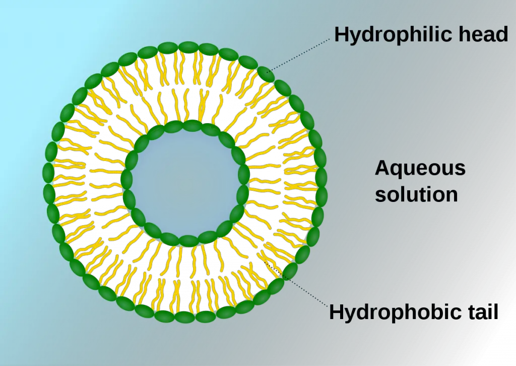
Functions of Vesicles
Vesicles are essential structures within cells that perform a variety of functions related to storage, transport, and communication. The functions of vesicles can be described as follows:
- Storage: Vesicles play a crucial role in the storage of various substances within cells. They can store molecules such as proteins, lipids, ions, and neurotransmitters. By sequestering these molecules within their lumen, vesicles provide a controlled environment for their storage, protecting them from degradation or unwanted interactions with other cellular components.
- Transport: Vesicles are involved in the transport of materials within the cell and between different cellular compartments. They act as vehicles for the movement of molecules from one location to another. Vesicles can transport substances such as proteins, lipids, and carbohydrates, ensuring their delivery to specific organelles or extracellular spaces. This transport function is crucial for maintaining cellular processes, including the synthesis and degradation of molecules.
- Exchange: Vesicles facilitate the exchange of molecules between cells. They can fuse with the plasma membrane, releasing their contents into the extracellular space or taking up molecules from the surrounding environment. This allows for communication and exchange of materials between adjacent cells or between cells and their environment. Vesicles are involved in processes such as cell signaling, immune responses, and the release of neurotransmitters in neuronal communication.
- Metabolism and Enzyme Storage: Vesicles are involved in cellular metabolism and the storage of enzymes. They can contain specific enzymes that participate in metabolic reactions. These enzymes may be involved in processes such as the breakdown of macromolecules, synthesis of cellular components, or detoxification of harmful substances. Vesicles provide a compartmentalized environment for these enzymes, ensuring their availability and proper functioning.
- Temporary Storage and Buoyancy Control: Vesicles can serve as temporary storage compartments within cells. They can store food reserves, such as starch or lipids, for later use by the cell. Additionally, in certain specialized cells, vesicles can help control the buoyancy of the cell. For example, in certain aquatic organisms, gas-filled vesicles allow the cell to adjust its position within the water column.
Overall, vesicles are versatile structures that contribute to the storage, transport, and communication functions within cells. They play vital roles in maintaining cellular homeostasis, regulating cellular processes, and enabling interactions between cells. The diverse functions of vesicles make them essential components of cellular function and organization.
A Brief Summary on Cell Organelles
| Cell Organelles | Structure | Functions |
| Cell membrane | A double membrane composed of lipids and proteins. Present both in plant and animal cell. | Provides shape, protects the inner organelle of the cell and acts as a selectively permeable membrane. |
| Centrosomes | Composed of Centrioles and found only in the animal cells. | It plays a major role in organizing the microtubule and Cell division. |
| Chloroplasts | Present only in plant cells and contains a green-coloured pigment known as chlorophyll. | Sites of photosynthesis. |
| Cytoplasm | A jelly-like substance, which consists of water, dissolved nutrients and waste products of the cell. | Responsible for the cell’s metabolic activities. |
| Endoplasmic Reticulum | A network of membranous tubules, present within the cytoplasm of a cell. | Forms the skeletal framework of the cell, involved in the Detoxification, production of Lipids and proteins. |
| Golgi apparatus | Membrane-bound, sac-like organelles, present within the cytoplasm of the eukaryotic cells. | It is mainly involved in secretion and intracellular transport. |
| Lysosomes | A tiny, circular-shaped, single membrane-bound organelles, filled with digestive enzymes. | Helps in the digestion and removes wastes and digests dead and damaged cells. Therefore, it is also called as the “suicidal bags”. |
| Mitochondria | An oval-shaped, membrane-bound organelle, also called as the “Power House of The Cell”. | The main sites of cellular respiration and also involved in storing energy in the form of ATP molecules. |
| Nucleus | A largest, double membrane-bound organelles, which contains all the cell’s genetic information. | Controls the activity of the cell, helps in cell division and controls the hereditary characters. |
| Peroxisome | A membrane-bound cellular organelle present in the cytoplasm, which contains the reducing enzyme. | Involved in the metabolism of lipids and catabolism of long-chain fatty acids. |
| Plastids | Double membrane-bound organelles. There are 3 types of plastids:Leucoplast –Colourless plastids.Chromoplast–Blue, Red, and Yellow colour plastids.Chloroplast – Green coloured plastids. | Helps in the process of photosynthesis and pollination, Imparts colour for leaves, flowers and fruits and stores starch, proteins and fats. |
| Ribosomes | Non-membrane organelles, found floating freely in the cell’s cytoplasm or embedded within the endoplasmic reticulum. | Involved in the Synthesis of Proteins. |
| Vacuoles | A membrane-bound, fluid-filled organelle found within the cytoplasm. | Provide shape and rigidity to the plant cell and helps in digestion, excretion, and storage of substances. |
FAQ
What is a cell organelle?
A cell organelle is a specialized structure within a cell that performs a specific function.
What are some examples of cell organelles?
Some examples of cell organelles include the nucleus, mitochondria, ribosomes, endoplasmic reticulum, Golgi apparatus, lysosomes, and peroxisomes.
What is the function of the mitochondria?
The mitochondria are responsible for producing energy for the cell through the process of cellular respiration.
What is the function of the endoplasmic reticulum?
The endoplasmic reticulum is responsible for protein synthesis and lipid metabolism.
What is the function of the Golgi apparatus?
The Golgi apparatus is responsible for modifying, sorting, and packaging proteins and lipids for transport within or outside the cell.
What is the function of lysosomes?
Lysosomes are responsible for the breakdown of cellular waste and debris.
What is the function of peroxisomes?
Peroxisomes are responsible for breaking down toxic substances within the cell.
What is the function of ribosomes?
Ribosomes are responsible for protein synthesis.
How do cells maintain the proper functioning of their organelles?
Cells maintain the proper functioning of their organelles through a variety of mechanisms, including regulating gene expression, recycling damaged organelles, and targeting dysfunctional organelles for degradation.
What happens when organelles malfunction or are damaged?
When organelles malfunction or are damaged, it can lead to cellular dysfunction and disease. Cells have various mechanisms for repairing or replacing damaged organelles, but severe or chronic damage can lead to irreversible cellular damage or death.
