Table of Contents
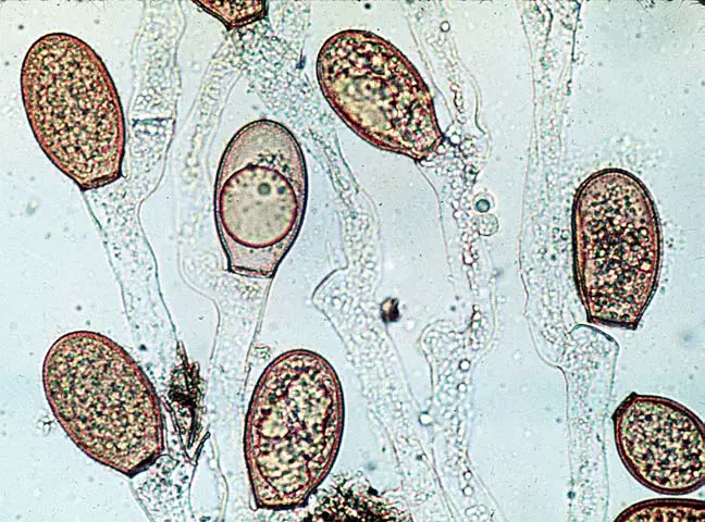
Chytridiomycota is a sub-group of zoosporic organisms from the kingdom Fungi. They are also known as chytrids. Named after the Ancient Greek word khutridion, which means “little pot”, it refers to the structure that contains unreleased zoospores. The earliest diverging fungal lineages are the Chytrids. Their membership in kingdom Fungi can be seen with chitin cell wall, posterior whiplash flagellum and absorptive nutrition. They also use glycogen to store energy and produce lysine through the a-amino acid (AAA).
The chytrids can be saprobic and degrade refractory materials like chitin or keratin. They also sometimes act as parasites. Since the discovery of Batrachochytrium, the causal agent in chytridiomycosis, there has been a marked increase in research on chytrids.
Definition of Chytridiomycota
Chytridiomycota is a phylum (kingdom Fungi), that has zoospores, or motile cells, with a whiplash structure (flagellum) at the end. Most species are microscopic and can be found in either freshwater or moist soils. Many species are parasites of animals and algae, or live on organic matter (as saprobes). A few species of the order Chytridiales can cause plant disease. One species, Batrachochytrium desndrobatidis has been proven to cause disease in frogs, amphibians, and other animals.
- However, chytrids are more than “first” due to the age of their fossils. Study of the evolutionary relationships among chytrids, fungi and other fungal groups has shown that they are either the sister group to the rest of the fungal groups or a paraphyletic base assemblage. This suggests that chytrids could provide a useful picture of the ancestors and descendants of fungi.
- The aquatic habitat of chytrids is not terrestrial, but they are primarily aquatic. This suggests that fungi, like plants and vertebrates, probably started in water.
- Flagellated gametes are a characteristic of Chytrids — the flagellum allows them to swim and their reproductive cells have flagellums. Flagella is not a characteristic of other fungi, so it seems that this trait was lost at some time in the evolution of other fungi. This is consistent with the information we have about close relatives of the fungi that also have flagellae.
- Chitin strengthens chytrids’ cell walls like other fungi. One subgroup, Hyphochytrids, has cellulose, which is a unique trait among living fungi. Chitin is an important characteristic of fungi.
- Chytrid ecology and morphology can vary greatly. Some are freshwater and some are marine. Others are parasites of plants and dipterans. While others live off decaying plants or insect parts, some chytrids are both freshwater and marine.
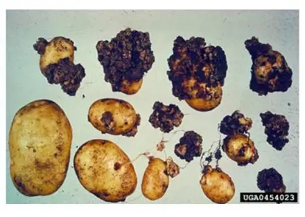
- Some are unicellular while others coenocytic. Others produce mycelium that is similar to other fungi. With the exception of those that parasitize algae and cause potato wart (Synchytrium Endoboticum) or those used in experimental research (e.g. Allomyces).
- Chytridiomycota eat both living and dead organisms. They are heterotrophic.

Habitats of Chytridiomycota
Although Chytrids can be classified as aquatic fungi they are often considered terrestrial. However, those that live in soil particles’ capillary network are known to thrive. The chytrids can be isolated from many aquatic habitats including rivers, lakes, bogs and springs. They also thrive in terrestrial habitats such as soils that are acidic, alkaline, temperate forest soils and rainforest soils. Many chytrid species are widespread and widely distributed, resulting in the belief that they are diverse and multi-species.
Periglacial soils are one of the most unlikely terrestrial environments where chytrids can thrive. Because of the high water content and the pollen rising from below the timberline, the population of Chytridiomycota species can be sustained.
Chytridiomycota characteristics

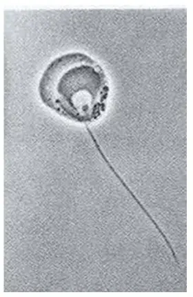
- Chytridiomycota, a phylum of fungi (kingdomFungi)distinguished by having zoospores (motile cells) witha single, posterior, whiplash structure (flagellum).
- Most species are microscopic and can be found in freshwater or soil. Many species are parasites of animals and algae, or live off organic matter (as saprobes).
- Chitin is the main component of cell wall.
- Use an absorptive eating style.
- These cells are not uniflagalated.

- The zoosporangium allows for sexual reproduction through zoopsore production.
- Zoospore has one posterior flagellum.
- Variable Vegetative Thalus – can range from multinucleated, globose to hyphal forms.
- Growth can be either indeterminate or determinate.
- The cell’s flagellum is attached within the cell to the blepharoplast.
- Some species have a nuclear cap made up of RNA.
- It protects the nucleus located at the anterior end the cell. The majority of these members are found in water.
- They can be found in the soil. Many of them are dependent on plants with higher economic value.
- These primitive members are microscopic.
- They contain a vegetative body that is coenocytic and acellular.
- This class has the main characteristic of producing uniflagellate reproductive cell (zoospores or planogametes).
- The single flagellum, which is posteriorly inserted, is of whiplash type. Opisthocont is a zoospore that has a posteriorly placed flagellum.
- The members of this division have unicellular redumentary and mycelial-thalli.
- The majority of their cell walls are composed of chitin.
- There are two types of flagellated spores: gametes and flagellated spores. The posterior whiplash flagellum is the same for gametes and zoospores.
- Variable sexual reproduction can occur. It may be anisogamous, isogamous, or oogamous.
- Chytridiomycota’s distinctive characteristic is the ultrastructure of zoospore.
- This division includes a single class, Chytridiomycetes. There are also three orders: Monoblepharidales, Blastocadiales and Chytridiales.

- It does not have a cell wall in its earlier stages (Olpidium).
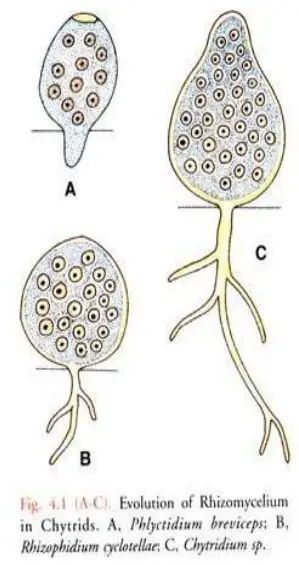
- The unicellular thallus of advanced species is divided at one point into fine, branching, hairs called rhizoids, which aid in anchorage as well as intake of nutrients (Rhizophidium).
- A branched form of rhizomycelium is found in more complex members. They are eucarpic.
- Even more advanced forms have a sparse mycelium with a few filamentous hyphae.
- Advanced members have a mycelium that consists of typical hyphae interwoven into a eucarpic meshwork. The hyphe are coenocytic.
- Chitin is the main constituent of the cell that forms the hyphal wall.
- A glucan is also available.
- The septa are normally kept inactive during vegetative phases, but they can be seen as solid plates that delimit reproductive organs.
- The sporangia are the asexual reproductive organs. Each produces a small number of tiny, uninucelate, and uniflagellate, opisthocont-zoospores.
- For a brief time, the liberated zoospores can swim. Each zoospore retracts its flagellum, and then undergoes encystment. The encysted spore germinates after a brief rest.
- It is possible for sexual reproduction to be anisogamous, or isogamous. Some people are oogamous.
Chytridiomycota Examples
- Orpinomyces joyonii- found in the intestines of cattle.
- Allomyces anomalus- can withstand extreme temperatures.
- Synchytrium endobioticum- a species that causes the “potato wart”

Thallus Forms of Chytridiomycota
- The thallus forms of Chytrids are microscopic and can be used to adapt for their life as biotrophs, necrotrophs, or saprotrophs of organic material.
- The simplest form of thallus is the holocarphic, which only contains the sporangium (without the rhizoids), and occurs within a substrate or host.
- The eucarpic thallus consists both of a sporangium as well as root-like structures called rhizoids.
- A thallus with only one sporangium is monocentric. However, it can be considered polycentric if there’s more than one.
- The diameter of sporangia may vary from less than 10mm (Longcore and al. 2016) and over 100 mm in Sparrow 1960.
- The eucarpic thallus is either internal or external to the food source. When epibiotic, the rhizoids anchor it to the substrate.
- Rhizoids are thought to play a role in the uptake of nutrients. They can be small and delicate, or large and coarse, which increases food absorption.
- Although Rhizoids have high levels of mitochondria, endoplasmicreticulum, vacuoles and vesicles, they lack nuclei.
- The thallus is still developing and there is an open space between the rhizoids, sporangium, and the rhizoids. However, as the sporangium matures, and gets closer to zoospore production, a septum containing the plasmodesmata develops.
- This means that although there is still a pathway for nutrient uptake, the portal for organellar movement has been blocked.
- The eucarpicmonocentric thallus’s size determines its extent. Therefore, the monocentric thallus tends to have a localized growth rate and has a determinate growth. The number of thalli it produces will determine its ability to colonize a substrate and compete with filamentous species.
- Some chytrids have a more complex thallus, with multiple sporangia and polycentric segments joined by mycelial/rhizoidal segmentes, the Rhizomycelium.
- Polycentric rhizomycelial thallus growth, in contrast to monocentric thallus growth, is indeterminate. This means that growth can be extensive branched and efficient in radiating into the substrate.
- Although chytrids with Rhizomycelia are more common on cellulosic substrates, they can also be found on chitin or keratin.
- Monoblephs often produce a filamentous, hyphal-like structure with a basal holdfast.
- The cytoplasm is very vacuolated and gives the hypha an unusual foamy appearance. These filaments have sporangia at their apex.
- Harpochytrium or Oedogoniomyces has a significantly reduced thallus, which consists of sporangium and a holdfast cells.
- Hyaloraphidium curvatum is a newly recognized monobleph. It was previously thought to be a colorless relative to algae. However, its sporangia produce autospores instead of spores.
Sexual Reproduction of Chytridiomycota
Monoblephs have oogamous sexual reproduction. In three of six monobleph genera, fusion occurs between a nonmotile embryo and a motile fertilized sperm. Single flagella sperms are found in Antheridia. Oospheres can be produced in Oogonia. Sperms fertilize the oospheres. The zygote forms a thick wall, which is used in perennation.
Monoblepharis zygote becomes motile when fertilization is complete. The sperm’s flagellum propels the zygote. However, zygotes from Monoblepharella or Gonapodya produce thick walls immediately and become oospores upon fertilization. The reproductive cycle is controlled by the preferred temperature and light regimes. This includes asexual reproduction at lower temperatures (8-15 C), and sexual at higher temperatures (25-35 C).
Additional evidence has been provided by light microscopic examinations of Siphonaria variabilis, Polyphagus Euglenae and Polyphagus euglenae that nuclear fusion is preceded by resting spore formation in the chytrids. There are many strategies that can be used to recommend genetics, including:
- As in Synchytrium, plant pathogenic species, Fusion between motile gamestes
- Gametangial copulation is where one gametangium transmits its protoplasm to another, as described by Sporophlyctis.
- Gametangial contact is where the contents of one gametangium move through a conjugation tube to the other gametangium. The zygotic restingspore forms as in Zygorhizidium.
- Somatogamy involving fusion of rhizoidal filaments as in Chytriomyces variabilis and Siphonaria hyalinus.
Systematics of Chytridiomycota
New orders for monophyletic linesages within the Chytridiomycetes have been identified based on ultrastructural and molecular analyses. The Rhizophlyctidales have been seperated from the Spizellomycetales.
The current class Chytridiomycetes includes ten orders. These were delineated based upon molecular and comparative character analyses.
- Chytridiales
- Cladochytriales
- Rhizophydiales
- Lobulomycetales
- Rhizophlyctidales
- Spizellomycetales
- Polychytriales
- Gromochytriales
- Mesochytriales
- Synchytriales
Structure of Chytridiomycota
- Chytridiomycota has unicellular and mycelial Thalli.
- Although cell walls are made from chitin and one group contains walls made from cellulose, the majority of them are cell walls.
- Cell growth can take place in unicellular or multicellular myceliums of aseptate-hyphae.
- The thallus is typically unicellular, and may have limited hyphal growth.
- It is not mycelial.
- Although hyphal cells are coenocytic (although this is not true where there are reproductive structures),
- The ultrastructure of the Zoospore is a distinctive characteristic of Chytridiomycota.
- The structure shows ribosomes aggregating around a nucule, which is not enclosed in nuclear caps.
- A nuclear cap is an extension to the nuclear membrane.
- Chytridiomycota has one or two flagella.
Life cycle of Chytridiomycota
Chytridiomycota can reproduce sexually by using zoospores. Zoospores can swim until they find a suitable substrate for asexual reproduction. The zoospore attaches to itself and feeds on its host. Cytoplasm grows, meiotic divisions take place, and a cell-wall forms around the original. As the cell develops, protoplasm grows. Finaly, the protoplasm is cleaved, which results in individual zoospores, which are released through a porous membrane. The dominant haploid method of sexual reproduction is the haploid dominant. It is dependent on the isomorphic alternation between generations. The gametothallus is a haploid thallus that produces male and female gametes. They are both terminal and subterminal and occur in pairs. Female gametes are more orange-colored than male gametes. Female gametes are also larger than males. Females are attracted by males who produce sirenin. Males will be attracted to females if they produce parisin.
The sporothallus is a diploid thallus. Two types of zoosporgia are produced by the sporothallus: meitosporangium (zoosporgangium) and resistantsporangium (meiosporangium). Zoosporangia can produce diploid zoospores that can be used for asexual reproduction. It is possible to have sexual reproduction that is isogamous or anisogamous. Allomyces macrogynus is one species that has a sporic cycle. This occurs in plants, but is uncommon for fungi. An anisogamous species is A. macrogynus.
Ecological functions of Chytridiomycota
- Batrachochytrium Dendrobatidis: This is the chytrid Batrachochytrium, which causes chytridiomycosis. It is a disease that affects amphibians. This disease was discovered in Australia and Panama in 1998. It is believed to be the main cause of global amphibian decline. The fungus was responsible for the extinction in 1989 of the golden toad and the death of a large portion of the Kihansi spray toad population in Tanzania.
- Other parasites: Other parasites include the chytrids, which mainly infect algae as well as other eukaryotic or prokaryotic microbes. The infection can cause severe damage to the primary production of the lake. Parasitic chytrids may have a significant impact on the food webs of lakes and ponds, according to some reports. Plant species may also be infected by chytrids; Synchytrium endobioticum, a key potato pathogen, is one example.
- Saprobes: The most important ecological function that chytrids can perform is their decomposition. These ubiquitous, global organisms are responsible to the decomposition of refractory substances, such as pollen and cellulose, chitin and keratin. Chytrids can also live on pollen and attach threadlike structures called rhizoids to the pollen grains.
References
- Taylor, Thomas N. (2015). Fossil Fungi || Chytridiomycota. , (), 41–67. doi:10.1016/b978-0-12-387731-4.00004-9
- Money, Nicholas P. (2016). The Fungi || Fungal Diversity. , (), 1–36. doi:10.1016/b978-0-12-382034-1.00001-3
- Letcher, Peter & Longcore, Joyce & Standridge, Sharon & Porter, David & Powell, Martha & Griffith, G.W. & Vilgalys, Rytas. (2006). A molecular phylogeny of the flagellated fungi (Chytridiomycota) and description of a new phylum (Blastocladiomycota). Mycologia. 98. 860-71. 10.3852/mycologia.98.6.860.
- https://ucmp.berkeley.edu/fungi/chytrids.html
- https://microbewiki.kenyon.edu/index.php/Chytridiomycota
- https://www.sciencedirect.com/topics/immunology-and-microbiology/chytridiomycota
- https://www.britannica.com/science/morel
- https://en.wikipedia.org/wiki/Chytridiomycota
- http://website.nbm-mnb.ca/mycologywebpages/NaturalHistoryOfFungi/Chytridiomycota.html
- https://www.cambridge.org/core/books/abs/introduction-to-fungi/chytridiomycota/EF320FEFF4DAD274CB74EBEA64CCC4C1
- http://www.botany.hawaii.edu/faculty/wong/Bot201/Chytridiomycota/Chytridiomycota.htm
- http://www.vpscience.org/materials/Fungi-1.pdf
- https://link.springer.com/content/pdf/10.1007%2F978-3-319-28149-0_18.pdf
- https://milnepublishing.geneseo.edu/botany/chapter/chytrids/
- https://cdnsciencepub.com/doi/10.1139/b00-009
