Table of Contents
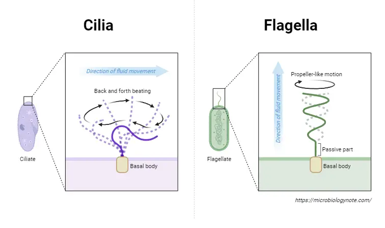
Cilia as well as Flagella can be described as tiny structures that attach to prokaryotic and eukaryotic cells. They are elongated from the outer surface on their cell. They aid in the movement for unicellular living organisms. They are mainly composed of microtubules and basal bodies. For single-celled organisms both flagella and cilia are crucial for motion. However multicellular organisms, flagella and cilia assist in the flow of fluids as well as other materials within the body tubes as well as carrying out the task of moving cells or the cells of a group. There is a distinction between flagella and cilia, and they differ by their length, size appearance and types of cells they are connected.
What are Flagella?
Flagella are whip-like , unbranched extensions that extend from the cell’s body. The structure of a flagellum is three major parts to the flagellum: filament, hook, and basal body. They are also longer than cilia and each cell has a number of flagella. The number of flagella differs from one to eight in the cells. Additionally, they are found in prokaryotic cells, like bacteria. In humans, they are plentiful in gametes.
Usually, flagella exit out of the cell at one location and display unruly or wave-like movements. However, in bacteria the number of flagella and their arrangement are different. In relation to number and arrangement certain bacteria are monotrichous whereas others are amphitrichous as well as lophotrichous and peritrichous.

Types of flagella
There are three kinds of flagella: bacterial, archaeal , and eukaryotic.
- Bacterial filaments are made of helical material which are able to rotate as screws. They are present inside E. coli, Salmonella Typhimurium and E. coli. There could have one, two, or many flagella within a cell. These flagella allow for the movement of bacteria.
- The archaeal flagella look similar to bacterial flagella , however they have an unique structure that is devoid of central channels.
- Eukaryotic flagella can be described as complex cell projections that whip back and back. (e.g. the sperm cells, which makes use of its flagellum for propelling itself along in the reproductive tract of females.
What are Cilia?
Cilia are hairy extensions that extend from the cell’s body. They are less long than flagella. Additionally, they exist in huge numbers per cell. Additionally, they are found primarily in eukaryotic cells like protozoa, yeasts, macrophages and fungi and sperm cells, as well as white blood cells and even in the respiratory tracts of human beings as well as other. Humans’ respiratory tract is awash with these , which prevent the entry of smog, dust and other harmful substances to the lungs.
Additionally the two types of cilia, nonmotile and motile (primary) the cilia. Nonmotile cilia perform a sensorial function, while the motile cilia assist in the process of locomotion.
Different types of cilia
There are two kinds of cilia: primary and non-motile the cilia.
- Primary or non-motile cilia are present in virtually every single cell of mammals, and as their name suggests, they are not beatable. They are found in the sensory organs of humans, like the eyes and nose.
- Motile cilia are located on the cell’s surface and beat in a rhythmic way. They are located inside the lining of trachea (windpipe) and clean dirt and mucus out of the lung. Female mammals also experience the beating of the cilia within the fallopian tubes transports the ovum out of the uterus to the Ovarian duct.
Diseases
The inability to function properly of flagella and cilia can result in a myriad of issues for human beings. For instance,
- If the cilia inside the fallopian tubes aren’t functioning correctly, then fertilized ovum won’t be able to reach the uterus, and will can result in an an ectopic pregnancy.
- A defect in the primary cilium inside the renal tube cells may result in polycystic renal illness (PKD).
- The condition of flagellum may also cause male infertility since the sperm cells are not mobile and therefore cannot reach the ovum.
What are the Similarities Between cilia and flagella
- The flagella and the cilia are tiny structures that are tiny.
- Cell appendages are cells and are made up of microtubules.
- In the first place, they are composed of proteins.
- They are also thread-like structures which protrude away from the cells.
- The primary purpose of both is to aid the process of locomotion.
- In addition, flagella and the cilia can also be considered organelles.
- They also have similar structures, which are arranged in 9+2.
What is the difference between cilia and flagella?
| Character | Cilia | Flagella |
| Definition | Cilia are hairy, short like appendages that grow out from the top of living cells. | Flagella are thread-like, long appendages that are found on the outside of living cells. |
| Etymology | Originated from Latin word meaning “eyelash”. | Originated from Latin word meaning “whip”. |
| Single form | Cilium | Flagellum |
| Found in | Eukaryotic cells | Prokaryotic and eukaryotic cells |
| Distribution | In protozoans belonging to the class ciliate and ciliated epitheliums of the metazoan larvae of some platyhelminthes, the echinodermata, mollusc, and the an annelid. | In certain bacterial cells, protozoans belonging to the class Flagellata Choanocytes of sponges, spermatzoa, and spermatzoa of metazoan as well as among plants in the gamete and algae cells. |
| Length | Short and hairy like organelle (5-10u) | Long wipe like organelle (150u) |
| Thickness | More size than flagella. They measure between 0.3 up to 0.5 millimeters thick. | The flagella attached to the edge of the bacteria is about 20-25 millimeters (0.02 up to 0.025 millimeters) thin. |
| Number | Numerous | Lesser in Number.Prokaryotes may have multiple flagella. |
| Density | Numerous (hundreds) per Cell | A few (less that 10, if any)) per cell |
| The position of the cell | The cell’s surface is affected by this. | The presence at one or two ends, or all across the surface. |
| Organization | Have a central bundle of microtubules referred to as the axoneme. It is comprised of nine doublet microtubules on the outer edges surround one central pair of singlet microtubules. The distinctive “9 + 2” arrangement of microtubules can be seen when the axoneme can be observed in cross-section with an electron microscope. | Eukaryotic flagella are extremely similar in the way they are organized to the cilia.Flagella prokaryotic are simpler in structure comprised by flagellin (53KDa subunit). |
| Beating synchronization | Cilia beats in a coordinated rhythm, either at the same time (synchronous) or one following one (metachronic). | They are able to beat one another. |
| Motion type | It is a rotational motion, similar to a motor that is very swiftly moving. | Prokaryotes’ movements in the rotary direction.Bending movement in eukaryotes. |
| Motion of swimming | Cilia moves in the same way as the stroke of breasts | Flagella move with an oar-like look. |
| Energy Production | Cilia utilize kinesin, which is an ATPase-related enzyme that generates energy to carry out the movements. | They are powered through protons-motive force that is generated by the plasma membrane in prokaryotes.ATP-driven in eukaryotes. |
| Types | Two kinds of cilia can be present in eukaryotic cells: primary and non-motile cilia as well as motile cilia. | Three types of flagella can be identified: archaeal, bacterial and Eukaryotic. |
| Purpose | Aids in locomotion, or moving substances on the outside that the cells (for instance the cilia in cells that line the fallopian tubes which move the ovum towards the uterus, or the cilia that line the respiratory tract’s cells which move particles of matter towards the throat, where mucus has been kept in) as well as Aeration, feeding circulation etc. | Assistance is mainly for locomotion. |
| Functions | Other than the sperms, cilia are mammalian systems is not used for locomotion. | Extend the plasma membrane, and are utilized to move the entire cell. |
| Examples | Cilia are present inside Paramecium | Flagella present in Salmonella |


