Table of Contents
Digestive System Of Frog
- The digestive system of a frog is a complex structure designed specifically for its dietary needs and lifestyle. This system is essential for breaking down food, absorbing nutrients, and eliminating waste. Therefore, understanding its components and functions is crucial for a comprehensive knowledge of frog biology.
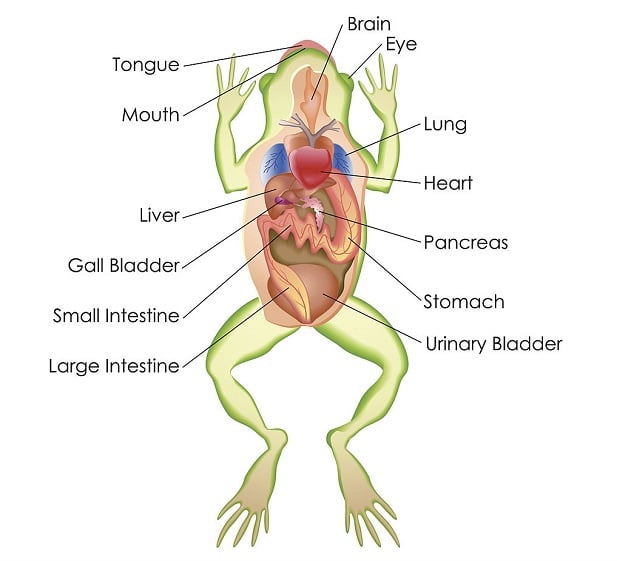
- The frog’s digestive system primarily consists of the alimentary canal and the associated digestive glands. The alimentary canal is a continuous tube that runs from the mouth to the cloaca, facilitating various processes such as digestion, absorption, and mastication. Besides the alimentary canal, the digestive glands play a pivotal role by producing enzymes essential for the digestion of consumed food.
- Starting at the beginning of the alimentary canal, the wide mouth opens into the buccal cavity. This cavity houses a large, muscular, and sticky tongue, which is attached at the front and free at the back, ending in a forked edge. When a frog spots an insect, it swiftly flicks out its tongue, capturing the insect with its sticky surface. Then, the tongue retracts, bringing the insect into the mouth for further processing.
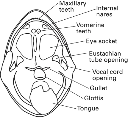
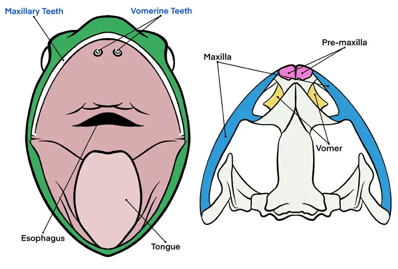
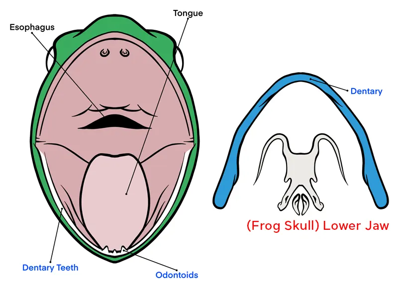
- Inside the buccal cavity, there are specific dental structures that aid in the initial breakdown of food. A row of pointed maxillary teeth can be found in the upper jaw. Additionally, two groups of vomerine teeth are located near the internal side of the nostrils. However, it’s essential to note that the lower jaws lack any teeth.
- Following the buccal cavity, the food moves through the pharynx and into the oesophagus, a narrow tube responsible for transporting food to the stomach. The oesophagus then leads to the stomach, where the primary digestion occurs. From the stomach, the digested food progresses into the intestine, where nutrients are absorbed. The remaining undigested food then moves into the rectum and is eventually expelled through the cloaca.
- In conclusion, the frog’s digestive system is a marvel of biological engineering, tailored to its specific needs. Every component, from the sticky tongue to the cloaca, has a distinct function, ensuring the efficient processing of food. The system’s emphasis on function, combined with its clear and concise design, showcases the intricate balance of nature.
Features of Digestive system of Frog
The digestive system of a frog comprises several key features that are specialized for its mode of life and diet. Here are the main features:
- Mouth: The mouth of a frog is wide, allowing for the consumption of large prey relative to its size. It is the entry point to the alimentary canal.
- Teeth: Frogs have teeth, but they are not used for chewing. Instead, the teeth hold prey and prevent it from escaping. The teeth are homodont and acrodont, meaning they are all similar in shape and are not set in sockets but rather are attached directly to the jaw bone.
- Tongue: The tongue is large, muscular, and sticky, capable of being rapidly extended to capture prey. It is bifurcated at the end and is attached at the front of the mouth, allowing for quick protrusion and retraction.
- Buccal Cavity: The buccal cavity is lined with ciliated columnar epithelial cells and contains mucous glands that secrete mucus to lubricate the food. Frogs lack salivary glands, so the mucus also aids in the initial stages of digestion.
- Pharynx: The pharynx is a muscular part of the throat that aids in swallowing. Frogs can depress their eyeballs into the roof of their buccal cavity to help push food down into the pharynx.
- Esophagus: This is a short tube that transports food from the mouth to the stomach. It is relatively narrow and serves mainly as a conduit.
- Stomach: The stomach is the primary site for the breakdown of food using digestive enzymes and acids. It churns the food to mix it with digestive juices.
- Small Intestine: The small intestine is where most nutrient absorption occurs. It is lined with villi that increase the surface area for absorption.
- Large Intestine: Also known as the colon, the large intestine absorbs water and electrolytes from the remaining indigestible food matter.
- Cloaca: The cloaca is a common chamber into which the digestive, urinary, and reproductive tracts all open. It is the final site before waste is expelled from the body.
- Liver and Pancreas: These are the main digestive glands in frogs. The liver produces bile, which aids in fat digestion, while the pancreas produces a variety of enzymes that further break down carbohydrates, proteins, and fats.
- Gallbladder: Frogs have a gallbladder that stores bile produced by the liver until it is needed in the small intestine.
These features work in concert to ensure that the frog can efficiently capture, ingest, digest, absorb nutrients from, and finally expel the remains of its food.
Alimentary Canal
- The alimentary canal of a frog is a critical component of its digestive system, serving as the pathway through which food is ingested, digested, and excreted. This canal is a complete, long, and coiled tube with varying diameters, extending from the mouth to the cloaca. It is comprised of several distinct sections, each with a specific function in the digestive process.
- It consists of:
- Buccal cavity
- Pharynx
- Oesophagus
- Stomach
- Small intestine
- Large intestine
- Cloaca
- Beginning with the buccal cavity, this area serves as the entry point for food intake. The buccal cavity is equipped to capture and initiate the breakdown of food, preparing it for further digestion. Then, the food passes through the pharynx, a muscular funnel that aids in the swallowing process.
- Subsequently, the oesophagus, a narrow tube, transports the food from the pharynx to the stomach. The oesophagus’s primary function is to serve as a conduit, moving food efficiently to the next stage of digestion without any significant digestion occurring within it.
- The stomach is a muscular organ that uses both mechanical and chemical processes to further break down the food. Enzymes and gastric juices mix with the food to create a semi-liquid mixture called chyme. After sufficient processing in the stomach, the chyme moves into the small intestine.
- The small intestine is a narrow, coiled tube where the majority of nutrient absorption occurs. It is lined with villi, small finger-like projections that increase the surface area for absorption. The nutrients from the digested food are absorbed through the walls of the small intestine into the bloodstream.
- Following the small intestine is the large intestine, which is shorter and wider. The large intestine’s primary role is to absorb water and electrolytes from the remaining indigestible food matter and to compact the waste into feces.
- Finally, the waste material enters the cloaca, which is a common chamber for the digestive, urinary, and reproductive tracts in frogs. The cloaca expels the waste from the body through the cloacal aperture.
- In summary, the alimentary canal of a frog is a specialized system designed for the efficient processing of food. Each section, from the buccal cavity to the cloaca, plays a vital role in the frog’s overall digestion and nutrient absorption. This sequential and detailed explanation underscores the importance of each part of the alimentary canal in maintaining the frog’s digestive health.
1. Mouth
- The mouth of a frog is a vital component of its digestive system, serving as the initial point of entry for food. It is characterized by a very wide gap that spans from one side of the snout to the other. Structurally, the mouth is bounded by two bony jaws. These jaws are enveloped by immovable lips, which play a role in the frog’s ability to capture and hold onto its prey.
- The upper jaw of the frog is fixed and does not exhibit any movement. In contrast, the lower jaw is flexible, allowing it to move up and down. This movement of the lower jaw facilitates the opening and closing of the mouth, enabling the frog to consume food efficiently.
- Furthermore, the mouth’s primary function is to aid in the ingestion of food. This process, known as ingestion, is the first step in the frog’s digestive journey. Once the food is captured and ingested through the mouth, it then proceeds to the next stages of the alimentary canal for further digestion and nutrient absorption.
- In summary, the mouth of a frog is not just a simple opening but a complex structure equipped with specific features that optimize its feeding habits. From the wide gap that allows for the consumption of larger prey to the flexible lower jaw that aids in capturing and holding onto food, every aspect of the frog’s mouth is purposefully designed to support its dietary needs.
2. Buccal cavity of frog
The buccal cavity, also known as the oral cavity or mouth, is an essential component of the frog’s digestive system. It serves as the initial point of entry for food and plays a crucial role in the frog’s feeding mechanism.
Structure and Components of the Buccal Cavity:
- Size and Shape: The buccal cavity of a frog is large, wide, and shallow, allowing it to accommodate and capture sizable prey.
- Lining: The inner lining of the buccal cavity consists of ciliated columnar epithelial cells. These cells play a role in moving mucus and trapped particles.
- Mucous Glands: Embedded within the lining are mucous glands. These glands secrete mucus, which serves to lubricate the food, facilitating its movement down the alimentary canal. This lubrication is especially vital because frogs lack salivary glands.
- Teeth: The frog’s teeth are not designed for chewing. Instead, they function to grasp and hold onto prey. The teeth are homodont, meaning they are all similar in shape, and acrodont, indicating they are directly attached to the jaw bone without sockets. The upper jaw possesses teeth, while the lower jaw does not. Additionally, the roof of the mouth contains vomerine teeth, which aid in holding prey.
- Tongue: The frog’s tongue is a remarkable organ. It is large, muscular, and sticky, with the ability to be rapidly extended out of the mouth to capture prey. The anterior end of the tongue is fixed to the lower jaw, while the posterior end is free and bifurcated. This unique arrangement allows the frog to flick its tongue out with precision, adhering to insects and other prey items.
- Internal Nostrils: Located at the front of the buccal cavity, near the vomerine teeth, are the internal nares or nostrils. These openings connect the nasal cavities to the buccal cavity and play a role in respiration.
- Bulging of Orbits: Behind the vomerine teeth, the roof of the buccal cavity exhibits two large oval areas. These areas represent the inward bulging of the eyeballs. When swallowing food, the frog depresses its eyes, causing these orbits to push inward, which in turn helps push the food towards the pharynx.
Function of the Buccal Cavity:
The primary function of the buccal cavity is to receive and hold food. The mucus secreted by the mucous glands aids in the initial stages of digestion and helps in swallowing. The teeth and tongue work in tandem to capture and hold prey, preventing it from escaping. The internal nostrils facilitate the passage of air, allowing the frog to breathe even when its mouth is closed.
3. Pharynx
- The pharynx, situated posteriorly to the buccal cavity, serves as a crucial segment in the frog’s digestive and respiratory systems. There isn’t a distinct boundary separating the buccal cavity and the pharynx, leading some experts to refer to them collectively as the bucco-pharyngeal cavity. This region is characterized by several openings that facilitate various functions.
- Central to the pharynx’s floor is a prominent median elevation, which houses the glottis. The glottis is a longitudinal, slit-like aperture that plays a pivotal role in respiration. It provides a passage to the laryngo-tracheal chamber, ensuring the efficient exchange of gases. Besides the glottis, the roof of the pharynx features a broad eustachian aperture on each lateral side. These apertures connect to the middle ear, playing a role in auditory functions.
- Furthermore, in male frogs, the pharynx’s floor exhibits additional features. Specifically, there are small openings of vocal sacs located on either side, near the junction of the two jaws. These vocal sacs are instrumental in producing mating calls and other vocalizations.
- Transitioning from the pharynx, there is a tapering structure known as the gullet. The gullet is a broad opening that serves as a conduit to the esophagus, ensuring the smooth passage of food particles from the pharynx to the subsequent stages of digestion.
- Therefore, the pharynx, with its intricate structure and multiple apertures, plays a multifaceted role in the frog’s physiology. From facilitating respiration to aiding in digestion and vocalization, its functions are diverse and essential for the frog’s survival.
4. Oesophagus
- The oesophagus, a vital component of the digestive system, is characterized as a short, broad, muscular, and highly flexible tube. Its structure is specifically designed to facilitate the smooth passage of food from the mouth to the stomach. The inner surface of the oesophagus is lined with mucous epithelial tissue, which is folded in a longitudinal manner. These longitudinal foldings play a crucial role in the digestive process. When food passes through, these folds allow for the expansion of the oesophagus, ensuring that the food moves seamlessly without causing any damage or obstruction.
- Besides its structural features, the oesophagus also has a functional aspect. Embedded within its mucous epithelial lining are mucous glands. These glands are responsible for secreting an alkaline digestive juice. This secretion aids in the initial breakdown of food, preparing it for further digestion in the subsequent stages of the digestive tract.
- Furthermore, as the oesophagus extends downwards, it experiences a noticeable enlargement. This enlargement signifies the point where the oesophagus merges with the stomach, located within the peritoneal cavity. Therefore, the oesophagus serves as a bridge, ensuring that ingested food is efficiently and safely transported from the oral cavity to the stomach for further digestion.
5. Stomach
- The stomach, a pivotal organ in the digestive system, is strategically positioned on the left side of the body cavity. Anchored to the dorsal body wall by a mesentery known as the mesogaster, the stomach exhibits a unique structure tailored for its multifaceted functions. This organ can be described as a broad, slightly curved tube or bag, approximately 4 cm in length, fortified with thick muscular walls.
- Diving deeper into its anatomy, the stomach is bifurcated into two distinct sections. The anterior segment, expansive and broad, is termed the cardiac stomach. Conversely, the posterior segment, which is shorter and narrower, is referred to as the pyloric stomach. A noteworthy feature of the stomach’s inner surface is the presence of several prominent longitudinal folds. These folds are not mere anatomical embellishments; they play a crucial role in allowing the stomach to distend when it receives food, ensuring optimal storage and processing.
- Furthermore, the stomach’s mucous epithelium is a hub of activity. It houses multicellular gastric glands that are responsible for secreting the enzyme pepsinogen. In addition to these, there are unicellular oxyntic glands that produce hydrochloric acid, a vital component for digestion. As we move towards the pyloric end of the stomach, there is a noticeable constriction. This region is safeguarded by the pyloric valve, which serves as a gateway into the small intestine. This valve is not just a passive barrier; it is a dynamic circular ring-like sphincter muscle that meticulously regulates the flow of digested food from the stomach to the intestine.
- Therefore, the stomach is not just a storage unit for food. It is an active site where rigorous digestion takes place, facilitated by a symphony of enzymes and acids. Besides its role in digestion, the stomach’s design, from its longitudinal folds to its specialized glands, underscores its importance in ensuring that the food we consume is broken down efficiently, paving the way for nutrient absorption in subsequent stages of the digestive tract. In essence, the stomach, with its intricate structure and functions, stands as a testament to the marvels of biological engineering.
6. Intestine
The intestine, following the stomach, is a crucial segment of the alimentary canal, tasked with the continuation of the digestive process and the absorption of nutrients. It is a tubular, elongated, and coiled structure, affixed to the dorsal body wall by mesentery, ensuring its secure placement within the body cavity.
The intestine is divided into two main parts:
- Small Intestine: The small intestine is the primary site for the absorption of nutrients. It is here that the majority of the digestive process is completed. The inner lining of the small intestine is characterized by numerous folds, known as villi and microvilli, which significantly increase the surface area for absorption. These structures facilitate the transfer of nutrients into the bloodstream.
- Large Intestine: The large intestine, though shorter in length compared to the small intestine, is wider in diameter. Its primary function is to absorb water and electrolytes from the remaining indigestible food matter and to transport the useless waste material out of the body. The large intestine culminates in the cloaca, which serves as a common passage for the excretory, digestive, and reproductive systems.
The transition from the stomach to the intestine is marked by a sphincter that regulates the passage of chyme – the partially digested food mixed with stomach acids. As the chyme enters the small intestine, it encounters digestive enzymes and bile, which continue the breakdown of proteins, carbohydrates, and fats.
The small intestine is further subdivided into three sections: the duodenum, jejunum, and ileum. Each section plays a specific role in digestion and absorption. The duodenum receives bile and pancreatic juices that aid in digestion, the jejunum is primarily involved in nutrient absorption, and the ileum absorbs vitamin B12 and bile salts, and whatever products of digestion were not absorbed earlier.
a. Small intestine of Frog
The small intestine is a vital component of the digestive system, responsible for the absorption of nutrients and the continuation of the digestive process initiated in the stomach. Structurally, it is a long, coiled, and narrow tube, measuring approximately 30cm in length. The small intestine is anchored mid-dorsally to the body wall by mesenteries, ensuring its stability within the body cavity.
The small intestine can be further divided into two distinct parts:
- Duodenum: This is the anterior section of the small intestine. It is characterized by its U-shaped structure, running parallel to the stomach. The duodenum receives digestive juices from both the liver and the pancreas via a common hepatopancreatic duct. These juices, namely bile and pancreatic juice, play a crucial role in the breakdown of food. The internal mucous lining of the duodenum forms low transverse folds, enhancing its surface area for efficient digestion.
- Ileum: Representing the posterior section, the ileum is the longest part of the alimentary canal. It forms several loops before enlarging posteriorly to join the rectum. Unlike the duodenum, the internal mucous lining of the ileum forms many longitudinal folds. However, it is essential to note that in higher vertebrates, the ileum does not possess true villi or definite glands and crypts.
The mucosal lining of the small intestine, apart from housing the intestinal glands, comprises two primary cell types:
- Goblet Cells: These are large cells characterized by the presence of oval vacuoles and granular substances. Their primary function is to produce mucus, which aids in the smooth passage of food. The nucleus of goblet cells is located near the base of the cell.
- Absorbing Cells: In contrast to goblet cells, absorbing cells are smaller in size. They play a pivotal role in the absorption of digested nutrients. Similar to goblet cells, their nuclei are also situated near the base.
In terms of functionality, the small intestine is the primary site for both the digestion of food and the absorption of digested nutrients. The presence of various digestive enzymes and a large surface area, courtesy of the mucosal folds, ensures that the food is broken down efficiently and the nutrients are effectively absorbed into the bloodstream.
b. Large intestine of Frog
- The large intestine, also known as the rectum in frogs, is a relatively short and wide tube, approximately 4cm in length. It extends straight from the small intestine and culminates at the cloaca, which is the common exit point for the intestinal and urinary tracts. The transition from the small intestine to the large intestine is marked by the ileo-rectal valve, which functions to regulate the passage of digested material.
- The large intestine’s primary function is the re-absorption of water from the remaining indigestible food matter, effectively concentrating waste before it is excreted. This process is facilitated by the numerous low longitudinal folds in the inner lining of the large intestine, which increase the surface area for water absorption.
- The terminal opening of the large intestine, the anus, is guarded by a muscular structure known as the anal sphincter. This sphincter muscle maintains control over the expulsion of feces, ensuring that waste is excreted at an appropriate time.
- In summary, the large intestine or rectum in frogs serves as the final segment of the digestive tract, where the last stages of water re-absorption occur, and feces are stored prior to defecation. Its structural features, such as the longitudinal folds and the anal sphincter, are critical to its function in maintaining water balance and controlling waste excretion.
7. Cloaca
- The cloaca is a small, sac-like terminal part located at the posterior end of the body. It serves as a common chamber into which the digestive, urinary, and reproductive tracts open. Specifically, both the anus and the urinogenital apertures open into the cloaca. The cloaca itself opens to the outside environment through the vent or cloacal aperture.
- Functionally, the cloaca plays a vital role in the excretion and reproductive processes. It collects waste products from the digestive and urinary systems and facilitates their expulsion from the body. Additionally, in many animals, the cloaca is involved in the reproductive process, serving as the site for the transfer of sperm during mating.
- In summary, the cloaca is an essential anatomical structure that manages waste excretion and reproductive functions in certain animals. Its presence ensures that these processes are carried out efficiently and effectively.
Histological Structure of the Alimentary Canal
The alimentary canal’s wall is histologically organized into four concentric layers. From the innermost to the outermost, these layers are:
- Mucosa:
- The innermost layer, also known as the mucous membrane.
- It is characterized by numerous folds, pits, and glands.
- Its primary functions are digestion and absorption of digested foods.
- The mucosa is further subdivided into:
- Epithelium: The outermost layer composed of simple columnar epithelium, both glandular and ciliated, resting on a thin basement membrane.
- Lamina Propria: A delicate layer of connective tissue housing blood capillaries, lymph, and nerves.
- Muscularis Mucosae: A thin layer of smooth muscle containing both outer and inner circular muscles.
- Submucosa:
- A protective layer composed of coarse connective tissue, elastic fibers, fat, lymph vessels, blood vessels, and nerve cells.
- In mammals, it houses glands.
- The Meissner plexus, comprising nerve fibers and cells, is also present here.
- Muscularis:
- Composed of outer longitudinal and inner circular smooth muscles arranged in spirals.
- A connective tissue layer with a network of nerve cells and nerve fibers from the Auerbach plexus is situated between these muscle layers.
- Serosa or Visceral Peritoneum:
- The outermost layer, absent in the esophagus.
- Comprises a thin layer of connective tissue and an outermost layer of flattened cells.
- It is attached to the mesentery, the lining of the body cavity.
Histological Variations in Different Parts of the Alimentary Canal
The aforementioned layers, especially the mucosa, undergo significant changes in different parts of the alimentary canal, adapting to the specific functions of each segment.
a. Oesophagus:
- Lacks the visceral peritoneum as it lies outside the coelom.
- Its wall contains both involuntary and voluntary muscle fibers, with striated muscle fibers present in its upper part.
- The mucous membrane lining is made of stratified epithelial cells, not columnar cells.
- Goblet cells produce mucus, facilitating the smooth passage of food. The mucous epithelium invaginates into the submucosa, forming tubular branched glands that secrete pepsin, an enzyme essential for protein digestion.
b. Stomach:
- Possesses a thick wall comprising all typical layers of the alimentary canal.
- The stomach wall is longitudinally folded internally, which unfolds when the stomach expands.
- The mucous epithelium consists of columnar mucous-secreting gland cells. These glands, embedded in the lamina propria, are invaginations of the mucous epithelium and are known as gastric glands. These glands differ in structure and function in various parts of the stomach.
c. Small Intestine or Duodenum:
- Covered by all four typical layers.
- Its mucosa is thick and forms irregular transverse folds, enhancing the absorptive surface area.
- The mucosa comprises columnar epithelial cells with mucus-secreting glands. However, there are no villi, definite glands, or crypts as in higher vertebrates.
d. Ileum:
- Also possesses the standard four layers.
- The mucosa is divided into multiple folds of varying sizes, projecting into the intestinal lumen.
- The mucosa consists of tall columnar epithelial cells interspersed with absorptive and goblet cells.
e. Large Intestine or Rectum:
- Comprises all four layers of the alimentary canal.
- The muscularis mucosa is less developed, while the muscular coat is thick and populated with voluntary muscle fibers.
- The mucosa is dense with longitudinal folds and contains numerous tubular goblet cells that produce mucus. Near the anus, the mucosa epithelium becomes stratified.
In conclusion, the alimentary canal’s histological structure is intricate, with each layer and segment tailored to perform specific functions. Understanding these details provides insights into the complex processes of digestion and absorption in vertebrates.
Digestive glands of frog
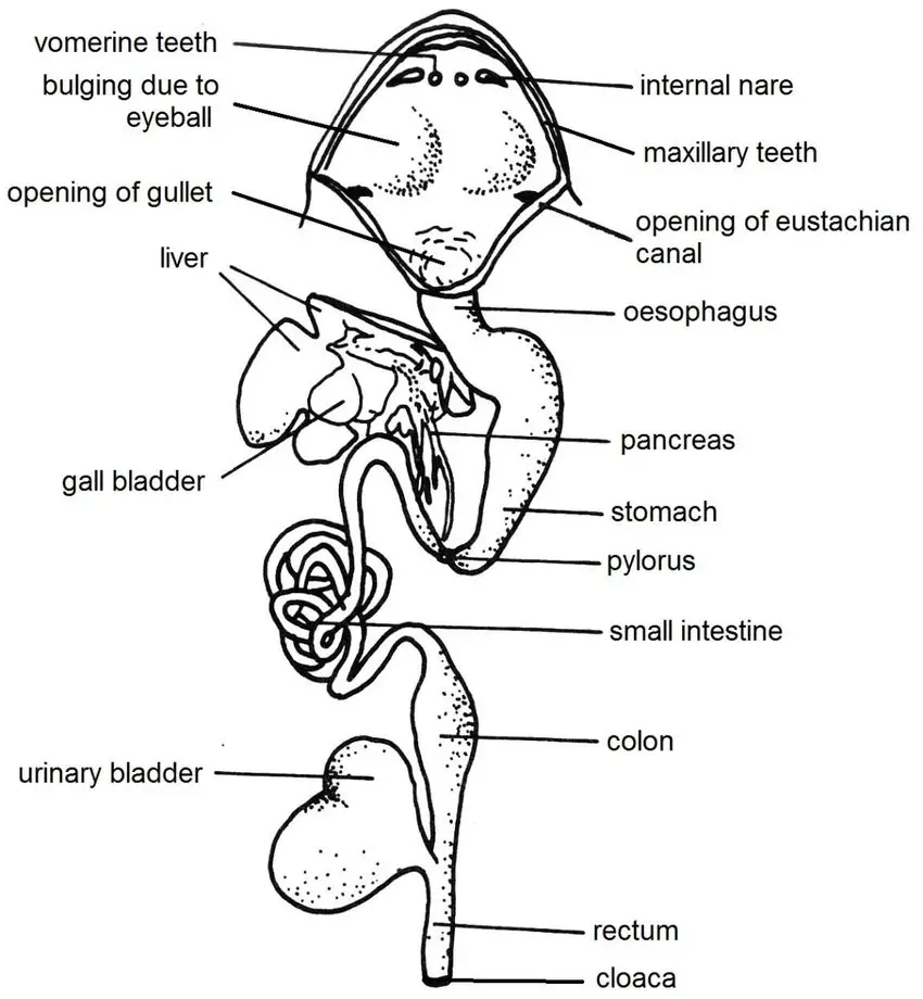
The digestive system of the frog is equipped with specialized glands that play a pivotal role in the digestion and metabolism of food. Two primary glands associated with the alimentary canal of the frog are the liver and the pancreas. This article provides a detailed and sequential explanation of these glands, emphasizing their structure, histology, and functions.
1. Liver:
The liver, a vital organ in the vertebrate body, plays a multifaceted role in various physiological processes. This article delves into the structure, histology, and functions of the liver, providing a comprehensive understanding of its significance in the body.
a. Structure of the Liver
- The liver is the largest gland in the body, characterized by its reddish-brown color. It is strategically positioned in the anterior part of the abdominal cavity, adjacent to the lungs and heart. Structurally, the liver is divided into two primary lobes: the left and the right. These lobes are interconnected through a narrow liver connection. Furthermore, the left lobe is bifurcated into two subsections. Nestled between these lobes is a thin-walled, greenish sac known as the gallbladder. This sac serves as a storage unit for the bile produced by the hepatic cells of the liver.
- Bile, an essential secretion of the liver, travels from the liver to the gallbladder through cystic ducts. Additionally, it can directly enter the bile duct via small liver channels. The convergence of the cystic and hepatic ducts gives rise to the common bile duct, which traverses through the pancreas before emptying into the duodenum. This duct, due to its connection with the pancreatic ducts, is often referred to as the hepatopancreatic duct.
- It’s crucial to note that bile, while essential, does not contain digestive enzymes. Its primary function is to emulsify fats, making the liver a non-digestive organ in a strict sense.
b. Histology of the Liver
Histologically, the liver is a complex organ composed of numerous tubules or lobules. These lobules interconnect, forming a reticulated structure, leading to its designation as the retinal gland. Each lobule is demarcated by connective tissue, which houses liver ducts, bile capillaries, and blood sinuses and capillaries.
The fundamental units of these lobules are the polyhedral, glandular hepatic cells. These cells are rich in nuclei, cytoplasm, protein granules, glycogen droplets, fats, and occasionally, dark brown pigment particles. These hepatic cells are systematically arranged in columns, separated by bile capillaries, which eventually coalesce to form larger hepatic ducts. These ducts subsequently connect to the gallbladder through cystic ducts or merge to form the primary bile duct.
For its sustenance and functionality, the liver receives blood via the hepatic arterial vein and the hepatic portal vein. These vessels supply the essential materials required for bile production.
c. Functions of the Liver
The liver, being a multifunctional organ, is responsible for a plethora of physiological processes:
- Bile Production: The liver secretes an alkaline, watery bile rich in bile salts, bile pigments, cholesterol, lecithin, and water. Bile salts, such as sodium bicarbonate, taurocholate, and glycocholate, play a pivotal role in digestion. Sodium bicarbonate modulates the pH of intestinal food, while the other salts activate pancreatic lipase and reduce fat friction for better emulsification.
- Metabolic Functions: The liver is instrumental in carbohydrate metabolism. It stores excess sugar as glycogen through a process called glycogenesis. Conversely, when blood sugar levels dip, the liver converts this glycogen back to glucose via glycogenolysis.
- Protein Regulation: The liver maintains blood protein levels. Unused amino acids are converted to ammonia in the liver, which then reacts with carbon dioxide to form urea and other nitrogenous wastes. These wastes are subsequently excreted by the kidneys.
- Detoxification: The liver is the body’s primary detoxification center. It eliminates various excretory materials, which are then excreted through feces. Additionally, it neutralizes hazardous substances like prussic acid, converting them to non-toxic compounds like potassium sulphocyanide.
- Blood Functions: In embryonic stages, the liver is responsible for red blood cell production. In adults, it aids in the destruction of old erythrocytes. Furthermore, it produces vital blood-clotting agents like prothrombin and fibrinogen and secretes heparin, an anticoagulant.
- Storage and Other Functions: The liver stores essential minerals like iron and copper and synthesizes vitamin A. It also plays a role in immune defense by eliminating bacteria and foreign substances from the blood.
2. Pancreas:
The pancreas, a vital gland in the vertebrate body, plays a dual role in endocrine and exocrine functions. This article delves into the structure, histology, and functions of the pancreas, providing a comprehensive understanding of its significance in the body.
a. Structure of the Pancreas
- The pancreas is an intricately branched, flattened gland, exhibiting a pale hue. It is strategically positioned in the mesentery, nestled between the stomach and the duodenum. The pancreas is connected to the common bile duct, into which its pancreatic ducts open. This connection is often referred to as the hepatopancreatic duct due to its association with both the liver and the pancreas.
b. Exocrine Function of the Pancreas
- The exocrine part of the pancreas is segmented into various parts and lobules, held together by connective tissue. This section houses the pancreatic ducts, lymph vessels, blood vessels, and nerves. The lobules are characterized by numerous branching tubules or alveoli, also known as acini. Each alveolus is structured with pancreatic glands that have a pyramidal configuration surrounding a central cavity. These alveoli are interconnected by ductules, which subsequently merge to form larger ducts, culminating in the formation of the primary pancreatic ducts. As these ducts traverse the pancreas, they expand and merge with the bile duct.
- The cells of the pancreas, characterized by large nuclei and non-granular cytoplasm, are responsible for secreting pancreatic juice. This juice is enriched with a plethora of enzymes that play a pivotal role in the digestion of carbohydrates, proteins, and fats present in food.
c. Endocrine Function of the Pancreas
Within the acini, enveloped by connective tissue, are minuscule clusters of cells known as pancreatic islets or islets of Langerhans. These islets, which exhibit a spherical shape and a compact arrangement, are lightly stained. They are composed of three distinct types of cells, each separated by capillaries:
- Alpha Cells: These cells, which possess achromatic nuclei and large acidophilic granules, are responsible for producing the hormone glucagon. Glucagon plays a crucial role in elevating blood sugar levels. A deficiency in this hormone can lead to hypoglycemia.
- Beta Cells: Characterized by small, rounded structures with deeply stained nuclei and orange-brown granules, these cells produce the hormone insulin. Insulin is indispensable for carbohydrate metabolism, regulating glycogen storage in muscles and the liver, maintaining blood sugar levels, and enhancing the ability of tissues to oxidize glucose for energy.
- D Cells: These cells, distinguished by vesicular nuclei and basophilic granules, play a role in the endocrine functions of the pancreas.
Insulin is paramount for the body’s metabolic processes. It regulates the storage of glycogen in muscles and the liver, controls blood sugar levels, and augments the capacity of tissues to oxidize glucose, providing a vital energy source. A deficiency in insulin production or function can lead to a condition known as diabetes or hyperglycemia.
Physiology of digestion in frog
The physiology of digestion in a frog is a sequential process that begins with the capture and ingestion of food and ends with the absorption and assimilation of nutrients. Frogs, being strictly carnivorous, consume a diet consisting of insects, worms, crustaceans, molluscs, small fish, and occasionally other small amphibians. The process of digestion in frogs is devoid of mastication; instead, prey is captured with a rapid flick of the tongue and swallowed whole.
1. Ingestion and Bucco-Pharyngeal Phase:
The absence of salivary glands in frogs means that lubrication of food is facilitated by mucus secreted from the lining of the bucco-pharyngeal cavity and esophagus. The food is then propelled down the esophagus by peristalsis, a wave-like contraction of the muscular wall.
2. Gastric Digestion:
Upon reaching the stomach, the food is subjected to gastric digestion for up to 2-3 hours. Gastric glands in the stomach wall secrete gastric juice, which contains hydrochloric acid and the inactive pre-enzyme pepsinogen. The acidic environment converts pepsinogen into active pepsin, which then catalyzes the hydrolysis of proteins into peptones and proteases. The acidic medium softens the food, provides an optimal pH for enzyme activity, and serves as a defense mechanism by killing potential pathogens.
3. Hormonal Regulation:
The presence of food in the stomach triggers the secretion of the hormone gastrin, which in turn stimulates the production of hydrochloric acid. The resulting semi-digested acidic food is referred to as chyme. When the chyme reaches an appropriate consistency, the pyloric sphincter relaxes, allowing the chyme to enter the duodenum.
4. Intestinal Digestion:
The entry of acidic chyme into the duodenum prompts the release of several intestinal hormones, each with specific roles:
- Enterogastrone: This hormone circulates back to the stomach to inhibit further secretion of gastric juice.
- Cholecystokinin: It induces the gallbladder to contract, releasing bile into the duodenum via the hepatopancreatic duct.
- Secretin and Pancreozymin: These hormones stimulate the pancreas to release pancreatic juices into the duodenum.
- Enterocrinin: It activates the secretion of intestinal juice, known as succus entericus.
5. Digestive Juices and Enzymatic Action:
The digestion in the intestine is facilitated by the combined action of bile, pancreatic juice, and intestinal juice:
- Bile: Secreted by the liver, bile is an alkaline fluid that emulsifies fats, neutralizes the acidity of chyme, and stimulates intestinal peristalsis.
- Pancreatic Juice: This alkaline juice contains enzymes that act on all three classes of food, breaking down proteins into amino acids, starch into maltose, and emulsified fats into fatty acids and glycerol.
- Succus Entericus: The intestinal juice contains a variety of enzymes that further break down polypeptides, disaccharides, and fats into their absorbable monomers.
6. Egestion, Absorption, and Assimilation:
- Egestion: The undigested material is moved into the rectum by peristalsis, stored temporarily, and then egested through the cloacal aperture.
- Absorption: The final products of digestion are absorbed through the walls of the small intestine, which is structurally adapted with folds and villi to increase the surface area. The mechanisms of absorption involve osmotic forces among other factors.
- Assimilation: The absorbed nutrients are utilized for energy production or assimilated into the body’s structures. Excess glucose may be stored as glycogen, and amino acids may be used for protein synthesis or converted into waste products like urea.
Throughout this process, the frog’s digestive system efficiently converts prey into vital nutrients, ensuring the animal’s survival and energy needs are met.
Functions of Digestive system of Frog
- Ingestion: The process begins with the frog capturing its prey, often insects, using its sticky, protrusible tongue. The food is then taken into the mouth.
- Mechanical Digestion: Once inside the mouth, the food is held by the backwardly pointed teeth present on the upper jaw. These teeth are not meant for chewing but to prevent the prey from slipping out.
- Transportation: The lubrication provided by the mucus secreted by the mucous glands in the buccal cavity aids in the smooth passage of food. The food is then pushed towards the pharynx, aided by the inward bulging of the eye orbits during swallowing.
- Chemical Digestion: The food then enters the stomach, where it is acted upon by gastric juices containing enzymes that break down proteins. The acidic environment of the stomach also helps in killing harmful bacteria.
- Absorption: The partially digested food moves from the stomach to the small intestine. The small intestine is the primary site for the absorption of nutrients. Here, enzymes from the pancreas and liver further break down the food, and the nutrients are absorbed into the bloodstream through the walls of the intestine.
- Water Absorption: The large intestine or colon primarily absorbs water and some minerals from the remaining undigested food matter.
- Excretion: The waste products, now in a semi-solid form, are passed into the rectum and are eventually expelled out of the body through the cloaca and vent.
- Role of Liver and Pancreas: Besides the primary digestive tract, the liver and pancreas play crucial roles in digestion. The liver produces bile, which aids in the emulsification of fats, while the pancreas secretes digestive enzymes into the small intestine.
- Respiration through the Buccal Cavity: Besides digestion, the buccal cavity in frogs also plays a role in respiration. The internal nostrils, or nares, allow for the passage of air from the nasal cavities to the buccal cavity and then to the lungs.
