Table of Contents
What is a Digital Microscope?
Digital microscopes are modern microscope which does not have an eyepiece. This is a huge contrast from an optical microscope. Digital microscopes have an electronic camera that acts as the detector as well as the imaging output gadget. It displays the images takes place via a computer’s screen or monitor, which defines the scope of the microscope’s digital.
The light source of this microscope is an internal LED source when compared to an optical microscope where the light source is located outside the microscope using an eyepiece. Thus, with this digital microscope human optical access is eliminated because the entire instrument is equipped with an image monitoring system.
There are various variations of the digital Microscope including the USB digitalized microscopes, which are expensive, industrialized digital microscopes including the Kohler illumination as well as the Phase-contrast Illumination. They come equipped with webcams, as well as a macro lenses.
A first digital Microscope was manufactured in 1986 in Tokyo, Japan which constituted the control box and lens that was attached with the camera. It is now known as The Hirox Company. LTD. Because of its computer connection, it is able to handle large amounts of digital information taken from the digital camera inside the microscope. In 2005, an sophisticated digital microscope was developed that did not require a computer. Instead it was an in-built unit that served as the monitor and computer. However, in 2015, the new digital microscope was built, equipped with an external computer with an USB connection that grew the life and performance that the machine. This reduced cables connecting to the external computer and the size of the computer was decreased.
They are equipped with an image processing software which alters the brightness of the image and contrast, size, and also crop the image.
Digital Microscope Principle
Digital Microscopes are equipped with the hardware and software capabilities necessary for specimen focusing and picture extraction. Using installed software, the specimen’s image is taken and displayed on the computer monitor. Depending on the software, visualised images may be captured as still photographs or moving films, as well as recorded, altered, cropped, labelled, and saved. The software can also be used to measure image sizes, enlarge them, and modify them in a variety of ways.
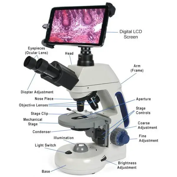
Parts of a Digital Microscope
The evolution of the Digital Microscope is based on two of its main features. The functions of input and out and in this respect the digital microscope composed of two components:

1. The hardware
The hardware is mostly the analog portion of the microscope. It comprises an illumination source, an analog microscope, the camera, and camera components. Cameras replace the eyepiece on the conventional microscope. The image taken from the specimen is focused by the camera. It is projected onto the computer’s screen, which is later saved and then processed.
2. The software
The software is the part of the microscope which contains cameras and drivers as well as the image processing software. It includes organized units that comprise the viewer unit, the brightness adjustment unit, the histogram equalization unit as well as the image scaling unit. the unit for cropping images. This unit constantly displays the specimen, and also captures the image taken by the microscope, which is then stored and processed based on the preferences of the user. The unit for adjusting the brightness of images increases the brightness of the image, based on how much light is focused onto the specimen. It adjusts the brightness of light that is reflected in the pixels. The brightness adjustment for the image is made by transforming the grayscale with an equation.
Jo = Ji + C, (whereby Jo is the output image, Ji is the input image, and C is the constant for brightness adjustment). C is set to a positive value to increase brightness, and in a negative value , it reduces the brightness.
The unit for image contrast enhancement is used to adjust the contrast of an image. Lower contracts display a narrow histogram whereas high contrast represented by a wide histogram. It is meant to increase the image’s contrast.
3. Objective lens
The objective lens is the lens located at the bottom of the microscope, closest to the specimen. It is used to collect light from the specimen and focus it onto the eyepiece or the digital camera.
4. Digital camera
The digital camera is the main component of a digital microscope. It captures images of the specimen and sends them to a computer or other display device for viewing.
5. Eyepiece
Some digital microscopes have an eyepiece, which is a lens that allows users to view the magnified image of the specimen directly through the microscope.
6. Stage
The stage is the platform on which the specimen is placed. It can be moved up and down or side to side to position the specimen in the field of view.
7. Illumination system
The illumination system is the source of light used to illuminate the specimen. It may consist of a lamp or LED light source, as well as various filters and diffusers to control the intensity and quality of the light.
8. Focus knob
The focus knob is used to adjust the focus of the image by moving the objective lens or the stage up and down.
9. Digital LCD Display
A digital LCD (liquid crystal display) is a type of display commonly used in digital microscopes to display the magnified images of a specimen. The LCD display is a flat panel display that uses liquid crystals to produce images. It is thin, lightweight, and energy-efficient, making it well-suited for use in portable and handheld digital microscopes.
The LCD display in a digital microscope is typically connected to the digital camera, which captures images of the specimen and sends them to the display for viewing. The user can adjust the magnification, focus, and lighting of the image using the controls on the microscope or the software. The LCD display allows users to view the magnified image of the specimen in real-time, as well as to store and share the images for further analysis or documentation.
Some digital microscopes also have an eyepiece, which is a lens that allows users to view the magnified image of the specimen directly through the microscope. However, many modern digital microscopes rely solely on the LCD display for viewing the image, as it provides a larger and more detailed view of the specimen.
Characteristics of a Digital Microscope
Digital microscopes are advanced imaging tools that use digital cameras to capture and display magnified images of small objects or samples. Some of the key characteristics of digital microscopes include:
- High magnification: Digital microscopes can provide high magnification, allowing users to view small details and features of a specimen at a very high resolution.
- Digital camera: Digital microscopes use a digital camera to capture images of the specimen, which can be displayed on a computer or other display device.
- User-friendly interface: Most digital microscopes have an intuitive user interface that allows users to easily adjust the magnification, focus, and lighting of the image, as well as to analyze and measure features of the specimen.
- Software: Digital microscopes often come with software that allows users to manipulate and analyze the images they capture. This can include features such as image processing, measurement tools, and annotation tools.
- Versatility: Digital microscopes can be used to view a wide range of specimens, including biological samples, microelectronic devices, and other materials. They can also be used for a variety of applications, including research, quality control, and medical diagnosis and treatment.
- Cost-effectiveness: Digital microscopes are generally more cost-effective than traditional optical microscopes, particularly for large-scale applications. They require less maintenance and have a longer lifespan, which can help to reduce overall costs.
Types of Digital Microscopes
Based on the use of the microscope as well as the user, various digital microscopes are manufactured and labelled, with the majority of them being lightweight and user-friendly. They can be used for anything from industrial and biomedical functions. The microscopes are used in conjunction with high-end digital microscope cameras and computer monitors.
- Biological Digital Microscopes: They are high-magnification microscopes with the ability to light sources beneath the mechanical stage. It has an objective of 4x-100x and either LED or halogen light sources.
- Fluorescence Digital Microscopes: These are optical microscopes which use fluorescence and phosphorescence as sources of light to create an image.
- Inverted digital Microscopes: They are trinocular microscopes where the sources of light and condenser are located on high on the stage, and the objective is located below the stage.
- Metallurgical digital Microscopes: Digital Microscopes with a Metallurgical Design can be used to observe metal surfaces or wire circuits, as well as transparent surface.
- Phase digital Microscopes: These are upright or inverted microscopes that are used to examine objects that aren’t stained and could be dead or live.
- Stereo Digital Microscopes: Stereo Digital Microscopes are able to reflect light from of the object, and can be utilized to see electrical components, artifacts Circuitry, plants, and art.
- Polarizing Digital Microscopes: These are a particular kind of digital video camera, which has an extremely magnifying lens and a number of ultra-bright LEDs. They are able to analyze 3D structures and the compositions of anisotropic objects, by with polarized light which creates waves of light that move in one direction.
- Digital USB Microscopes: These are microscopes that connect to computers through the USB connector. They feature a permanent connected camera, which is not the case with most digital microscopes, that have cameras connected to C-mount adapters and are able to be removed.
- Handheld Digital Microscopes: These are extremely modern digital microscopes which are built into an handheld microscope system. They can be is used to inspect surfaces as well as for forensics.
- Portable digital Microscopes: They are tiny digital microscopes that have built-in wireless technology. They are utilized to observe surfaces that are difficult to reach. They are mostly utilized for medical surface such as field inspections, as well as for dermatological research.
Operating Procedure of digital microscope
- Place the sample specimen on the microscope stage in the first step.
- Illuminate the sample with the proper light intensity and angle.
- Select the desired objective magnification in the third step.
- Using the coarse and fine focus adjustment knobs, bring the specimen sample into optimum focus.
- Observe the image of the specimen on the computer monitor and take the necessary measurements (width, length, diagonal, and circular measurements).
How does a digital microscopy work?
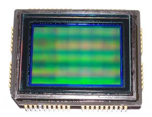
In standard digital microscopy, the specimen is initially positioned on the stage. The specimen is then lit according to the required light levels. The light rays reflected from the specimen pass through the objective lens of the microscope to generate an upright image within the microscope tube. Where the CCD (charge-coupled device) is located is where the actual image is created. The CCD processes light rays from this spot to create an enlarged image of the specimen on the monitor screen. If the microscope employs a digital camera as opposed to a CCD, the point at which the intermediate image is created coincides with the focus point of the camera lens.

By expanding the size of the monitor screen, the magnification of the picture produced by the CCD can be enhanced. The specimen’s width, length, diagonal, and circumference can be acquired from the screen itself. Some sophisticated digital microscopes have software for performing these measurements. Digital microscopy aids in acquiring images of various sorts of living and nonliving sample specimens that are crystal clear and of high quality.
Resolution of a digital microscope
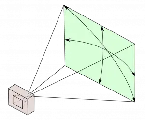
A digital microscope having a conventional 2 megapixel CCD generates a picture of 1600×1200 pixels. In this instance, the image resolution relies on the field of view of the camera lens. When the horizontal field of view is divided by 1600, the approximate pixel resolution can be calculated. The resolution of an image can be increased by creating a sub-pixel image.

The Pixel Shift Method is utilised to obtain photographs with a better resolution. In this technology, the CCD is physically moved using an actuator in order to take many overlapping images simultaneously. These numerous images are merged with photos captured by the microscope to provide images with sub-pixel resolution. This method provides sub-pixel information, and averaging a standard image is an established method for obtaining sub-pixel information. Certain software is built specifically to improve the visual resolution displayed on the monitor.
2D measurement
Digital microscopic systems, particularly high-end digital microscopic systems, typically measure the specimen in two dimensions. These measurements are performed on-screen by measuring the distance between two pixels. This is able to provide width, length, diagonal, and circle dimensions as well as additional structural data. Some digital microscopy systems are also capable of particle counting.
3D measurement
Some modern digital microscopy equipment can currently measure in three dimensions. This is accomplished using image stacking. Using a step motor, the microscopic system captures photos of the specimen from the lowest to the highest focal plane inside the field of view of the camera lens. These photos are then reconstructed to generate a 3D model based on image contrast in order to generate a 3D colour image of the specimen.
These 3D models can subsequently be utilised for calculations, although their accuracy is dependent on the step motor and depth of field of the camera lens. Digital microscopy is extensively utilised for a variety of material research applications, including tests with dynamic materials.
Uses of Digital Microscopes
Digital microscopes are advanced imaging tools that use digital cameras to capture and display magnified images of small objects or samples. They are widely used in various fields, including scientific research, medicine, engineering, and quality control. Some of the common applications of digital microscopes include:
- Microscopy: Digital microscopes are used to study and analyze the microstructures of various materials and biological specimens at high magnification. They can be used to identify cells, tissues, and other microstructures in biological samples, as well as to study the properties and characteristics of microelectronic devices, minerals, and other materials.
- Quality control: Digital microscopes are used to inspect and evaluate the quality and defects of various products, such as electronics, automotive parts, and medical devices. They can be used to identify and measure surface defects, such as cracks, scratches, and contamination, and to ensure that products meet specific quality standards.
- Research and development: Digital microscopes are used in various research and development (R&D) activities, such as material science, biology, and engineering. They can be used to study the properties and characteristics of materials and biological specimens, as well as to develop new technologies and products.
- Education and training: Digital microscopes are widely used in educational and training institutions to teach students about microscopy and to allow them to observe and study various specimens at high magnification.
- Medical diagnosis and treatment: Digital microscopes are used in medicine to diagnose and treat various conditions and diseases. For example, they can be used to examine tissue samples in a laboratory setting to diagnose cancer, or to perform microsurgeries, such as removing small tumors or repairing damaged tissues.
Advantages of Digital Microscopes
There are several advantages to using digital microscopes over traditional optical microscopes, which use lenses and light to magnify and view specimens. Some of the key benefits of digital microscopes include:
- High resolution: Digital microscopes use high-resolution digital cameras to capture detailed images of small objects or samples. This allows for more accurate and precise analysis and measurement of microstructures and features.
- Easy to use: Digital microscopes are relatively easy to use, with intuitive controls and user-friendly interfaces. They often come with software that allows users to adjust the magnification, focus, and lighting of the image, as well as to analyze and measure features of the specimen.
- Easy to share: Digital microscopes allow users to easily capture, store, and share images and videos of their observations. This can be useful for collaboration, education, and communication with colleagues or students.
- Versatility: Digital microscopes can be used to view a wide range of specimens, including biological samples, microelectronic devices, and other materials. They can also be used for a variety of applications, including research, quality control, and medical diagnosis and treatment.
- Cost-effective: Digital microscopes are generally more cost-effective than traditional optical microscopes, particularly for large-scale applications. They require less maintenance and have a longer lifespan, which can help to reduce overall costs.
Limitations of Digital Microscopes
Although digital microscopes offer many advantages over traditional optical microscopes, they also have some limitations that users should be aware of. Some of the potential drawbacks of digital microscopes include:
- Dependence on computer: Digital microscopes require a computer to display and analyze the images they capture. This can be inconvenient for users who do not have access to a computer or who prefer to view their observations directly through the eyepieces of a traditional microscope.
- Limited depth of field: Digital microscopes often have a limited depth of field, which means that only a small portion of the specimen is in focus at any given time. This can make it difficult to get a clear and detailed image of a three-dimensional specimen.
- Lower contrast: Digital microscopes can sometimes produce images with lower contrast than traditional optical microscopes, particularly when viewing transparent or low-contrast specimens. This can make it more difficult to see small or subtle features in the specimen.
- Sensitivity to light: Digital microscopes are sensitive to light, which can affect the quality of the images they capture. Users may need to use special lighting techniques or filters to reduce glare or reflections, or to enhance the contrast of the specimen.
- Vulnerability to damage: Digital microscopes are vulnerable to damage from spills, drops, or other accidents, which can cause costly repairs or replacement. They also require regular maintenance to ensure optimal performance.
5 Things You Should Know About Digital Microscopes
- One can create a digital microscope by incorporating a camera inside the microscope or by attaching a digital microscope camera to a conventional microscope.
- With an external digital microscope camera, you can utilise a variety of cameras with the same microscope and vice versa.
- The majority of digital microscopes and microscope cameras come equipped with picture capture and documentation software.
- Verify that the shop can provide assistance for the software; occasionally, after-sale support is useful.
- Since most computer monitors have a maximum resolution of 2 megapixels, you may not require a higher resolution than 2.0 megapixels if you do not intend to print in high quality frequently.
Digital microscope cameras
Digital microscope cameras are specialized digital cameras that are designed for use with microscopes. They are used to capture and display magnified images of small objects or specimens, and are an essential component of digital microscopes.
Digital microscope cameras are typically connected to the microscope via a cable or wireless connection, and are controlled using software on a computer or other display device. They have a high resolution and sensitivity, which allows them to capture detailed and accurate images of the specimen at high magnification.
Digital microscope cameras are used in a wide range of applications, including scientific research, medicine, engineering, and quality control. They can be used to view and analyze a wide range of specimens, including biological samples, microelectronic devices, and other materials.
Some features of digital microscope cameras include:
- High resolution: Digital microscope cameras have a high resolution, which allows them to capture detailed and accurate images of the specimen at high magnification.
- Sensitivity: Digital microscope cameras are sensitive to light, which allows them to capture images of specimens that may be difficult to see with the naked eye.
- Versatility: Digital microscope cameras can be used to view a wide range of specimens and can be used for a variety of applications.
- Compatibility: Digital microscope cameras are typically compatible with a wide range of microscopes, including traditional optical microscopes and digital microscopes.
- Software: Digital microscope cameras often come with software that allows users to control the camera, adjust the settings, and analyze and measure features of the specimen.
Difference between a stereo microscope and a digital microscope
A stereo microscope, also known as a binocular microscope, is a type of microscope that uses two eyepieces to provide a three-dimensional view of a specimen. It is commonly used for observing and analyzing small, solid objects, such as rocks, coins, and circuit boards, as well as for performing simple tasks, such as soldering or dissection.
A digital microscope, on the other hand, is a microscope that uses a digital camera to capture and display magnified images of a specimen on a computer or other display device. It can be used to view a wide range of specimens, including biological samples, microelectronic devices, and other materials.
One key difference between a stereo microscope and a digital microscope is the way they provide a magnified view of the specimen. A stereo microscope uses lenses and light to magnify the image and present it through the eyepieces, while a digital microscope uses a digital camera to capture and display a magnified image on a computer or other display device.
Another difference is the level of magnification and resolution. Digital microscopes often have higher magnification and resolution than stereo microscopes, which allows for more detailed and precise analysis of the specimen. However, stereo microscopes have a wider field of view and a greater depth of field, which can make it easier to observe and manipulate larger or three-dimensional specimens.
In general, stereo microscopes are more suitable for simple tasks and observations, while digital microscopes are better suited for more complex analysis and measurement of small specimens.
Examples of Digital microscope – Best digital microscope
Eyeclops digital microscope
The Eyeclops Digital Microscope is a portable and handheld digital microscope that allows users to view and capture magnified images of small objects or specimens. It is designed for use by both children and adults, and is marketed as a educational and scientific toy.
The Eyeclops Digital Microscope has a high magnification range, from 20x to 200x, and a built-in digital camera that captures and stores images of the specimen on a memory card. It also has an LCD display that allows users to view the magnified image in real-time. The microscope is powered by batteries and can be used for a variety of applications, including science education, hobbies, and quality control.
Some features of the Eyeclops Digital Microscope include:
- High magnification: The microscope has a magnification range of 20x to 200x, allowing users to view small details and features of a specimen at a very high resolution.
- Built-in digital camera: The built-in digital camera captures and stores images of the specimen on a memory card, which can be viewed on a computer or other display device.
- LCD display: The LCD display allows users to view the magnified image of the specimen in real-time, as well as to store and share the images for further analysis or documentation.
- Portable and lightweight: The Eyeclops Digital Microscope is portable and lightweight, making it easy to take it with you wherever you go.
- Educational and fun: The microscope is marketed as an educational and scientific toy, and is designed to be fun and engaging for children and adults alike.

About this item
- Magnify objects up to 800x!
- Built in 2.4” Color Screen!
- Turn off the Zoom and use as a regular Camera or video recorder!
- Take pictures and videoUse indoors & outdoors!
- Download via USB or MicroSD
To buy this product click Here: Buy Now
Celestron digital microscope
Celestron is a company that produces a range of digital microscopes for various applications, including scientific research, education, and quality control. Celestron digital microscopes are designed to be user-friendly and easy to use, and are suitable for a wide range of users, including students, educators, and professionals.
Some features of Celestron digital microscopes include:
- High magnification: Celestron digital microscopes have a high magnification range, allowing users to view small details and features of a specimen at a very high resolution.
- Digital camera: Celestron digital microscopes have a built-in digital camera that captures and stores images of the specimen on a memory card, which can be viewed on a computer or other display device.
- LCD display: Many Celestron digital microscopes have an LCD display that allows users to view the magnified image of the specimen in real-time, as well as to store and share the images for further analysis or documentation.
- Software: Celestron digital microscopes often come with software that allows users to control the microscope, adjust the settings, and analyze and measure features of the specimen.
- Versatility: Celestron digital microscopes can be used to view a wide range of specimens and can be used for a variety of applications, including scientific research, education, and quality control.
- Compatibility: Celestron digital microscopes are typically compatible with a wide range of computers and operating systems, making it easy to use them with various software and devices.

About this item
- True 5MP sensor to capture and save high resolution images and videos of your specimens
- 5 Element IR cut high quality glass lens ensures sharper images. Shutter Speed 1 second to 1 by 1000 second
- 20x to 200x powers, great for low power observation of 3D specimens (Note: Final magnification determined by monitor size)
- 4 foot USB 2.0 cable for easy maneuverability when viewing large surfaces
- Intuitive software with measuring features
- Windows and Mac compatible SOFTWARE and FIRMWARE: MicroCapturePro (Model B), Windows 7 (update 2.4.1) MicroCapturePro (Model B), MAC OS (update 2.4.1) MicroCapture Pro, Windows (update 2.3) MicoCapturePro (Model B), Windows 8 Windows 10 (update 2.4.1) MicroCapturePro , MAC OS (update 2.3)
- Software CD not included
To buy this product click Here: Buy Now
wifi digital microscope
A WiFi digital microscope is a type of digital microscope that uses a WiFi connection to transmit images of the specimen to a computer or other display device. It allows users to view and analyze the magnified image of the specimen remotely, without the need for a physical connection to the microscope.
WiFi digital microscopes are useful for applications where it is inconvenient or impractical to view the magnified image directly through the eyepieces of the microscope. They are also useful for collaboration, as they allow multiple users to view and analyze the specimen simultaneously from different locations.
Some features of WiFi digital microscopes include:
- WiFi connectivity: WiFi digital microscopes use a WiFi connection to transmit images of the specimen to a computer or other display device, allowing users to view the magnified image remotely.
- Digital camera: WiFi digital microscopes have a built-in digital camera that captures and stores images of the specimen on a memory card, which can be accessed and viewed via the WiFi connection.
- High magnification: WiFi digital microscopes have a high magnification range, allowing users to view small details and features of a specimen at a very high resolution.
- Software: WiFi digital microscopes often come with software that allows users to control the microscope, adjust the settings, and analyze and measure features of the specimen.
- Versatility: WiFi digital microscopes can be used to view a wide range of specimens and can be used for a variety of applications, including scientific research, education, and quality control.
- Compatibility: WiFi digital microscopes are typically compatible with a wide range of computers and operating systems, making it easy to use them with various software and devices.

About this item
🔍 STPCTOU WIRELESS DIGITAL MICROSCOPE – 50X-1000X magnification zoom, this digital microscope offers you a clear view of objects’ details and enables capture pictures, record video and save these wonderful moment. Nice tool to help your curiosity kids to explore the wonderful micro world.
🔍 8 ADJUSTABLE LED LIGHT – Built-in 8 LED lights and adjustable illumination with CMOS sensor image processing technology provides great image and video quality with 640×480 resolution. Flexible metal tripod offers an optimal and comfortable observation experience for you. Note: please remove the plastic protective cover before use.
🔍EASY TO USE – Only need to download the “Max-see” from APP store or Google play store and connect your phone to the microscope via WIFI, you can use the microscope easily. Taking an image or video simply just tap the Image/Video button of your device or press related APP trigger. Also support windows PC/mac OS by USB connection.
🔍MINI SIZE & RECHARGEBLE – With the portable mini size, USB rechargeable design, more than 3 hours of effective usage time, easy to put this mini microscope in your pocket and take it wherever you go. allows you to take it on your trips for children to study plants, minerals, insects, or have fun outdoor activities.
🔍GREAT COMPATIBILITY – The handheld digital zoom microscope magnifier included user-friendly software compatible with Windows XP/Vista/7/8, Mac & software, and tools for multiple purposes.
🔍WIFI CONNECTION FOR ANDROID AND IOS – 1.Download the software “Max-see” from Google Play or APP Store. 2. Long press the power button to turn on the microscope. 3. Connect the wifi “Max-see”(no password) which emits from the microscope. 4. Run the app and it is easy to use.
🔍USB CONNECTION FOR COMPUTER – 1. For win 10, you can directly plug into the USB port and search CAMERA in WINDOWS to find it and click on. 2. WIN 7/8, downloading an Amcap will solve this issue. 3. For MacBook, please use Macbooks’ bundled software Photo Booth or Quick Time Player directly. 4. Note: Please disable the default laptop camera in Windows! And you have to change your privacy settings for the camera, which needed to be changed to permit access.
🔍A FUNNY TOOL – This is an electron microscope, not a traditional microscope, Not suitable for professional serious biologists! This is definitely a very interesting thing for parents, adults, teachers, students, kids, children, collectors, testers, electronics’ repair folks, and inquisitive folks who interested in exploring the microscopic world.
🔍STPCTOU’S BRAND QUALITY ASSURANCE – We provide 12-Month Warranty for any quality issues. If you’re not 100% completely satisfied with your Digital Microscope, just simply contact us and we will refund or replace your product immediately. Please contact us first if you have any question about our Digital Microscope, our support team will try our best to help you solve all the issues. email reply within 24hs.
🔍WHAT YOU GET – 1x wireless digital microscope, 1x USB cable, 1x metal stand, 1x plastic base, 1x user manual, our 24-hour professional after-sales service.
To buy this product click Here: Buy Now
Jiusion digital microscope
Jiusion is a company that produces a range of digital microscopes for various applications, including scientific research, education, and quality control. Jiusion digital microscopes are designed to be user-friendly and easy to use, and are suitable for a wide range of users, including students, educators, and professionals.
Some features of Jiusion digital microscopes include:
- High magnification: Jiusion digital microscopes have a high magnification range, allowing users to view small details and features of a specimen at a very high resolution.
- Digital camera: Jiusion digital microscopes have a built-in digital camera that captures and stores images of the specimen on a memory card, which can be viewed on a computer or other display device.
- LCD display: Many Jiusion digital microscopes have an LCD display that allows users to view the magnified image of the specimen in real-time, as well as to store and share the images for further analysis or documentation.
- Software: Jiusion digital microscopes often come with software that allows users to control the microscope, adjust the settings, and analyze and measure features of the specimen.
- Versatility: Jiusion digital microscopes can be used to view a wide range of specimens and can be used for a variety of applications, including scientific research, education, and quality control.
- Compatibility: Jiusion digital microscopes are typically compatible with a wide range of computers and operating systems, making it easy to use them with various software and devices.

About this item
- Jiusion portable magnification is a useful and funny microscope for students, engineers, inventors, and others who need to magnify and explore the micro things.
- Be compatible with Mac, Window XP and above, Linux. This microscope isn’t compatible with iPhone/iPad.
- This magnification only support Android smartphone which has OTG function.(How to Check OTG? Download the free app”USB OTG Checker”)
- Built-in 8 LED lights, digital microscope’s 2 adjusting knob can change the focus and brightness.
- Connected to the devices, you can use the software to record the micro world, capture screenshot and record video. Besides you can use the Windows software’s measurement function to measure the least bit.
To buy this product click Here: Buy Now
Tomlov digital microscope
Tomlov is a brand of digital microscopes produced by the company Shenzhen Tomlov Science & Technology Co., Ltd. Tomlov digital microscopes are designed for use in a variety of applications, including scientific research, education, and quality control. They are known for their high magnification, high resolution, and user-friendly interfaces.
Some features of Tomlov digital microscopes include:
- High magnification: Tomlov digital microscopes have a high magnification range, allowing users to view small details and features of a specimen at a very high resolution.
- Digital camera: Tomlov digital microscopes have a built-in digital camera that captures and stores images of the specimen on a memory card, which can be viewed on a computer or other display device.
- LCD display: Many Tomlov digital microscopes have an LCD display that allows users to view the magnified image of the specimen in real-time, as well as to store and share the images for further analysis or documentation.
- Software: Tomlov digital microscopes often come with software that allows users to control the microscope, adjust the settings, and analyze and measure features of the specimen.
- Versatility: Tomlov digital microscopes can be used to view a wide range of specimens and can be used for a variety of applications, including scientific research, education, and quality control.
- Compatibility: Tomlov digital microscopes are typically compatible with a wide range of computers and operating systems, making it easy to use them with various software and devices.
USB digital microscope
A USB digital microscope is a type of digital microscope that uses a USB (Universal Serial Bus) connection to transmit images of the specimen to a computer or other display device. It allows users to view and analyze the magnified image of the specimen on a computer, as well as to store and share the images for further analysis or documentation.
USB digital microscopes are convenient and easy to use, as they do not require any specialized software or drivers to operate. They can be plugged into any computer with a USB port, making them a popular choice for users who do not have access to more advanced imaging equipment.
Some features of USB digital microscopes include:
- USB connectivity: USB digital microscopes use a USB connection to transmit images of the specimen to a computer or other display device, allowing users to view the magnified image on a computer.
- Digital camera: USB digital microscopes have a built-in digital camera that captures and stores images of the specimen on a memory card, which can be accessed and viewed via the USB connection.
- High magnification: USB digital microscopes have a high magnification range, allowing users to view small details and features of a specimen at a very high resolution.
- Software: USB digital microscopes often come with software that allows users to control the microscope, adjust the settings, and analyze and measure features of the specimen.
- Versatility: USB digital microscopes can be used to view a wide range of specimens and can be used for a variety of applications, including scientific research, education, and quality control.
- Compatibility: USB digital microscopes are typically compatible with a wide range of computers and operating systems, making it easy to use them with various software and devices.
Handheld digital microscope
A handheld digital microscope is a portable and compact digital microscope that can be carried and used easily in the field or in a laboratory. It is designed to be lightweight and easy to hold, with a compact size that allows it to be carried in a pocket or a bag.
Handheld digital microscopes are useful for applications where it is necessary to view and analyze small objects or specimens in the field or on the go. They are also useful for tasks that require a portable and lightweight microscope, such as quality control, maintenance, or repair.
Some features of handheld digital microscopes include:
- Portability: Handheld digital microscopes are portable and lightweight, making them easy to carry and use in the field or on the go.
- Digital camera: Handheld digital microscopes have a built-in digital camera that captures and stores images of the specimen on a memory card, which can be viewed on a computer or other display device.
- LCD display: Many handheld digital microscopes have an LCD display that allows users to view the magnified image of the specimen in real-time, as well as to store and share the images for further analysis or documentation.
- Software: Handheld digital microscopes often come with software that allows users to control the microscope, adjust the settings, and analyze and measure features of the specimen.
- Versatility: Handheld digital microscopes can be used to view a wide range of specimens and can be used for a variety of applications, including scientific research, education, and quality control.
- Compatibility: Handheld digital microscopes are typically compatible with a wide range of computers and operating systems, making it easy to use them with various software and devices.
Dinolite digital microscope
A Dinolite digital microscope is a type of microscope that uses digital imaging technology to capture and display magnified images of objects. It typically consists of a microscope body with an objective lens and eyepiece, a digital camera that captures images of the specimen, and a display screen or computer interface for viewing the images.
Digital microscopes are often used in a variety of applications, including scientific research, education, and quality control. They offer several advantages over traditional optical microscopes, including the ability to capture and store high-resolution images, share images with others easily, and manipulate and analyze images using computer software. Some digital microscopes also have additional features such as built-in lighting and the ability to adjust focus and magnification digitally.
If you are interested in purchasing a Dinolite digital microscope, you can find a variety of models available for purchase online or through scientific equipment retailers. It is important to consider the features and specifications of different models to choose the one that best meets your needs.
More Digital Microscope
- Dino-Lite USB Digital Microscope AF4115ZT – 1.3MP, 20x – 220x Optical Magnification, Measurement, Polarized Light, WF-20 Compatible
- Bysameyee USB Digital Microscope 40X to 1000X, 8 LED Magnification Endoscope Camera with Carrying Case & Metal Stand, Compatible for Android Windows 7 8 10 Linux Mac
- LCD Digital Microscope,ANNLOV 4.3 inch Handheld USB Microscope 50X-1000X Magnification Coin Microscope Video Camera with 8 Adjustable LED Lights for Adults PCB Soldering Kids Outside Use
- TOMLOV DM9 7″ LCD Digital Microscope 1200X, 1080P Video Microscope with Metal Stand, 12MP Ultra-Precise Focusing, LED Fill Lights, PC View, Windows/Mac OS Compatible, with SD Card
- Plugable USB 2.0 Digital Microscope with Flexible Arm Observation Stand Compatible with Windows, Mac, Linux (2MP, 250x Magnification)
