Table of Contents
| Test Name | Esculin Hydrolysis test |
| Purpose | This test is used to try to figure out what kind of Enterobacteriaceae it is and how it differs from other kinds. To see if the microbe can use esculin as a source of carbon by breaking it down with water. |
| Media Required | Esculin Hydrolysis Agar |
| Result | Positive: Blackened medium, which would also show a loss of fluorescence under the Wood’s lamp. Negative: No blackening and no loss of fluorescence under the Wood’s lamp, or slight blackening with no loss of fluorescence under the Wood’s lamp. |
| Control organisms | Positive: Enterococcus faecalis Negative: Escherichia coli |
| Uses | Used to identify Enterobacteriaceae, Streptococcus, Listeria, gram-negative bacilli that don’t ferment, and anaerobes. |
- Esculin Hydrolysis test is suggested for growing and separating bacteria that can break down esculin and make H2S.
- Esculin is a glycoside that is used to help identify different types of organisms, such as Enterobacteriaceae, Enterococci, and anaerobes.
- Esculin Agar is based on the formula recommended for growing bacteria and telling them apart based on how well they can break down esculin and make H2S.
- Long wave UV light at 360 nm can be used to find the unhydrolyzed esculin because it will stay the same and glow under UV light.
- Esculin that has been broken down won’t fluoresce, and medium will turn black.
- Tryptone and HM infusion solids B provide amino acids and other nitrogenous substances that help bacteria grow.
- Esculin is a differentiating agent that can be used to find organisms that make esculin. Esculin is broken down into dextrose and esculetin, which, when mixed with iron salt, form a brown-black complex (ferric citrate). The agar medium turned black.

Purpose of Esculin Hydrolysis Test
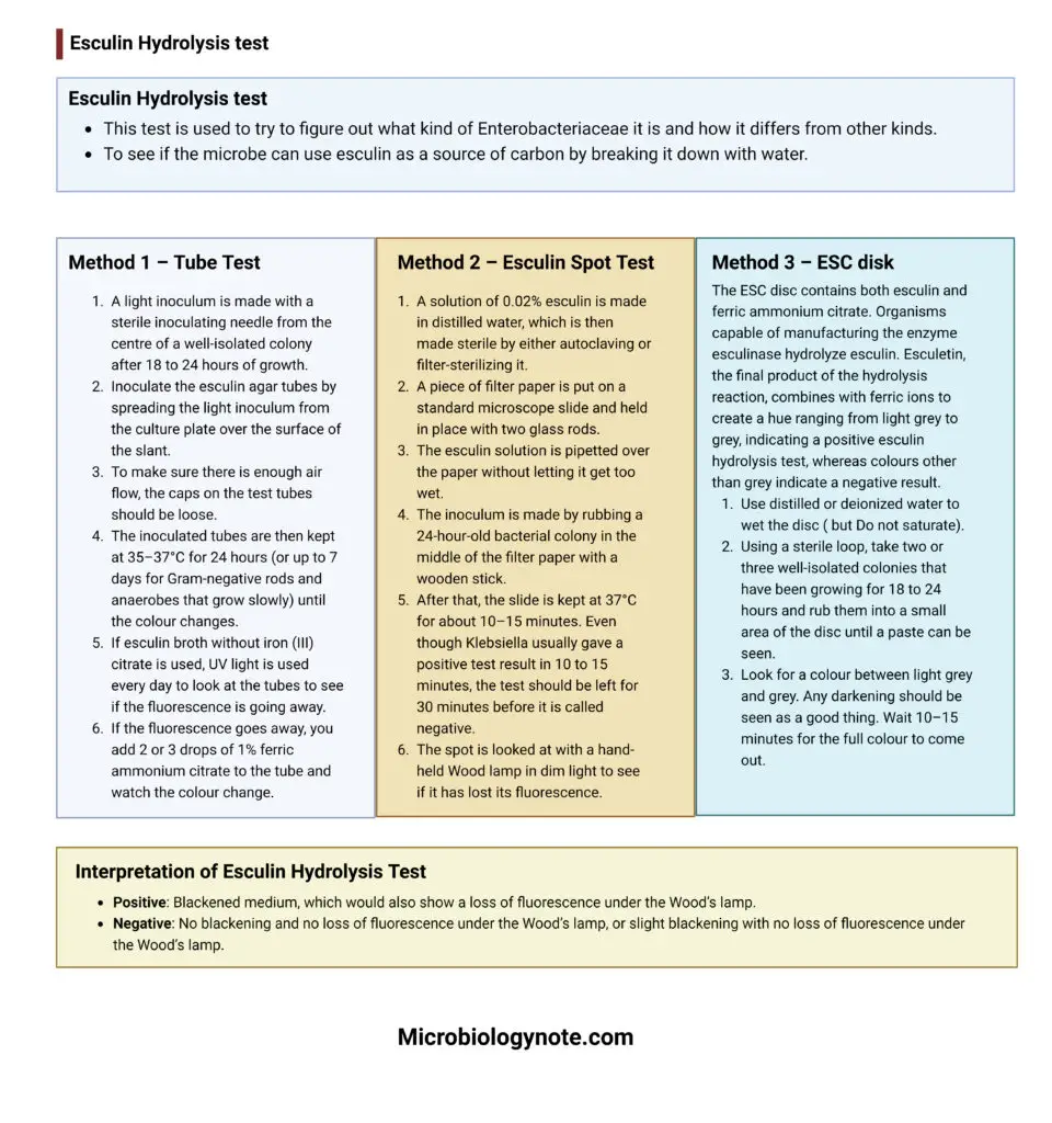
- This test is used to try to figure out what kind of Enterobacteriaceae it is and how it differs from other kinds.
- To see if the microbe can use esculin as a source of carbon by breaking it down with water.
Principle
- Microorganisms need nitrogenous compounds, amino acids, vitamins, and trace minerals to grow. Casein peptone, yeast extract, and beef heart infusion all have these things.
- Sodium chloride is a source of electrolytes that your body needs and keeps the osmotic balance.
- Vitamin K and hemin are both growth factors that make anaerobic bacteria grow faster.
- When ferric ammonium citrate is mixed with organisms that break down esculin in the medium, they make black ferric salts.
- This causes the medium to turn a colour between brown and black.
- Agar is used to make things solid.
- In the laboratory esculin hydrolysis test, the reaction between esculetin and ferric ions makes a black compound that can be seen on esculin agar or with the Esculin spot test.
Requirement
Esculin Hydrolysis Agar composition and preparation
Composition #1
- Casein Peptone ………………………………………………………. 13.0 g
- Ferric Ammonium Citrate ……………………………………………0.5 g
- Sodium Chloride ……………………………………………………….. 5.0 g
- Vitamin K ………………………………………………………………..10.0 mg
- Yeast Extract…………………………………………………………….. 5.0 g
- Hemin……………………………………………………………………….5.0 mg
- Beef Heart Infusion……………………………………………………. 2.0 g
- Agar………………………………………………………………………..15.0 g
- Esculin……………………………………………………………………… 1.0 g Demineralized
- Water…………………………………………… 1000.0 ml
Composition #2
| IngredientsGram/literCasein enzymic hydrolysate13.0Yeast extract5.0Beef heart infusion (solids)2.0Sodium chloride5.0Ferric citrate0.5Agar15.0 |
Final pH at 25°C: 7.3 ±0.2
- In a beaker, 1000 millilitres of distilled or deionized water is mixed with 41.5 grammes of the dehydrated powder or lab-made media.
- After mixing, the medium is completely dissolved by heating it and shaking it until it boils.
- The solution is then put into screw-capped tubes of about 3 ml each and sterilised in an autoclave for 15 minutes at 15 pounds of pressure (121°C).
- After being put in an autoclave, the tubes are taken out and cooled at an angle to about 40-45°C. The position should be kept at an angle in order to get butts that are 1.5 to 2 cm deep.
Other Requirement
- For the esculin spot test, distilled water is mixed with 0.02% esculin.
- UV light with a long wave (360 nm)
- Sticks, needles, or inoculation loops that are clean
- Pasteur pipettes or drinking straws
- A block of boiling heat
- At 35 and 30°C, incubators
Procedure
Method 1 – Tube Test
- A light inoculum is made with a sterile inoculating needle from the centre of a well-isolated colony after 18 to 24 hours of growth.
- Inoculate the esculin agar tubes by spreading the light inoculum from the culture plate over the surface of the slant.
- To make sure there is enough air flow, the caps on the test tubes should be loose.
- The inoculated tubes are then kept at 35–37°C for 24 hours (or up to 7 days for Gram-negative rods and anaerobes that grow slowly) until the colour changes.
- If esculin broth without iron (III) citrate is used, UV light is used every day to look at the tubes to see if the fluorescence is going away.
- If the fluorescence goes away, you add 2 or 3 drops of 1% ferric ammonium citrate to the tube and watch the colour change.
Method 2 – Esculin Spot Test
- A solution of 0.02% esculin is made in distilled water, which is then made sterile by either autoclaving or filter-sterilizing it.
- A piece of filter paper is put on a standard microscope slide and held in place with two glass rods.
- The esculin solution is pipetted over the paper without letting it get too wet.
- The inoculum is made by rubbing a 24-hour-old bacterial colony in the middle of the filter paper with a wooden stick.
- After that, the slide is kept at 37°C for about 10–15 minutes. Even though Klebsiella usually gave a positive test result in 10 to 15 minutes, the test should be left for 30 minutes before it is called negative.
- The spot is looked at with a hand-held Wood lamp in dim light to see if it has lost its fluorescence.
Method 3 – ESC disk
The ESC disc contains both esculin and ferric ammonium citrate. Organisms capable of manufacturing the enzyme esculinase hydrolyze esculin. Esculetin, the final product of the hydrolysis reaction, combines with ferric ions to create a hue ranging from light grey to grey, indicating a positive esculin hydrolysis test, whereas colours other than grey indicate a negative result.
- Use distilled or deionized water to wet the disc ( but Do not saturate).
- Using a sterile loop, take two or three well-isolated colonies that have been growing for 18 to 24 hours and rub them into a small area of the disc until a paste can be seen.
- Look for a colour between light grey and grey. Any darkening should be seen as a good thing. Wait 10–15 minutes for the full colour to come out.
Interpretation of Esculin Hydrolysis Test
- Positive: Blackened medium, which would also show a loss of fluorescence under the Wood’s lamp.
- Negative: No blackening and no loss of fluorescence under the Wood’s lamp, or slight blackening with no loss of fluorescence under the Wood’s lamp.

| Positive bacteria | Negative bacteria | Variable result |
| Enterococcus spp. | Gemella haemolysans | Aerococcus viridans |
| Streptococcus group D (non-Enterococci) | Gemella morbillorum | Aerococcus urinae |
| Pediococcus spp. | Streptococcus agalactiae (group B) |
*V(+) = Variable reactions where the majority of isolates are positive (>80%): Leuconostoc spp.
*(-) = Variable reactions where the majority of isolates are negative (>80%): Streptococcus pyogenes (group A), Streptococcus pneumoniae, Streptococcus viridans (non-group D)
Control organisms
- Positive: Enterococcus faecalis
- Negative: Escherichia coli
Uses
- Used to identify Enterobacteriaceae, Streptococcus, Listeria, gram-negative bacilli that don’t ferment, and anaerobes.
- Used to figure out if an organism can break down esculin or make the esculinase enzyme.
- By adding the bile solution, it can be used to separate out different types of Streptococcus.
Limitations
- For comprehensive identification, it is suggested that biochemical, immunological, molecular, or mass spectrometry tests be undertaken on pure culture colonies.
- To establish the presence of gram-positive, catalase-negative cocci, a Gram stain and catalase test should be done.
- Mixed cultures may produce false-positive results.
- Certain bacteria, including E. coli, contain an inducible -glucosidase and will only yield a positive result after a lengthy incubation period (up to 7 days). However, lengthy incubation should not be used if only constitutive -glucosidase is to be detected.
- If inadequate inoculum is used, any of the reactions may produce false-negative results.
- The esculin hydrolysis test does not assess bile tolerance, hence findings obtained with a disc may not correspond with those obtained with Bile Esculin medium.
- Several species create H2S during metabolism, which may react with iron to form a black complex that interferes with the esculin hydrolysis test’s interpretation. Therefore, for Gram-negative rods, examine under UV light tubes that have darkened after the addition of the reagent.
References
- https://www.vumicro.com/vumie/help/VUMICRO/Esculin_hydrolysis_Test.htm
- http://tools.thermofisher.com/content/sfs/manuals/IFU1440.pdf
- https://www.gov.uk/government/publications/smi-tp-2-aesculin-hydrolysis-test
- https://microbenotes.com/esculin-hydrolysis-test-principle-procedure-and-result-interpretation/
- https://himedialabs.com/TD/M1386.pdf
- https://universe84a.com/esculin-hydrolysis-test/
- https://assets.publishing.service.gov.uk/government/uploads/system/uploads/attachment_data/file/698994/TP_2dg_.pdf
- https://scholarlycommons.pacific.edu/cgi/viewcontent.cgi?article=3013&context=uop_etds
- http://www.apsi.it/public/ufiles/smi/tp2_3_en_141106.pdf
