Table of Contents
Urinary casts are small, cylinder-shaped, tube-shaped particles that can be detected by a microscopic analysis of urine. Casts are the only kidney-specific components discovered in the urine sediment. It is believed that Tamm-Horsfall mucoprotein (uromodulin) released by renal tubular epithelial cells forms the fundamental matrix of all casts. Any components present in the tube filtrate, including as organisms, bacteria, granules, colours, and crystals, can become imbedded in the cast matrix to create a variety of casts. Different forms of casts detected in urine sediment are indicative of distinct physiological and clinical situations. Casts of the urinary tract can be categorised into two categories: cellular and acellular. The hyaline cast is an example of an acellular cast.
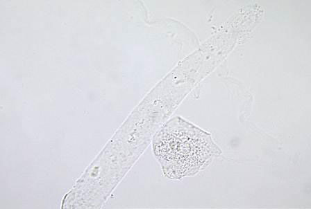
Hyaline casts are the most prevalent type of casts found in urine. They are predominantly constituted of Tamm-Horsfall mucoprotein. They have an extremely low refractive index and a smooth texture, similar to that of the urine in which they are suspended. Under a light field microscope, they seem colourless and nearly undetectable, with parallel sides and rounded ends. Infrequent granular inclusions may exist inside the matrix.
What are hyaline casts?
- Hyaline casts are small, tube-like structures that are formed by the aggregation of proteins and other substances within the tubules of the kidney. They are typically found in the urine and are an indication of kidney damage or dysfunction.
- Hyaline casts can be seen in a variety of conditions that affect the kidneys, such as inflammation, infection, or injury. They can also be seen in conditions that affect the tubules of the kidney, such as diabetes mellitus, hypercalcemia, or hyperuricemia.
- Hyaline casts are typically small, smooth, and transparent, and can be seen when the urine is examined under a microscope. They may be accompanied by other types of casts, such as granular, waxy, or fatty casts, depending on the underlying cause of the kidney damage or dysfunction.
- The presence of hyaline casts in the urine can be an indication of kidney damage or dysfunction, and may warrant further evaluation and treatment by a healthcare provider. It is important to follow the recommended treatment plan and to closely monitor kidney function in order to prevent further damage and to maintain overall health.
Formation of Urinary casts
Urinary casts are small, tube-like structures that are formed by the aggregation of proteins and other substances within the tubules of the kidney. They are typically found in the urine and are an indication of kidney damage or dysfunction.
There are several different types of urinary casts, including hyaline casts, granular casts, waxy casts, and fatty casts, each of which can be seen under a microscope when the urine is examined. The specific type of cast that is present can provide information about the underlying cause of the kidney damage or dysfunction.
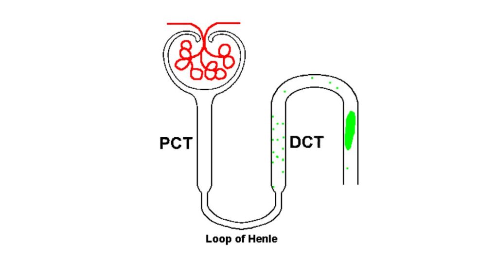
The formation of urinary casts occurs when substances that are normally filtered out of the blood by the kidneys become concentrated in the tubules of the kidney and aggregate to form the casts. This can occur due to a variety of factors, including inflammation, infection, injury, or certain underlying health conditions.
The presence of urinary casts in the urine can be an indication of kidney damage or dysfunction, and may warrant further evaluation and treatment by a healthcare provider. It is important to follow the recommended treatment plan and to closely monitor kidney function in order to prevent further damage and to maintain overall health.
Types of urinary Casts
Casts of the urinary tract can be categorised into two categories: acellular and cellular.
| Acellular Casts | Cellular Casts |
| Hyaline Casts | RBC Casts |
| Granular Casts | WBC Casts |
| Waxy Casts | Bacterial Casts |
| Fatty Casts | Epithelial Cell Casts |
Acellular Casts
1. Hyaline Casts
- The most prevalent variety of casts consisting of solidified Tamm-Horsfall mucoprotein is hyaline. They have a smooth texture and a refractive index that is nearly identical to the surrounding fluid.
- In most cases, hyaline casts have parallel sides with distinct edges and blunted ends. Even in healthy patients, hyaline casts are observable. They may be found in greater numbers during dehydration, physical exertion, or the use of diuretic medications.
2. Granular Casts
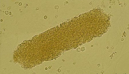
- Granular casts originate from either the degeneration of cellular casts or the direct aggregation of plasma proteins or immunoglobulin light chains. Characteristically, their surface texture ranges from fine to coarse.
- In general, they have a cigar-shaped look and a greater refractive index than hyaline casts. They are observed during strenuous physical activity, chronic renal illness, acute tubular necrosis, etc.
3. Waxy Casts
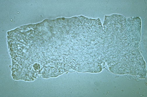
- Waxy casts reflect the final step of cellular casts’ deterioration. They are more visible than hyaline casts because they are more refractive.
- Typically, they are observed in tubular injuries of a more chronic nature than granular or cellular casts, such as in severe chronic renal illness and renal amyloidosis. These casts are also known as casts for renal failure.
4. Fatty Casts
- By decomposing lipid-rich epithelial cells, fatty casts are produced. These are characterised by the presence of refractile lipid droplets inside the protein matrix of the cast and contain lipid droplets.
- They are commonly observed in illnesses such as tubular degeneration, nephrotic syndrome, and hypothyroidism, among others.
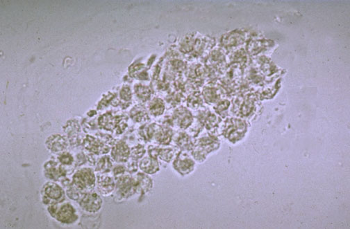
Cellular Casts
A cellular cast may consist of any of the cells present in the urine sediment, including RBC, WBC, and renal tubular epithelial cell. The cellular cast appears to be composed of clumps of cells embedded in a protein matrix.
1. Red Blood Cell Casts
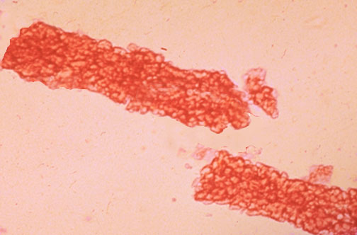
- Red blood cells may adhere and produce casts of red blood cells. These casts indicate glomerulonephritis with RBC leakage from glomeruli or extensive tubular injury.
2. White Blood Cell Casts
- There are white blood cells (often neutrophils) within or on casts. These casts are characteristic of acute pyelonephritis, but they may also be observed in glomerulonephritis.
- In addition to acute interstitial nephritis, lupus nephritis, and acute papillary necrosis, they may also be observed.
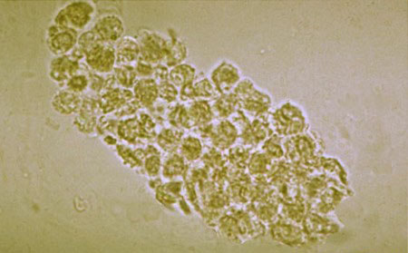
3. Renal Tubular Epithelial Cell Casts
- These casts are made of epithelial cells from the kidneys. In situations such as renal tubular necrosis, viral illness (such as CMV nephritis), and kidney transplant rejection, these casts are observed.
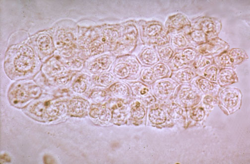
Bacterial Cell Casts
Bacterial casts consist of bacteria in a protein (hyaline) matrix. The presence of bacterial casts is indicative of acute pyelonephritis and kidney infection.
Clinical Significance
Normal is the presence of 0 to 2 hyaline casts per LPF. Certain physiological and pathological circumstances are related with an increase in hyaline casts in the urine.
Physiological Causes
Typically, more hyaline casts are associated with:
- Physical exercise
- Physiologic dehydration
- Exposure to heat
- Emotional stress
Pathological Causes
Typically, an increase in hyaline casts indicates a pathological condition.
- Acute glomerulonephritis
- Pyelonephritis
- Chronic renal disease
- Congestive heart failure
- Meningitis
- Diabetic nephropathy
What do hyaline casts in urine indicate?
Hyaline casts are small, tube-like structures that are formed by the aggregation of proteins and other substances within the tubules of the kidney. They are typically found in the urine and are an indication of kidney damage or dysfunction.
The presence of hyaline casts in the urine can be an indication of kidney damage or dysfunction, and may warrant further evaluation and treatment by a healthcare provider. It is important to follow the recommended treatment plan and to closely monitor kidney function in order to prevent further damage and to maintain overall health.
Hyaline casts can be seen in a variety of conditions that affect the kidneys, such as inflammation, infection, or injury. They can also be seen in conditions that affect the tubules of the kidney, such as diabetes mellitus, hypercalcemia, or hyperuricemia.
The formation of hyaline casts occurs when substances that are normally filtered out of the blood by the kidneys become concentrated in the tubules of the kidney and aggregate to form the casts. This can occur due to a variety of factors, including inflammation, infection, injury, or certain underlying health conditions.
It is important to seek medical attention if you notice the presence of hyaline casts in your urine, as this may be an indication of kidney damage or dysfunction. Your healthcare provider will be able to provide further evaluation and treatment to help prevent further damage and to maintain overall health.
Are hyaline casts dangerous?
- Typically, hyaline casts are not considered a worrisome finding. Other forms of urinary casts are typically associated with renal disease, unlike hyaline casts. For example, epithelial cell casts indicate significant damage and death of tubule cells, commonly known as tubular necrosis.
- In the majority of instances, red blood cell casts indicate glomerulonephritis or microscopic bleeding within the kidney.
- Typically, white blood cell casts are indicative of a kidney infection (such as pyelonephritis) or inflammatory processes. Individuals with nephrotic syndrome, which causes proteinuria (excess protein in the urine), as well as tissue edoema, low blood protein levels, and elevated blood cholesterol levels, frequently have fatty casts.
Are hyaline casts normal in urine?
- The presence of casts in the urine is typically regarded as an unusual discovery. However, small numbers of hyaline casts (between 0 and 2 casts per low power field of the microscope) can be observed in the urine of healthy people without necessarily indicating a significant problem such as renal disease. In reality, only hyaline casts should be seen in the urine in the absence of kidney or renal disease.
What causes hyaline casts?
- In the tubular lumen of the kidney, hyaline casts consist exclusively of aggregated Tamm–Horsfall protein.
- Tamm–Horsfall protein precipitation rises when the urine is acidic, has a reduced or sluggish flow, or is highly concentrated.
- Therefore, it is possible to identify a greater number of hyaline casts after vigorous exercise, after treatment with specific diuretic drugs, or in persons with acute vomiting or fever (indicative of dehydration).
- In certain clinical circumstances, such as acute renal injury, hyaline casts may occur alone or in conjunction with other types of casts.
How do you diagnose hyaline casts?
- Urinary sediment microscopy, which is conducted as part of the urine analysis, can diagnose hyaline casts (i.e. urinalysis).
- This test is performed by centrifuging a urine sample to separate the suspended particles in the urine (such as casts, cells, and pathogenic bacteria) from the fluid, also known as urine sediment. The urine sediment is then placed on a glass slide for microscopic examination.
- Hyaline casts emerge as transparent, tubule-shaped particles under bright field microscopy. Due to their low refractive index or the absence of contrast between the surrounding urine and the hyaline casts, they are frequently overlooked.
- In the majority of instances, the application of stains, decreased or dim lighting, and phase contrast microscopy enhance the detection of hyaline casts.
- Phase contrast microscopy is an optical microscopy technique that strengthens the contrast between two media with comparable refractive indices so that organisms and particles can be identified more precisely.
How do you treat hyaline casts?
- In healthy individuals, hyaline casts are not necessarily regarded as an abnormal sign and so may not necessitate any therapy.
- Nonetheless, if hyaline casts are seen in patients with kidney impairment, treatment may be required, depending on the exact reason of impaired renal function.
- For instance, the treatment of acute kidney injury may involve avoiding drugs that may impair kidney function, ensuring appropriate hydration through intravenous fluid therapy, and addressing the underlying cause of kidney damage.
What are the most important facts to know about hyaline casts?
- Hyaline casts are a form of urinary cast, which is a cluster of urinary particles, such as cells, fat bodies, or microbes, held together by a protein matrix and present in urine.
- Casts can be categorised into many varieties based on their content, including hyaline casts, granular casts, renal tubular epithelial casts, and red or white blood cell casts, among others.
- The presence of hyaline casts can be observed in both healthy and diseased persons. In the majority of instances, they suggest a decreased or sluggish urine flow, which can be caused by dehydration due to excessive exercise, diuretic drugs, or severe vomiting, or by decreased blood supply to the kidneys.
- It is possible to identify hyaline casts through microscopic examination of the urine sediment, in which they appear as transparent, tube-shaped particles that are difficult to distinguish from the surrounding fluid. Typically, no treatment is necessary unless kidney impairment is detected.
FAQ
What are hyaline casts in urine?
Hyaline casts are small, tube-like structures that are formed by the aggregation of proteins and other substances within the tubules of the kidney. They are typically found in the urine and are an indication of kidney damage or dysfunction.
Hyaline casts are small, smooth, and transparent, and can be seen when the urine is examined under a microscope. They may be accompanied by other types of casts, such as granular, waxy, or fatty casts, depending on the underlying cause of the kidney damage or dysfunction.
The presence of hyaline casts in the urine can be an indication of kidney damage or dysfunction, and may warrant further evaluation and treatment by a healthcare provider. It is important to follow the recommended treatment plan and to closely monitor kidney function in order to prevent further damage and to maintain overall health.
Hyaline casts can be seen in a variety of conditions that affect the kidneys, such as inflammation, infection, or injury. They can also be seen in conditions that affect the tubules of the kidney, such as diabetes mellitus, hypercalcemia, or hyperuricemia.
The formation of hyaline casts occurs when substances that are normally filtered out of the blood by the kidneys become concentrated in the tubules of the kidney and aggregate to form the casts. This can occur due to a variety of factors, including inflammation, infection, injury, or certain underlying health conditions.
What does hyaline casts in urine mean?
Typically, the development of hyaline casts suggests a reduced or sluggish urine flow, which may be caused by physical activity, diuretic drugs, acute vomiting, or fever.
What is normal hyaline casts?
Zero to two hyaline casts per low-power magnetic field are considered typical. Increased urine concentration, such as that caused by dehydration, and increased albumin concentration also influence the formation of hyaline urine casts.
Which condition promotes the formation of casts in the urine?
In renal tubular necrosis, viral illness (such as cytomegalovirus [CMV] nephritis), and kidney transplant rejection, these casts are observed. People with advanced renal disease and long-term (chronic) kidney failure can develop waxy casts.
Can dehydration cause casts in urine?
Low urine output, concentrated urine, or an acidic environment can all contribute to the creation of hyaline casts, and as a result, they can be observed in normal individuals who are dehydrated or who engage in severe activity.
