Advertisements
Table of Contents
KOH Test (Potassium hydroxide test)
- A KOH pre-test is a quick, non-invasive procedure to diagnose fungal infections that affect the nails or skin.
- The cells are collected from the affected region and put on a slide using an acid composed of potassium hydroxide. They are looked at under a microscope in order to determine if there are evidence of the presence of a mold.
- Also known as a potassium hydroxyide test for skin lesions fungal smear, scraping the skin test, the KOH preparation test is fast precise, precise, and almost non-invasive.
Purpose of Test KOH Test
- To identify the fungal component in clinical specimens.
- To examine the skin scrapings, hair flakes for any hyphae as well as arthroconidia in the case of suspected dermatophyte infections.
Principle of KOH Test
- Potassium hydroxide (KOH) can be utilized on specimens from clinical studies to remove cell debris and improve visualization of fungal components.
- KOH preparation is an often utilized method to determine the presence of superficial fungal infections as well as for rapid identification of fungal elements within an actual specimen. KOH is a powerful alkali.
- KOH is able to separate the fungal components from the cells because it breaks down the protein waste and dissolves cements that hold the cells keratinized around the fungi, so that conidia and hyphae (spores) from fungi are able to be observed under a microscope.
- The sample is then placed in small amounts of between 10 and 20% KOH, and then is incubated for 5 to 10 minutes , where gentle heating will clear samples faster.
- A coverslip is placed on top of the sample digested in KOH and slides are examined microscopically , without staining.
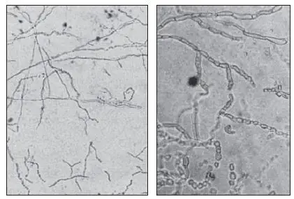
- Many fungal elements such as hypohyphae, pseudohyphae cells and spores, as well as spherules and sclerotic body parts can be observed clearly when using the KOH dry mount.
- In dermatophytosis arthrospores grow and grow as hyphae break apart , forming an elongated chain of tiny circular to rectangular extremely refractile structures.
- In the potassium hydroxide-based preparations of sputum fungus is seen as non-pigmented septate hyphae measuring 3-5 um in diameter, with distinctive distinct branching patterns and an irregular shape.
Procedure KOH test
Slide method
- Put the specimens such as epidermal scales or nail skin scraping, hair and tissue onto a clear glass slide.
- Pour a small amount 10 percent KOH onto the sample and then place the coverlip on top.
- The slide is heated gently on the flame.
- Allow the slide to rest for 5-10 minutes , or put the slide in a Petri dish, or in other containers that have lids, by a moist piece cotton wool or filter paper to prevent the prepared in drying.
- Examine the slide using the microscope with 10X and 40X objectives.
Tube test
- The homogenized tissue into a test tube, and add 10 percent KOH.
- The tube should be incubated overnight at 37°C.
- After incubation, put one drop of suspension on the slide that is clean and cover with a cover slip.
- Review the slide under the microscope with 10X and 40X objective.
Note: This method can be utilized for nail clippings as well as skin biopsies, which dissolve with difficulty, and it is possible that the amount of KOH can be increased.

Procedure to make 100 ml of KOH 10% w/v solution
- Ten grams of potassium hydroxide (KOH) pellets using the scale of weighing.
- Take care to transfer the weighted KOH pellets to a screw cap bottle.
- Now add 50 ml distilled water and mix to ensure that the KOH pellets have completely dissolved.
- Add the remaining amount of distilled water to make it 100 ml.
- Label the bottle with the 10% solution of KOH. mark it as with corrosive.
- Take note: Potassium hydroxide (KOH) is a highly acidic chemical, so handle it with care. Also, make sure that the bottle is marked as corrosive and seal it tightly.
- Keep it at the room temperatures. The reagent stays stable for approximately two years from the time of its preparation.
Uses KOH Test
- KOH preparation is utilized for the identification of ringworm infections. The diagnosis in a laboratory is based on the recognition of the organism using microscopically examining skin or nail scrapings using 10 20 up to 20 percent KOH during dry mount examination.
- KOH with blue-black preparation of ink is recommended when Malassezia furfur may be suspected.
- Calcofluor White-Potassium Hydroxide Preparation may also be used to aid in the study of Fungal disease because CW is not a specific stain and a keen understanding of the morphology of fungal elements on the spot is essential for a correct interpretation of the specimen.
- A KOH test can verify the presence of fungi including Dermatophytes. Dermatophytes are fungi which require the growth of keratin. The most common dermatophyte-related diseases are jock itch, athlete’s foot as well as nail infections and ringsworm. They are most often associated with skin infections on the feet, the genitals and, especially in children and scalps.
- The KOH test is performed in conjunction with a clinical exam as well as an Wood lamp examination. This makes use of ultraviolet light to take a close look to the surface.
| Suspected conditions | Specimen | Diagnostic characteristics |
| Aspergillus infection | Sputum | Septate hyphae with V-shaped branching |
| Dermatophytes (ringworm fungi) | skin scrapings, nails or hair | Depending on the cause, the yeast may have hyaline septate hyphae, arthroconida, or spherical yeast cells. |
| Blastomyces dermatitidis infection | Pus, sputum or skin specimens | Blastomyces dermatitidis yeast cells. These are large yeast cells that are growing and have a wide base. B.dermatitidis is a fungus that changes shape and has yeast cells in its tissue. |
| Mucormycosis | Exudates from infected lesions or tissue | Fungi that cause mucormycosis have hyphae that don’t branch. |
| Chromoblastomycosis | KOH preparation of scrapings from crusted lesions | Muriform cells, which are groups of dark brown cells that look like stones in a stone wall, or round, hard, brown bodies with fission planes that are 4–10 m in diameter. They look like pennies made of copper. |
Limitations KOH Test
- Potassium hydroxide can be a destructive deliquescent chemical, and it must be handled with care.
- Experience is required as background artifacts can be complicated.
- The removal of certain specimens, such as nail clippings, biopsy material could require an extended period of duration.
Modification in KOH Preparation method
- Use of the dimethylsulfoxide-KOH reagent: By adding dimethylsulfoxide (DMSO) to KOH, samples can be looked at right away or in just a few minutes.
- Adding blue-black fountain pen ink to KOH: The ink doesn’t just stain fungi because it also stains cells and other parts of the skin. When Malassezia furfur is suspected, adding ink is a good idea.
References
- Tille P.M (2014)Bailey and Scott’s diagnostic microbiology.
- Bilge Fettahlıoğlu Karaman B.F, Topal S.G, Aksungur V.L, Ünal L, İlkit M. 2017. Successive Potassium Hydroxide Testing for Improved Diagnosis of Tinea Pedis. 100(2):110-114.
- Cheesbrough M. 2006. Medical laboratory manual for tropical countries part 2. Second edition.
