Table of Contents
Definition of Monocot Leaves
- Monocotyledonous leaves are slender and elongated. They have parallel venation. It is commonly used to differentiate monocotyledonous species from dicots. Monocot leaves are bilateral since the surfaces of both leaves have the identical coloration.
- The initial monocot leaves comprise of a proximal base or hypophyll as well as the distal hyperphyll. The hyperphyll is the predominant component that makes up the dicot leaf but in monocots the hypophyll functions as the predominant structure.
- The leaves are slim and linear, with an outer sheath that covers the stem’s base However, there are many different monocots which do not have the same structures.
- The venation, which is mentioned is of the sort of striate, which is usually long-term striate. Sometimes, it is palmate-striate or even pinnate-striate.
- The veins that line the leaf surface begin from the bottom of the leaf, and then move towards the apex.
- The majority of monocotyledonous plants have only one leaf per node since the leaf’s base covers more than half of length of the stem.
- The existence of a bigger leaf base has been linked to differences in the development of the stem throughout zonal differentiation.

Definition of Dicot Leaves
- Dicotyledonous leaf blades are typically round with a reticulate venation. They is distinct from monocotyledonous leaves by their anatomy and structure. A typical dicot leaves consists from a blade that is sometimes referred to as the lamina. The lamina is by far the biggest portion of the leaf.
- The leaves of Dicot are dorsoventral, meaning that the ventral and dorsal portions of the leaves are distinguished based on the color in the leaves. The dorsal portion leaves are typically more pronounced than the ventral one.
- Dicot leaves attach to the stem using the petiole that differentiates dicots from the monocot leaves, which are attached directly on the stem.
- Stipules are small green appendages that could be found at the petiole’s bottom in dicot leaves.
- Dicot leaves are characterized by a midrib that is a part of the leaf blade, and continues along the length of the leaf. There are numerous branches that grow both sides of this midrib and give an Reticulate Venation.
- The number of leaves that can be found on the stem of a node is dependent on the species, however dicots generally contain two or more leaves that arise from a single node.
- Dicot leaves are further subdivided into various groups based on their leaf shape, since certain types of leaves are simple, while others are compound leaves.

Structure of Monocot and Dicot Leaves
The internal structure , or anatomy of both dicot and monocot leaves can be described by using the following patterns:
1. Epidermis
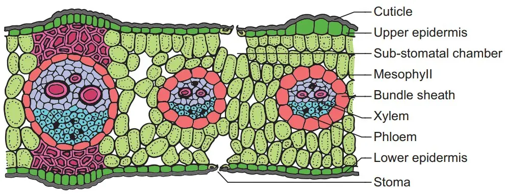
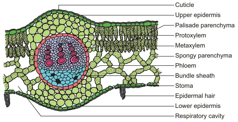
The epidermis is a layer of outer leaf tissue that is comprised of a dense layer of cells that are thin and barrel-shaped. The epidermis appears on the lower and upper portion on the leaves. The epidermis is enclosed by a waxy layer on the outside of the cuticle that serves to shield the leaf and to prevent loss of water.
The cuticle of the upper surface of the leaf appears more thick than the one on the lower side of dicot leaves since the leaves are dorsoventral. In monocots however, the epidermis layer is approximately the same across both sides. The epidermis is crucial since it helps to prevent the loss of water since it’s an elastomeric waterproof layer. Additionally, it is a key component in gas exchange because it has tiny pores known as stomata.
Stomata number on the surface is identical on both surfaces of monocot leaves. Stomata are located more in the lower epidermis and less on the epidermis of the upper part on dicot leaves. Stomata is a tiny opening that is located between bean-shaped cells known as the guard cells, which control the size of the stomata. Guard cells are dumb-bell formed in dicots. Epidermal cells don’t contain chloroplasts. However, the guard cells have chloroplasts, which give the distinctive green hue for the leaf.
In addition to the epidermal cells and these guard cells, the epidermis contains secondary cells, which usually reside in the vicinity of those guard cells. For dicot cells, the top epidermis is awash with large, thin-walled cells known as bulliform cells which are also called motor cells. These cells aid in rolling the leaves as a result of weather changing. In monocot leaves epidermal cells are stuffed with silica, also known as silica cells.
2. Mesophyll
The mesophyll forms the ground layer of leaves that is located between the lower and upper epidermis on the leaves. The mesophyll is distinguished into palisade parenchyma as well as the spongy parenchyma of dicot leaves. Neither differentiation is observed for monocot leaf. The palisade parenchyma lies just below the epidermis’s upper layer and is comprised of cylindrical cells that are vertically extended in layers of one or more. The cells are arranged in a compact manner without intercellular gaps.
Palisade Parenchyma cells are home to more chloroplasts than those of the parenchyma spongy. The spongy parenchyma can be found beneath the palisade parenchyma, where the cells are formed in an irregular manner. They have less chloroplasts that the palisade cells.
Cells of parenchyma spongy are loosely laid out with numerous spaces, hence the name. The air spaces allow for the exchange of gas between cells. The spaces located near the stomata are known as sub-stomatal or respiratory cavities. The mesophyll in dicot leaves are not separated into spongy or palisade parenchyma. It is comprised of thin-walled and isodiametric cells. Cells are organized compactly with air spaces interspersed.
3. Vascular Bundles
The thickest layer of tissue found in leaves of plants is the vascular bundle that is found beneath mesophyll as well as along the veins of leaves. Each vascular bundle is different in size, and the bigger bundles are found regularly along the veins. Large vascular bundles include two sclerenchyma cell patches between them. The vascular bundles in the leaves form part of the system of vascularization in the plant that runs through various organs, covering the entire plant.
The vascular bundles of leaves show extremely divergent patterns in terms of size and location. In dicot leaves veins, they are in different sizes, forming an extremely branched network. The vascular bundles beneath these veins are, therefore, different. In monocot leaves the longitudinal veins are parallel to the leaf blade. They are joined transversally by tiny commissural veins. This creates less distinct vessels.
Vascular bundles can be described as joint as well as collateral and are closed and are protected by a sheath. The bundle sheath on dicot leaves can be described as parenchymatous however the sheaths of dicot leaves that are sclerenchymatous. The xylem tissue of the vascular bundle is located toward the epidermis’s upper surface and the phloem located in an epidermis lower. The xylem is comprised of vessels and parenchyma xylem. The xylem of monocot leaves can be divided into protoxylem and metaxylem. The phloem is composed of sive cells and companion cells and the phloem parenchyma.
Functions of Monocot and Dicot Leaves
The roles of leaves are generally similar for monocot and dicot plants. The roles of leaves are contingent on the species of plant the environment they are in, as well as their age. Here are a few of the roles of dicot and monocot plants.
- The main purpose of leaves in green plants is the production of food via the photosynthesis process. The cells in the leaves contain chlorophyll as one-fifth of the cells in the mesophyll contain chlorophyll-containing chloroplasts. The wide, large leaf surface allows for the absorption of more of sunlight, which is vital to photosynthesis.
- The epidermis and the cuticle of the leaves block the loss of water during transpiration. This keeps plants from drying out.
- The leaves are composed of stomata that are essential for the flow of water out of the plant to the air through transpiration. This is necessary to pull water that contains mineral elements from soil through the roots.
- Alongside helping to transpire, stomata are associated with the movement of gases. Stomata absorb carbon dioxide out of the air and then give oxygen to photosynthesis.
- In certain plants, such as lettuce and cabbage the food produced by cells inside the leaves are stored within the leaves in various varieties.
Examples of Monocot Leaves
1. Maize leaves
Maize leaves are thought to be the most common monocot leaves with simple and well-organized structures. The typical maize plant has around 20 leaves which could exist in various phases of growth. A mature maize leaf measures approximately 70 cm long and 8cm wide. The maize leaf splits into 3 areas that are an upper blade, lower sheath, and an auricle. The cells of the leaf are directed and aligned in order to form parallel lines as well as an curvaceous shapes.
The blade of the maize leaves has adaxial and abaxial epidermal tissue , which covers mesophyll as well as vascular tissue. The epidermis cells of the leaf of maize are separated into two kinds; specialized cells and non-specialized intercostal cells. Specialized cells comprise complexes of stomatal function and three kinds of hair cells: large macrohairs, microbars as well as bicellular hairs. These cells perform a protective function.
2. Grass
The grass leaves are a singleton with an elongated shape which is formed by the node, which is comprised of a central cylindrical sheath surrounding the stem as well as younger leaves. The sheaths that cover the exterior are hollow cylinders which split down one way. They usually create overlapping structures. The leaf is auricle-shaped which could be present as an ear-like projection , or might become a hairy edge at the bottom on the blade of leaf.
The epidermis on a grass leaf is comprised of hair cells that shield the plant from diverse harmful agents. The texture, shape, and hairiness of leaves are commonly used as indicators to distinguish grass because they differ between species, or sometimes even inside the same species.
Examples of Dicot Leaves
1. Mustard leaves
Mustard is a dicot plant that is commonly used in research on dicotyledonous plants. The leaves of mustard begin to grow in just 4 weeks, when they reach 6-8 inches in size. A mature mustard leaf will measure approximately 15-18 inches within six weeks. The leaves are broad and green with a reticulate-venation. They are flattened dorsoventrally and the ventral portion is more slender than the dorsal one.
The epidermis of mustard leaves has some hair cells with specialized functions which protect against loss of water and damaging agents. Mustard leaves are utilized as vegetables because they possess an important nutritional value for the human body’s health.
2. Mint leaves
Mint is a plant that grows rapidly comprised of lanceolate, round leaves, which are placed in pairs that are opposite to each other around the stem. Mint leaves are small in size and range from 4 to 5 inches in length. They are small in size however, is contingent on the size of the leaves and their maturation.
The leaves are reticulate with an underlying midrib from which branching veins develop. The leaf’s surface is covered by tiny hairs, which appear both on the lower and upper surfaces of leaves. Mint leaves are fragrant and have a an odor that is distinctively mint. They are utilized in a variety of food items across different communities.
Differences between Monocot and Dicot Leaves (Monocot vs Dicot Leaves)
| Characteristics | Monocot leaves | Dicot leaves |
| Definition | Monocotyledonous leaves are slender and elongated, with the parallel venation that is frequently used to differentiate monocotyledonous species from dicots. | Dicotyledonous leaves typically have a rounded shape with reticulate veins that is distinct from monocotyledonous leaf in their anatomy and structure. |
| Shape | Monocot leaves are short thin, slimmer and more than dicot leaves. | Dicot leaves are wide and generally smaller than monocot leaves. |
| Symmetry | Monocot leaves are obilateral in the symmetry. | Dorsoventral leaves on Dicots are distinct because the lower and upper surfaces of the leaves can be clearly distinguished. |
| Venation | Monocot leaves feature parallel venations because the longitudinal veins are affixed across the leaf and are joined to each other by small commissural veins. | Dicot leaves are reticulate, comprised of veins of various sizes joined to form a network. |
| Stomata | A number of stomata found on the lower and upper surface of the leaves is identical, which is why monocot leaves can also be described as anmphistomatous. | Dicot leaves have more stomata located on the lower side than on the surface. Certain dicot leaves don’t have stomata on their upper surface. Such plants are referred to as hypostomatous. |
| Protect cells | The guard cells of the monocot leaves have dumb-bell shape. | The cells protecting dicot leaves have kidneys. |
| Intercellular spaces | Monocot leaves have smaller intercellular spaces because cells are arranged in a compact manner. | Dicot leaves are larger in intercellular spaces because the cells are packed loosely. |
| Massive bundles | Small and large vessels can be found within monocot leaves. The xylem in monocot leaves is divided into protoxylem and metaxylem. The bundle sheath of monocot’s leaves are sclerenchymatous. | Dicot leaves have larger vessels. The xylem of dicot leaves isn’t differentiated into protoxylem and metaxylem. The sheath around the bundle of dicot leaves are parenchymatous. |
| Epidermis | The epidermal cells of the monocot leaf are characterized by the heavy deposition of silica. The epidermis of monocot leaves contains motor cells that are bulliform. | Epidermal cells in dicot leaves are not able to deposit silica. The epidermis on dicot leaves doesn’t have motor or bulliform cells. |
| Mesophyll | The mesophyll of monocot leaf is distinguished into mesophyll spongy and mesophyll from palisade. | The mesophyll in dicot leaves isn’t differentiated. |
