Table of Contents
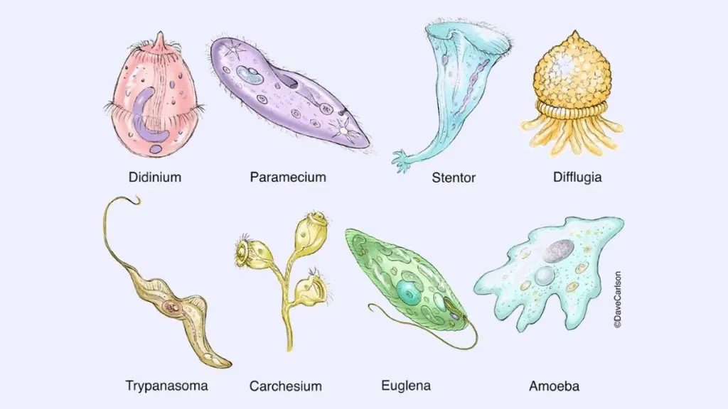
Protozoa don’t have any cell walls, instead they contain pellicle. The structure of Protozoa is similar to the eukaryotic cells. Some examples of protozoa are Trypanosoma, Trichonympha, Plasmodium, Paramecium.
Now, Protozoa is a strictly descriptive term, they are no longer recognized as a formal group in current biological classification systems.
Protozoa are ubiquitous, which means protozoa can be found from the South Pole to the North Pole, they are everywhere. Most of them are found in moist or aquatic habitats.
Their mode of nutrition is heterotrophic. Some protists also show mixotrophic characters, which means they show both heterotrophy and autotrophy characters. Chrysophytes is an example of protozoan mixotrophs.
Protozoa Definition
Protozoa are also known as a protozoan, they are one-celled, eukaryotic, microscopic animals which may be free-living or parasitic, and get their energy from the organic carbon.
In 1818, zoologist Georg August Goldfuss first coined the term “protozoa”. It comes from the Greek word prôtos which means “first” and zôon means “animal”.
All protozoa contain a “true,” or membrane-bound, nucleus and membrane-bounded organelles within their cytoplasm. Protozoa contain flagella, cilia, or pseudopodia means they are motile.
Example of Protozoa: Giardia, Trypanosoma, Trichonympha, Plasmodium, Paramecium, etc.
Characteristic of Protozoa
1. Size
- The size of protozoa is ranging from 1 micrometer to several millimeters, or more.
- The largest protozoa are found in deep-sea–dwellings known as xenophyophores, which can be 20 cm in diameter.
- The smallest one is Plasmodium falciparum, which size can be 1-2 micrometers in diameter.
2. Feeding
- They are heterotrophic in nature. Get their nutrients from other organisms.
- Some protozoa get their food by phagocytosis, where it engulfs the whole organic particles with the help of pseudopodia, such as amoebae. Some fungi get their food by cytostome, which is a specialized mouth-like aperture.
- Some fungi also get their food by absorbing the dissolved nutrients through their cell membranes, this process is known as osmotrophy.
- Plasmodium, during its immature trophozoite stage, take their foods by the pinocytosis. When they get matured, develop a dedicated feeding organelle known as cytostome.
- Some protozoa appear as mixotrophs which contain both heterotrophic and autotrophic nature.
- Protozoa also show a symbiotic relationship with photosynthetic algae, where they provide nutrients to the host in exchange for they live and grow within the host body.
- Some fungi are kleptoplasty, where they steal the chloroplast from algae and they produce nutrients by photosynthesis. For example, Mesodinium rubrum get plastids from the cryptophyte algae.
3. Habitat
- The protozoa can be found in fresh, brackish, and saltwater. Mainly they are lived in moist environments, for example, soils and mosses. They can survive in dry environments by forming resting cysts which is a dormant stage of protozoa. Until the conditions improve they remain in the dormant stage.
- Some protozoa can survive in extreme environments for example hot springs and hypersaline lakes and lagoons.
- The Parasitic and symbiotic protozoa live within or on the host body or other microorganisms such as vertebrates and invertebrates, plants, and other single-celled organisms.
4. Motility
- Protozoa are motile, they contain flagella, cilia, and pseudopodia. Which helps them in locomotion.
- The flagella is a whip-like structure, cilia is a hair-like structure and pseudopodia is a foot-like structure.
5. Pellicle
- The protozoa don’t contain any rigid cell wall, instead, they contain elastic structures of membranes that help them in movement.
- The ciliates and euglenozoans have a flexible and elastic to fairly rigid membranous envelope known as the “pellicle”. During the locomotion, it provides shape to the cell.
- In ciliates and Apicomplexa the pellicle is supported by the alveoli, which is a closely packed vesicle.
- In some protozoa, it is formed from protein strips which are arranged spirally along the length of the body, such as euglenids.
6. Life cycle of Protozoa
- Protozoa has two-stage in their life cycle such as proliferative stages (e.g., trophozoites) and dormant cysts.
- When protozoa are exposed to extreme environmental conditions such as high temperature, harmful chemicals, or lack of nutrients they form a dormant structure called cysts. The conversation of trophozoite to cyst is known as encystation. In this stage, they can transmit from one host to another.
- When the conditions are back to normal they form the trophozoite stage. In the trophozoite stage (Greek trophy = to nourish), they actively feed. The conversation of cyst to trophozoite is known as excystation.
- They reproduce by binary fission or multiple fission. Some protozoa also transfer their genetic material through conjugation.
Protozoa Structure
- Protozoa are eukaryotic cells.
- They are unicellular organisms.
- Their size ranges from 1 micrometer to 200000 or may be up to 200000 micrometres in diameter.
- The size of smaller protozoa is from 1 to 10 μm long.
- They contain membrane bounded organelles in their cytoplasm such as ribosome, Golgi apparatus.
- Protozoa contain a well organised nucleus which is covered with membrane.
- The types of organelles present in protozoa vary from species to species. They contain some characteristic organelles such as the Trichocysts of Paramecium, certain skeletal structures, Contractile vacuoles.
- The protozoa contain a vesicular nucleus. As such, the chromatic is scattered, the nucleus resulting a diffuse in look.
- The vesicular nucleus of Phylum Apicomplexa contains one or more nucleoli with DNA whereas the DNA is absent in the endosome of trypanosomes.
- They contain pseudopodia, flagella and cilia which help them in locomotion. These locomotory structures are covered by the plasma membrane.
- Some protozoa also contain a rigid structure known as pellicle which gives them shape and also helps in twisting and bending during locomotion.
Classification of Protozoa
Classification of Phylum Protozoa
There are present different phyla of protozoa such as;
(i). Phylum Euglenida
- They contain pellicle and flagella which help them in locomotion.
- The pellicle is located beneath the cell membrane and made of protein strips. It is the characteristic feature of Euglenida.
- Some Euglenidas are autotrophs, they contain chloroplasts which help them in photosynthesis to make their foods.
- Others use dissolved nutrients to get their foods. While few of them are parasitic.
(ii). Phylum Kinetoplastida
- They are protected by a pellicle which is made up of microtubules.
- They contain a single, much enlarged and elongated, mitochondrion.
- Some of them are parasitic such as leishmaniasis that cause disease in humans
(iii). Phylum Ciliophora
- They contain cilia for locomotion, which are much smaller structures than flagella.
- They act as parasites in the digestive tracts of larger organisms.
- The entire cell is covered with cilia, which propel the cell forward. Each cilia gives a forward-moving power stroke, then whips back to the starting position in the recovery stroke.
- Some ciliates are found at the bottom of marine environments, which are known as the benthic zone.
- The free-swimming organisms and sessile use cilia to filter food material from the water.
(iv). Phylum Apicomplexa
- These are parasitic organisms, which enter into their host cells by using apical complexes. They are much more resistant inside the cell and get better access to nutrients.
- They can hide from the immune system by changing the proteins exposed on their cell surface, that is why it is difficult to treat them by medicine.
(v). Phylum Dinoflagellata
- They contain flagella for locomotion and pellicle. The pellicle of Dinoflagellata is made up of a series of vesicles beneath the cell membrane which makes them rigid.
- Some of them protect their cells by filling their vesicles with polysaccharides and forming armor.
(vi). Phylum Stramenopila
- This phylum contains different varieties of organisms, from the shelled diatoms to brown and golden algae.
- They contain shells, scales, or tests that support the cell.
- The tests of diatoms are made of silicate, others use calcium carbonate or protein to make their shells.
(vii). Phylum Rhizopoda
- This phylum includes the amoebas. These are small, unicellular protozoa and don’t contain any hard covering.
- They extend their cytoplasm in the environment for locomotion. These extensions of amoeba are known as the pseudopodia.
(viii). Phylum Actinopoda
- They contain characteristic axopodia. These are sharp spines; extend from the cell; used for locomotion and feeding.
(ix). Phylum Granuloreticulosa
- Granuloreticulosa has an immense industrial value.
- They produce tests at the bottom of the ocean, where they fossilize together and form chalk, limestone, and marble.
The pyramids of Egypt were built from stones which originated from the shells of these protozoans.
(x). Phylum Diplomonodida
- They contain flagella (around 8)for their locomotion.
- An example of Diplomonodida is genus Giardia which causes flu-like symptoms and diarrhea in humans.
(xi). Phylum Parabasilida
- They contain thousands of flagella, and contain a fiber that attaches the Golgi apparatus to the base of the flagella.
- Some of them show symbiotic relationships with insects, mainly those insects eat wood.
- They release enzymes to break the cellulose.
Classification of Protozoa Based on the Mode of Existence
There are about 21,000 species of free-living protozoa and 11,000 species of parasitic microbes which are lives in both vertebrates and invertebrates.
The free-living protozoa live everywhere, they can be found in water, soil, etc. They cause disease in humans. Hence, based on the habitat of free-living protozoa they are classified into these following groups such as;
(a). Acanthamoeba
- They can be found in soil and water.
- They are responsible for chronic granulomatous amebic encephalitis, amebic keratitis, granulomatous skin as well as lung lesions.
(b). Naegleria fowleri
- They are mainly found in moist soil.
- They are responsible for the acute primary amebic meningoencephalitis.
(c). Balamuthia mandrillaris
- They are responsible for sub-acute to chronic granulomatous amebic encephalitis and also cause granulomatous skin and lung lesions.
(d). Sappinia diploidea
- They can be found in Elk and buffalo feces, Soil, water.
- The following symptoms can be observed in a Sappinia infected patient; Headache, Sensitivity to light, Nausea or upset stomach, Vomiting, Blurry vision, Loss of consciousness.
Classification of Protozoa based on the Mode of Nutrition
Protozoa are classified into three main category based on their mode of nutrition such as;
(a). Autotrophs
- They produce carbohydrates or foods from carbon dioxide and water through photosynthesis.They contain chlorophyll.
- They use acetates, simple fatty acids and alcohols as a main source of carbon.
- In presence of light they act as autotrophs, while in dark they switch to heterotrophs.
- Some examples of Photo-autotrophic protozoa are Euglenida, Cryptomonadida, Volvocida (both autotrophy and heterotrophy).
(b). Heterotrophs
- Most of the protozoa comes under this category. They feed on bacteria (microbivores) or algae(herbivores) or may be in both bacteria and algae (carnivorous ).
- They are divided into two distinct groups based on their entry point of food such as those that have a mouth/cytostome and those that lack a mouth or a definite point of entry for food.
(c). Chemoheterotrophic
- This group of protozoa needs energy and organic carbon sources.
Life Cycle of Protozoa

Parasitic Protozoa
The life cycle of parasitic protozoa occurs intracellular or in the lumen of given organs. Because of the diversity, different species follow different patterns of life cycle.
There is three most common pattern have been found in protozoa such as;
(i). First pattern
- This pattern is mainly found in phylum Apicomplexa. In this method the alteration occurs between asexual and sexual reproductive stages.
- The cycel is started with the asexual reproduction. In the first stage the population of host’s tissue is increased by the schizogony (involving mitosis and cytokinesis).
- After that the population undergoes gametogony(a sexual process) and develops gametes.
- These gametes undergo sporogony (asexual process) and form sporozoites. These sporozoites have a capability to infect a new host cell.
- When the sporozoite entered the host cell they started the reproduction cycle again.
- Apicomplexa required two hosts(vertebrate and invertebrate) to complete their life cycle. Inside the vertebrate they undergo the schizogony and gametogony. Inside the invertebrate the gametes unite and sporogony occurs in the tissues.
(ii) Second Pattern
- It is the most common pattern among the flagellates, where asexual reproduction is involved.
- In this cycle a number of morphological transformations occur. All of them reproduced by the binary fission.
- Some of them use a vertebrate host to complete their life cycle, where they transmit from one host to another through cysts.
(iii). Third pattern
- This is mainly found in amoebas and completed through the asexual reproduction.
- It required a single host to complete their reproduction. For example; trophozoites live in the lumen of the gut where they continuously increase their number by binary fission.
- Here, under certain conditions, the trophozoites are stimulated to encyst as they undergo nuclear division within the cyst. The cycle continues, When another host ingest the cyst.
Free Living Protozoa
- The life cycle of Free Living Protozoa is followed by the growth and increase the size of organism which is then followed by binary fission (or other forms of asexual reproduction).
- Under unfavorable conditions (unfavorable temperature, or reduced food supplies etc) the Free Living Protozoa start their sexual reproduction.
- The growth and division cycle of the free-living protozoa is completed by different phase such as
- First division phase
- End of division phase and beginning of DNA synthesis
- DNA synthesis
- End of DNA synthesis and beginning of next division
Protozoan Disease
Malaria
- The malaria disease in humans is caused by the Six Plasmodium species, but among them two are most significant such as Plasmodium falciparum and Plasmodium vivax.
- This disease is mainly found in tropical and subtropical regions of the world.
- The symptoms of this disease includes chills, fatigue, fever, night sweats, shivering, or sweating, pain in abdomen or muscles, diarrhoea, nausea, or vomiting, etc.
Toxoplasmosis
- This disease is caused by the Crytoptosporidium parvum and Cryptosporidium hominis.
- The symptoms are watery diarrhea,stomach cramps or pain, dehydration, nausea, vomiting, fever, weight loss.
African trypanosomiasis
- This disease is also known as the African sleeping sickness. This disease is caused by the genus Trypanosoma.
- Trypanosoma brucei gambiense and Trypanosoma brucei rhodesiense, respectively T. b. gambiense are responsible for the West African and East African trypanosomiasis.
- The symptoms of this disease include pain in joints or muscles, insomnia or sleepiness, muscle loss and weakness or weight loss, fever, headache, itching, mental confusion, personality change, problems with coordination, skin rash, or swollen lymph nodes.
Chagas disease
- It is also known as American trypanosomiasis. Trypanosoma cruzi is responsible for this disease in Latin America.
- The symptoms of this disease include pain in abdomen or muscles, fever or body ache, headache, painless swelling around eye, palpitations, or skin rash.
Leishmaniasis
- This disease is caused by the Leishmania parasites.
- The symptoms are fever, weight loss, enlargement (swelling) of the spleen and liver, and abnormal blood tests (a low red blood cell count (anemia), a low white blood cell count (leukopenia), and a low platelet count (thrombocytopenia)).
Examples of Protozoa

(i) Giardia : It is an intestinal parasite and responsible for diarrhoeal diseases in humans. They can be found in the small intestine of humans and other animals.
(ii) Trypanosoma: They can thrive in the circulatory system of the host and are responsible for the disease trypanosomiasis.
(iii) Trichonympha: These are multi-flagellate symbiotic protozoans and they inhabit the intestines of termites.
(iv) Leishmania: They are responsible for the leishmaniasis in humans. They can transmit from one host to another host through the bites of sand fly (Phlebotomus spp.).
(v) Entamoeba: It is a non-flagellated amoebae and responsible for the disease amoebiasis in humans.
(vi) Plasmodium: there are present different species of the genus Plasmodium, such as P. vivax, P. ovale, P. malariae and P. falciparum. They are responsible for the disease malaria that affects 200 to 300 million people yearly.
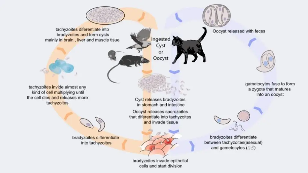
(vii) Toxoplasma: They are responsible for the disease of Toxoplasmosis. The symptoms of this disease are ever, sore throat and enlargement of spleen, liver and lymph nodes. They transmitted through the domestic cats.
(viii) Paramecium: They are looks like slippers. They contain a large number of cilia all over the surface. The cilia help to collect the solid food through the mouth. They also contain pellicle, which covers the cytoplasm.
(ix) Tetrahymena: They are the genus of free-living ciliates. Tetrahymena can switch from commensalistic to pathogenic modes of survival. They can be found in freshwater ponds.
Protozoa Cell Wall
Almost all eukaryotic cells contain cell walls and each of them contain different compositions. The protozoa don’t contain cell walls; instead they contain a flexible, proteinaceous covering known as pellicle.
