Table of Contents
What is Rocket Immunoelectrophoresis?
- Rocket Immunoelectrophoresis, also referred to as electro-immunodiffusion, is a technique used for quantitatively determining the concentration of an antigen in a given sample. It is a straightforward and reliable method that involves comparing the antigen of unknown concentration with a series of dilutions containing a known concentration of the antigen. To perform this analysis, a monospecific antibody specific to the antigen under investigation is required.
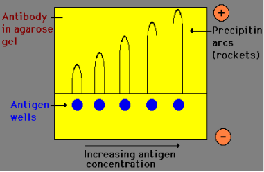
- The procedure of Rocket Immunoelectrophoresis entails the migration of the antigen through an agarose gel containing antiserum. As the antigen moves from the well, it forms distinct precipitin peaks resembling the shape of rockets. The height of these peaks is directly proportional to the concentration of the antigen loaded into the respective well. By measuring the height of the rocket-shaped precipitin bands, the concentration of the antigen in the sample can be determined.
- Rocket Immunoelectrophoresis is essentially an adaptation of radial immunodiffusion, which was originally developed by Laurell. It is also known by other names such as electroimmunoassay or electroimmunodiffusion. The term “rocket electrophoresis” is derived from the appearance of cone-like structures formed by the precipitin bands at the end of the reaction.
- In the process of Rocket Immunoelectrophoresis, the antigen migrates in an electric field within a layer of agarose that contains a suitable antibody. As the antigen moves toward the anode, it creates distinct patterns of precipitation in the shape of rockets. The area beneath the rocket-shaped structure is directly proportional to the concentration of the antigen being analyzed.
- Overall, Rocket Immunoelectrophoresis is a valuable technique for quantifying the concentration of antigens in unknown samples. Its simplicity, quickness, and reproducibility make it an attractive method in various research and diagnostic applications. By utilizing the rocket-shaped precipitin peaks, scientists and healthcare professionals can obtain quantitative data about antigen concentrations, aiding in further analysis and decision-making processes.
Objective of Rocket Immunoelectrophoresis Test
To study the technique of Rocket Immunoelectrophoresis for determination of the concentration of antigen in the unknown sample.
Principle of Rocket Immunoelectrophoresis
The principle of Rocket Immunoelectrophoresis revolves around the migration of negatively charged antigen samples through an agarose gel containing a specific antibody. As the antigen moves from the well into the agarose gel, it binds with the antibody, resulting in the formation of visible white precipitin arcs. This process occurs due to the application of an electric current, which drives the migration of the antigen in one direction.
Initially, there is an excess of antigen compared to the antibody, and no visible precipitation occurs. However, as the antigen sample continues to migrate through the agarose gel, it encounters more antibody molecules, leading to the formation of larger immune complexes. These complexes become large enough to be retained within the gel, causing the movement of the antigen to cease. The resulting precipitin area takes on the shape of a rocket, with the height of the rocket being directly proportional to the concentration of antigen in the corresponding well.
Rocket immunoelectrophoresis is a quantitative one-dimensional single electro-immunodiffusion technique. In this method, the antibody is incorporated into the gel at a pH value that immobilizes the antibodies. The antigen is loaded into wells created in the gel, and an electric current is passed through the gel, facilitating the migration of negatively charged antigens into the agar. As the antigen moves into the agarose gel, it forms immune complexes with the antibody, becoming visible as a precipitate. Initially, when there is an excess of antigen, no visible precipitation occurs. However, as the antigen migrates further into the gel, it encounters more antibody molecules, resulting in the formation of immune complexes. This leads to the formation of a conical precipitin line resembling a rocket.
The height of the rocket-shaped precipitin line and the area underneath it are directly proportional to the amount of antigen present in the sample. A larger amount of antigen loaded into a well requires the antigen to travel a greater distance through the gel before interacting with enough antibody to form a precipitate. Thus, by measuring the height of the rocket from the well to the apex and the area beneath it, the quantity of antigen in the sample can be determined.

gel before it interacts with sufficient antibody to form precipitin peaks
Material Required
To perform Rocket Immunoelectrophoresis, the following materials are required:
- Glass wares: Conical flask, measuring cylinder, beaker – These glass containers are essential for preparing and measuring reagents, as well as holding the agarose gel and samples during the procedure.
- Reagents: a. Sterile distilled water – Used for preparing the agarose gel and diluting reagents. b. Alcohol – Used for sterilizing the glassware and equipment to maintain aseptic conditions.
- Other requirements: a. Incubator (37°C) – An incubator with a controlled temperature of 37°C is necessary for maintaining optimal conditions for the reaction to occur. b. Microwave or Bunsen burner – Required for heating and melting the agarose gel to prepare the gel matrix. c. Vortex mixer – Used to mix and homogenize the samples and reagents thoroughly. d. Spatula – Used for transferring and manipulating solid reagents or gel materials. e. Micropipettes – Instruments used for accurate and precise measurement of small volumes of reagents and sample loading. f. Tips – Disposable tips used with micropipettes for preventing cross-contamination between samples. g. Moist chamber (box with wet cotton) – A chamber or container with moistened cotton is employed to create a humid environment, which helps prevent the agarose gel from drying out during the procedure.
These materials are necessary for the preparation of the agarose gel, dilution of samples and reagents, and maintaining the appropriate conditions for the Rocket Immunoelectrophoresis technique. Each item plays a specific role in facilitating the accurate measurement, mixing, and incubation required for the successful execution of the procedure.
Important Instructions
When performing the Rocket Immunoelectrophoresis experiment, it is crucial to follow these important instructions:
- Carefully read the entire procedure before starting the experiment to ensure a clear understanding of the steps involved.
- Always wear gloves throughout the experiment to maintain aseptic conditions and prevent contamination.
- Preparation of 1X TBE: a. Take the 20 ml tablet of 50X TBE from the pouch and transfer it to a 250 ml glass beaker. b. Add 20 ml of sterile distilled water to the beaker. c. Heat the beaker in a microwave for 3-5 seconds until the tablet dissolves and the solution turns into liquid form. d. Transfer the 50X TBE solution to a 1000 ml cylinder. e. Rinse the beaker with sterile distilled water to collect any remaining solution and pour it into the cylinder. f. Make up the volume to 1000 ml with sterile distilled water to obtain 1X TBE buffer.
- Preparation of 1% Agarose gel: a. Add 0.15 g of agarose powder to 15 ml of 1X TBE buffer to prepare 15 ml of agarose gel. b. Boil the mixture to dissolve the agarose completely.
- Add the antiserum to the agarose gel only after it cools down to 55°C. Higher temperatures can inactivate the antibody, affecting the experiment’s accuracy.
- Ensure the glass plates used for the gel have been wiped with cotton and made grease-free using alcohol. This step promotes even spreading of the agarose gel.
- Neatly cut the wells in the agarose gel, ensuring they have smooth edges without any rugged margins. This facilitates proper loading of the samples.
- Verify that the moist chamber contains enough wet cotton to maintain a humid atmosphere. This helps prevent the agarose gel from drying out during the experiment.
Note: It is recommended to use Molecular Biology Grade Water (Product code: ML064) for optimal results in the experiment.
Adhering to these important instructions ensures accurate and reliable outcomes while conducting Rocket Immunoelectrophoresis. These instructions cover various aspects such as reagent preparation, gel handling, and maintaining appropriate experimental conditions, all of which are crucial for a successful experiment.
Procedure of Rocket Immunoelectrophoresis
The procedure for Rocket Immunoelectrophoresis is as follows:
- Prepare 15 ml of 1% agarose solution following the instructions provided in the previous section.
- Allow the agarose solution to cool down to a temperature of 55-60°C. Once cooled, add 250 μl of antiserum to 13 ml of the agarose solution. Mix the solution thoroughly to ensure a uniform distribution of the antibody.
- Pour the agarose solution containing the antiserum onto a glass plate that has been made grease-free. Place the glass plate on a horizontal surface and allow the gel to set for approximately 30 minutes.
- Position the glass plate on the provided template, ensuring it aligns correctly.
- Using a gel puncher, gently punch wells into the gel. Take care to apply gentle suction to avoid creating rugged wells that may affect the experiment.
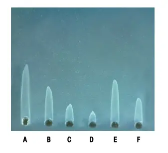
- Add 10 μl of the standard antigen samples and the test antigen samples into the respective wells. The standard antigens include:
- A. Standard Antigen A (1.87 mg/ml)
- B. Standard Antigen B (0.94 mg/ml)
- C. Standard Antigen C (0.47 mg/ml)
- D. Standard Antigen D (0.23 mg/ml)
- E. Test Antigen 1 F. Test Antigen 2
- Pour 1X TBE buffer into the electrophoresis tank, ensuring that it covers the gel completely. Any remaining 1X TBE buffer can be stored at room temperature for future use.
- Set the electrophoresis conditions to 80-120 volts and 60-70 mA. Allow the electrophoresis process to continue until the blue dye marker travels approximately 3-4 cm from the well. It is important not to exceed 3 hours of electrophoresis, as prolonged time may generate heat that can affect the results.
- After completing the electrophoresis, incubate the glass plate in a moist chamber overnight at a temperature of 37°C. The moist chamber helps maintain the humidity required for proper reaction and precipitation.
Following this step-by-step procedure ensures the accurate execution of Rocket Immunoelectrophoresis, allowing for the detection and quantification of antigen concentration in the samples.
Note: The remaining 1X TBE buffer can be stored at room temperature.
Observation and Result
Upon conducting the Rocket Immunoelectrophoresis experiment, the following observations and results can be noted:
- Observation: Look for the formation of precipitin peaks in the shape of ‘rockets’ spreading out from the loading well. These rocket-shaped precipitin peaks indicate a positive reaction or a specific antigen-antibody interaction, which occurs when there is an antibody present that is specific to the antigen being tested.
- Observation: If no precipitation or rocket-shaped formations are observed, it indicates a negative result, indicating no reaction or the absence of the corresponding antibody-antigen interaction.
- Result interpretation: The height of the rocket-shaped precipitin peak and its area directly correlate with the amount of antigen present in the sample. This means that the concentration of antigens loaded in the corresponding wells determines the height of the precipitin peak. Higher concentrations of antigens lead to taller rocket-shaped peaks.
In summary, the presence of a spreading rocket-shaped precipitin peak indicates a positive reaction, demonstrating the presence of a specific antigen-antibody interaction. Conversely, the absence of such precipitation indicates a negative result, suggesting no reaction or the absence of the corresponding antibody-antigen interaction. The height of the rocket-shaped precipitin peak serves as a quantitative measure of the antigen concentration, with taller peaks indicating higher levels of antigens in the sample.

Applications of Rocket Immunoelectrophoresis
Rocket Immunoelectrophoresis has several applications in various fields. The method is primarily utilized for the quantitative estimation of antigens in serum samples. Before automated methods became widely available, Rocket Immunoelectrophoresis played a crucial role in quantizing human serum proteins.
Some of the key applications of Rocket Immunoelectrophoresis include:
- Quantification of Specific Proteins: Rocket Immunoelectrophoresis allows for the determination of the concentration of a specific protein within a mixture of proteins. By comparing the rocket-shaped precipitin peaks formed by the antigen of interest with known standards, the concentration of the protein can be accurately measured.
- Estimation of Immunoglobulin Protease Activity: This method can be employed to assess the protease activity of immunoglobulins. By incorporating specific substrates into the agarose gel, the presence and activity of immunoglobulin proteases can be visualized based on the formation of rocket-shaped precipitin peaks.
- Studies on Antigenic Relationships: Rocket Immunoelectrophoresis is also used in studies that focus on antigenic relationships between different organisms. By comparing the rocket-shaped precipitin patterns obtained from different antigen samples, researchers can gain insights into the relatedness and cross-reactivity of antigens among various organisms.
- Enzyme Activity Electrophoresis: This technique finds application in the assessment of enzyme activity. By incorporating specific enzyme substrates into the gel, the presence and activity of enzymes can be detected and quantified based on the resulting rocket-shaped precipitin peaks.
Advantages of Rocket Immunoelectrophoresis
Rocket Immunoelectrophoresis offers several advantages as a method for analyzing antigens and proteins. These advantages include:
- Simplicity, Speed, and Reproducibility: Rocket Immunoelectrophoresis is a straightforward and easy-to-follow technique. It does not involve complex equipment or extensive preparation. The method is quick, providing results within a relatively short period. Moreover, it offers high reproducibility, ensuring consistent and reliable outcomes with repeated experiments.
- Multiple Sample Analysis: One of the notable advantages of Rocket Immunoelectrophoresis is the ability to analyze several unknown samples simultaneously on a single plate. This feature enhances efficiency and reduces the time and effort required for sample analysis. Multiple samples can be loaded into separate wells on the gel, allowing for efficient comparison and assessment of antigen concentrations.
- Sensitivity and Small Sample Requirement: Rocket Immunoelectrophoresis exhibits remarkable sensitivity, enabling the measurement of protein concentrations as low as 1 µg/mL. This sensitivity makes it suitable for the detection and quantification of even minute amounts of proteins. Additionally, the method requires as little as 20 ng of protein to be loaded in a well, minimizing the amount of sample material needed for analysis. This advantage is particularly valuable when working with limited or precious samples.
Overall, Rocket Immunoelectrophoresis provides a simple, quick, and reproducible approach for antigen and protein analysis. Its ability to accommodate multiple samples on a single plate, along with its high sensitivity and minimal sample requirement, makes it a valuable tool in research, diagnostics, and various laboratory applications.
Disadvantages of Rocket Immunoelectrophoresis
While Rocket Immunoelectrophoresis offers several advantages, it is important to consider its limitations. Some of the limitations of this technique include:
- Limited Applicability to Complex Mixtures: Rocket Immunoelectrophoresis is primarily designed for the quantitative analysis of antigens. However, it is not suitable for complex mixtures containing numerous proteins or antigens. The method relies on the migration and interaction of specific antigens with corresponding antibodies, and in complex mixtures, the interpretation of results becomes challenging due to overlapping precipitin peaks.
- Semi-Quantitative Nature: Although Rocket Immunoelectrophoresis allows for quantitative analysis of antigens, it should be noted that the method is semi-quantitative in nature. The height of the precipitin peak is used as an indicator of antigen concentration, but it does not provide precise and absolute measurements. The method provides relative comparisons between samples rather than exact quantification.
- Limited Resolution: Rocket Immunoelectrophoresis has limited resolution when compared to more advanced techniques such as chromatography or immunoassays. The separation of antigens in the gel is based on their migration under the influence of an electric field, which may result in overlapping bands or reduced separation efficiency. This limitation can affect the accuracy and precision of the analysis, particularly when dealing with closely related antigens.
- Subject to Interpretation: The interpretation of Rocket Immunoelectrophoresis results requires expertise and experience. Analyzing and accurately measuring the height of precipitin peaks can be subjective and may vary between individuals. It is important to have trained personnel who can interpret the results consistently and minimize potential errors.
In summary, while Rocket Immunoelectrophoresis allows for quantitative analysis of antigens, it has limitations when applied to complex mixtures, offers semi-quantitative measurements, has limited resolution, and requires expertise for result interpretation. Researchers should consider these limitations and explore alternative techniques when dealing with complex samples or when precise and absolute quantification is required.
Precautions
When conducting Rocket Immunoelectrophoresis, it is important to follow certain precautions to ensure accurate and reliable results. These precautions include:
- Familiarize Yourself with the Procedure: Before starting the experiment, carefully read and understand the entire procedure. This will help you to follow the steps correctly and avoid any mistakes or confusion during the process.
- Wear Gloves: Always wear gloves while performing the experiment. This precaution helps to maintain a sterile and contamination-free environment, ensuring the integrity of the samples and reagents.
- Preparation of 1X TBE Buffer: Follow the instructions provided for the preparation of 1X TBE buffer. Pay attention to the measurements and heating instructions to dissolve the tablet properly. Maintain sterility throughout the process by using sterile distilled water and clean glassware.
- Preparation of 1% Agarose Gel: When preparing the agarose gel, make sure to accurately measure the agarose powder and 1X TBE buffer. Follow the boiling instructions to dissolve the agarose completely. Allow the mixture to cool to approximately 55°C before adding the antiserum, as higher temperatures can inactivate the antibody.
- Grease-Free Glass Plates: Prior to pouring the agarose mixture, ensure that the glass plates are clean and grease-free. Use cotton and alcohol to wipe the plates, which helps in achieving an even spread of the agarose gel.
- Neat Wells: When cutting the wells on the gel, ensure that they are neat and free from rugged margins. Clean and well-defined wells help in accurately loading the antigen samples and prevent any diffusion or mixing during the electrophoresis process.
- Maintain Humidity in the Moist Chamber: To create a suitable environment for the experiment, make sure that the moist chamber contains enough wet cotton. This helps to keep the atmosphere humid, which is important for proper gel formation and antigen migration.
By adhering to these precautions, you can minimize potential errors and ensure the reliability and accuracy of your Rocket Immunoelectrophoresis experiment.
FAQ
What is Rocket Immunoelectrophoresis?
Rocket Immunoelectrophoresis, also known as electro-immunodiffusion, is a quantitative one-dimensional immunoelectrophoresis technique used to determine the concentration of an antigen in an unknown sample. It involves comparing the antigen sample with a series of dilutions of a known concentration of the antigen using a monospecific antibody.
How does Rocket Immunoelectrophoresis work?
In Rocket Immunoelectrophoresis, negatively charged antigen samples migrate through an agarose gel containing specific antibodies. As the antigen moves through the gel, it combines with the antibodies, forming immune complexes that precipitate. The height of the resulting precipitin peaks, which resemble rockets, is directly proportional to the antigen concentration in the corresponding well.
What are the applications of Rocket Immunoelectrophoresis?
Rocket Immunoelectrophoresis is primarily used for quantitative estimation of antigens in serum samples. It has been used to quantify human serum proteins, determine specific protein concentrations in mixtures, measure immunoglobulin protease activity, study antigenic relationships between organisms, and perform enzyme activity electrophoresis.
What are the advantages of Rocket Immunoelectrophoresis?
Rocket Immunoelectrophoresis offers several advantages, including its simplicity, quickness, and reproducibility. It allows multiple unknown samples to be analyzed on a single plate. Additionally, it can measure protein concentrations as low as 1 µg/mL, requiring only small amounts of protein to be loaded in the wells.
What are the limitations of Rocket Immunoelectrophoresis?
Rocket Immunoelectrophoresis is not suitable for analyzing complex mixtures. It is primarily used for quantitative analysis of specific antigens and may not be applicable when multiple antigens or a complex mixture of proteins is present.
What precautions should be taken during Rocket Immunoelectrophoresis?
It is important to read and understand the entire procedure before starting the experiment. Always wear gloves for protection. Follow the instructions carefully for preparing the TBE buffer and agarose gel. Maintain cleanliness and grease-free glass plates for even spreading of agarose. Cut neat wells without rugged margins. Ensure a humid atmosphere in the moist chamber.
How is the result observed in Rocket Immunoelectrophoresis?
The result of Rocket Immunoelectrophoresis is observed as precipitin peaks in the shape of rockets against a dark background. The height of the peak, measured from the upper edge of the well to the tip, is proportional to the antigen concentration in the corresponding well.
What does a positive reaction indicate in Rocket Immunoelectrophoresis?
A positive reaction in Rocket Immunoelectrophoresis indicates the presence of a specific antigen-antibody interaction. It is observed as a spreading precipitation rocket from the loading well. This indicates the presence of the antibody specific to the antigen being tested.
What does the absence of precipitation indicate in Rocket Immunoelectrophoresis?
The absence of precipitation in Rocket Immunoelectrophoresis indicates no reaction or the absence of any corresponding antigen-antibody interaction. It suggests that either the antigen of interest is not present in the sample or there is no specific antibody available to interact with the antigen.
Can Rocket Immunoelectrophoresis be used for protein quantification in automated methods?
Rocket Immunoelectrophoresis was used for protein quantification before automated methods became available. With advancements in technology, automated methods such as ELISA and other immunoassays have become more commonly used for protein quantification due to their higher throughput and efficiency.
References
- Walker JM. Rocket immunoelectrophoresis. Methods Mol Biol. 1984;1:317-23. doi: 10.1385/0-89603-062-8:317. PMID: 20512702.
- http://www.ispybio.com/search/protocols/RocketImmunoelectrophoresis.pdf
- http://www.ispybio.com/search/protocols/ie%20protocol8.pdf
- https://himedialabs.com/TD/HTI006.pdf
- https://microbenotes.com/rocket-immunoelectrophoresis/
