Table of Contents
The history of the simple microscope can be traced back to the 17th century, when it was first developed by Dutch mathematician and astronomer, Christiaan Huygens. Huygens designed a simple microscope that used a single converging lens to magnify the image of an object, similar to the design of a modern refracting simple microscope.
The simple microscope was not widely used at the time because it had a relatively low magnification and was not capable of resolving small structures or organisms. However, it was an important precursor to the compound microscope, which was developed a few decades later and used multiple lenses to achieve higher magnifications and better resolution.
The compound microscope was invented by Dutch scientist Antonie van Leeuwenhoek in the late 17th century. Van Leeuwenhoek used a small, powerful lens to focus the image of a sample onto an eyepiece, and he was able to achieve magnifications of up to 275x with his microscopes. Van Leeuwenhoek’s microscopes were the first to reveal the existence of microorganisms, and they played a crucial role in the development of modern biology and medicine.
Today, simple microscopes are still used in some educational and hobbyist applications, but they are not commonly used in scientific or medical research due to their limited magnification and resolution.
Definition of Simple Microscope
- A simple microscope is a type of microscope that has only one lens, which is used to magnify the image of an object. Simple microscopes are also known as monocular microscopes, and they are typically less expensive and easier to use than compound microscopes, which have multiple lenses and can achieve higher magnifications.
- Simple microscopes are typically used for low-magnification applications and are not suitable for studying small structures or organisms in great detail.
- Simple microscopes can be further divided into two types: refracting and reflecting. Refracting simple microscopes, also known as Galilean microscopes, use a converging lens to magnify the image of the sample. Reflecting simple microscopes, also known as Keplerian microscopes, use a concave mirror to focus the image onto an eyepiece.
- Both types of simple microscopes are limited in their magnification and resolution compared to compound microscopes, and they are not commonly used in scientific or medical research. However, they can be useful for certain educational or hobbyist applications, such as viewing small insects or examining the structure of leaves and flowers.
Principle of Simple Microscope
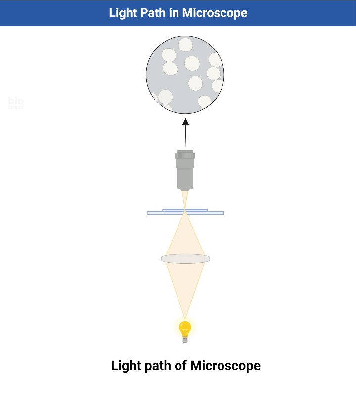
The principle of a simple microscope is based on the use of a single lens to magnify the image of an object. The lens is used to focus the light from the object onto an eyepiece, which is used to view the image. The magnification of the microscope is determined by the focal length of the lens and the distance between the lens and the eyepiece.
Simple microscopes can be further divided into two types: refracting and reflecting. Refracting simple microscopes, also known as Galilean microscopes, use a converging lens to magnify the image of the sample. Reflecting simple microscopes, also known as Keplerian microscopes, use a concave mirror to focus the image onto an eyepiece.
Both types of simple microscopes have a relatively low magnification and are not capable of resolving small structures or organisms in great detail. They are typically used for low-magnification applications and are not commonly used in scientific or medical research. However, they can be useful for certain educational or hobbyist applications, such as viewing small insects or examining the structure of leaves and flowers.
Working Mechanism of Simple Microscope
This ray diagram in below, explains how simple microscopes is working;
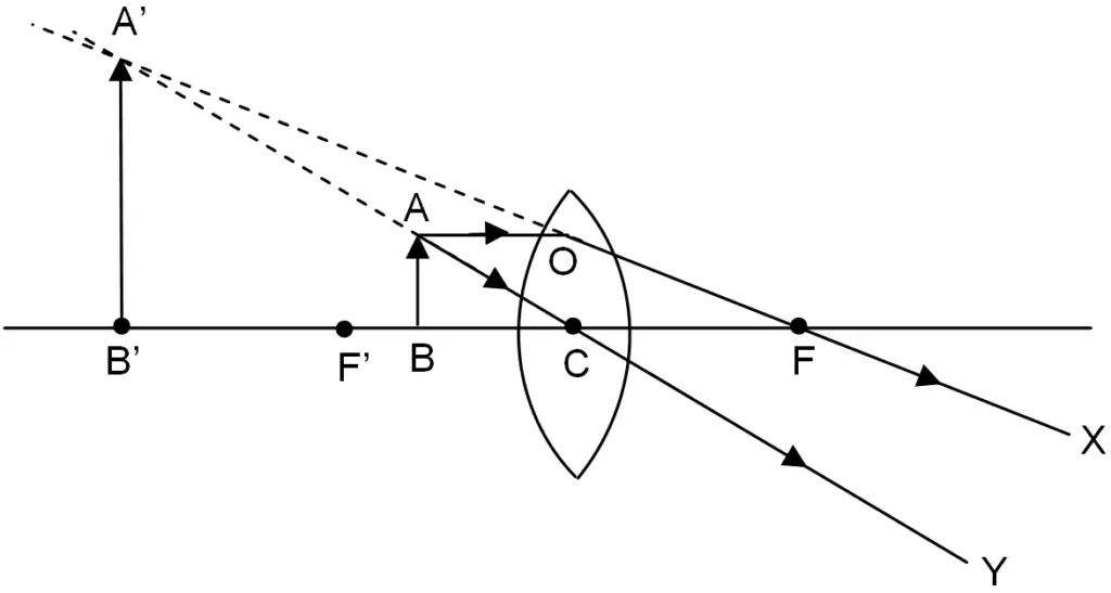
- A small object AB which is to be magnified is placed between the principal focus F’ and optical center C of the convex lens.
- Now, a ray of light AO parallel to a principal axis which is coming from point A of the object passes through the focus F along the straight line OX after getting refracted by the convex lens.
- A second ray of light AC coming from the point A of the object passes through the optical center C of the convex lens along the straight line CY.
- As is clear from the figure that the two rays i.e. OX and CY are diverging rays so these rays can intersect each other only at point A’ when produced backward.
- Now, on drawing A’B’ perpendicular from point A’ to the principal axis, we get the image A’B’ of the object which is virtual, erect, and magnified.
Magnification of a simple Microscope
The magnification of a simple microscope is determined by the focal length of the lens and the distance between the lens and the eyepiece. The magnification is calculated using the following formula:
Magnification = 1 + focal length of lens / distance between lens and eyepiece
Simple microscopes typically have a lower magnification than compound microscopes, which have multiple lenses and can achieve higher magnifications and better resolution. The magnification of a simple microscope is typically in the range of 5x to 50x, although some models may be able to achieve higher magnifications. The resolution, or the ability of the microscope to resolve fine details, is also typically lower for simple microscopes compared to compound microscopes.
Simple microscopes are not commonly used for high-resolution applications because they are limited in their magnification and resolution. However, they can be useful for certain educational or hobbyist applications, such as viewing small insects or examining the structure of leaves and flowers.
The magnifying power of simple microscopes is given by:
M = 1 + D/F
Where, D = the least distance of distinct vision
F = focal length of the convex lens
It should be noted,
- Smaller the focal length of the lens, greater will be its magnifying power.
- This microscope has a maximum magnification power of 10, which means the specimen will appear 10 times larger by using the simple microscopes of maximum magnification.
Parts of Simple Microscope with diagram
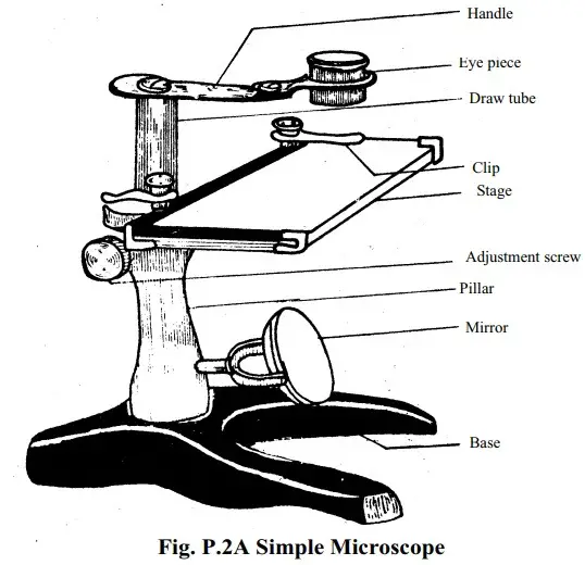
Simple microscopes are consist of two important parts, includes;
- The Mechanical Parts
- The Optical Parts
1. The Mechanical Parts of Simple Microscope
The Mechanical Parts support the optical parts and help in their adjustment for focusing the object. They include the following components;
a. Metal Stand
- The metal stand of a simple microscope is the support structure that holds the microscope in place and allows it to be positioned at the desired angle. The metal stand typically consists of a base, an upright support, and a horizontal support that connects the base to the microscope.
- The base of the metal stand is typically made of a heavy, sturdy material, such as cast iron or steel, to provide a stable foundation for the microscope.
- The upright support is a vertical rod that extends from the base and is used to hold the horizontal support at the desired height. The horizontal support is a bar that connects the upright support to the microscope and allows the microscope to be positioned at the desired angle.
- The metal stand of a simple microscope is an important component of the microscope because it provides stability and support for the instrument.
- It is typically adjustable, so that the microscope can be positioned at the desired height and angle for comfortable viewing. The metal stand also allows the microscope to be easily moved or transported if needed.
Function of Metal Stand
- Metal Stand provides support and stability to other parts of the microscope.
b. Stage
- It’s refers to a rectangular metal plate fitted to the vertical rod.
- The stage of a simple microscope is a platform that holds the sample being viewed. It is typically located above the base of the microscope and is adjustable to position the sample in the field of view. The stage is an important component of the microscope because it allows the user to precisely position the sample and ensure that it is in focus.
- The stage of a simple microscope is typically smaller and less complex than the stage of a compound microscope. It may have a simple mechanism for adjusting the position of the sample, such as a pair of X-Y knobs or a lever, or it may simply be a flat surface that the sample can be placed on. Some simple microscopes may also have a stage clip or other device to hold the sample in place.
- The stage of a simple microscope is an important component of the microscope because it allows the user to precisely position the sample and ensure that it is in focus. It is also typically used to control the amount of light that is allowed to pass through the sample, using a diaphragm or iris diaphragm, to enhance the contrast of the image.
- Stage also has a central hole for light to pass from below.
- Some simple microscopes have a pair of slanting wings projecting from both sides of the stage which provide support to hand for manipulating the object.
Function of stage
- The specimen slide place over it to be observed.
c. Base
- The base of a simple microscope is the lower part of the microscope that supports the rest of the instrument. It is typically made of a heavy, sturdy material, such as cast iron or steel, to provide a stable foundation for the microscope.
- The base often has a built-in light source or a holder for a lamp, and it may also have other features, such as a power switch or a fuse.
- The base of a simple microscope is an important component of the microscope because it provides stability and support for the instrument.
- It also typically houses the electrical components of the microscope, such as the light source and any additional features, such as a power switch or fuse.
- The base is typically located at the bottom of the microscope and is the part of the instrument that is in contact with the surface on which it is placed. It is typically a relatively simple component of the microscope, but it is an essential part of the overall design.
d. Stage clips
- Stage clips are a type of accessory that is used with a microscope to hold the sample being viewed in place on the stage. They are typically used with compound microscopes, but they may also be used with simple microscopes. Stage clips are used to secure the sample to the stage and prevent it from moving or sliding when the microscope is being used.
- Stage clips are typically made of a rigid material, such as metal or plastic, and are designed to grip the sample securely without damaging it.
- They are typically adjustable and can be positioned to hold the sample at different angles or in different locations on the stage. Stage clips are often used in conjunction with other microscope accessories, such as slides, to prepare and present the sample for viewing.
- Stage clips are an important accessory for microscopes because they help to ensure that the sample is stable and secure during use.
- This is particularly important when the microscope is being used at high magnifications, as even small movements of the sample can make it difficult to focus the image. Stage clips are also useful for holding the sample in place when the microscope is being moved or transported.
e. Adjustment screw
- It is used to adjust the focus on specimen.
2. The Optical Parts of Simple Microscope
The optical parts help in magnification and visualization of specimen. This part is consist of these following components;
1. Mirror
- It has a plano-convex mirror, which is located is below the stage to the vertical rod by means of a frame.
Function of Mirror
- The primary function is to focus the surrounding light on the object being examined.
2. Lens
- Simple microscopes has a biconvex lens which is located above the stage, to the vertical rod, by means of a frame.
- For proper focusing, the lens can be moved up and down by the frame.
Function
- It magnifies the size of the object and the enlarged virtual image formed is observed by keeping the eye above it.
A modern simple microscope contain these following parts;
- Eyepiece: A set of lenses, located at the top of microscope, which used to visualize the samples. It has a magnification power of 10X to 15X.
- Tube: It connect the eyepiece to the objective lenses.
- Revolving nose-piece: The Revolving nose-piece or turret holds the objective lenses and it can rotate during the study of sample.
- Fine adjustment knob: The Fine adjustment knob is used to focus on oil.
Types of simple microscope
There are two main types of simple microscopes: refracting and reflecting.
- Refracting simple microscopes, also known as Galilean microscopes, use a converging lens to magnify the image of the sample. The lens is located at the bottom of the microscope, near the sample being viewed, and is used to focus the light from the sample onto an eyepiece, which is used to view the image. Refracting simple microscopes are relatively simple in design and are often used for low-magnification applications, such as viewing small insects or examining the structure of leaves and flowers.
- Reflecting simple microscopes, also known as Keplerian microscopes, use a concave mirror to focus the image onto an eyepiece. The mirror is located at the bottom of the microscope, near the sample being viewed, and is used to reflect the light from the sample onto the eyepiece. Reflecting simple microscopes are similar in design to refracting simple microscopes and are also typically used for low-magnification applications.
Both types of simple microscopes are limited in their magnification and resolution compared to compound microscopes, and they are not commonly used in scientific or medical research. However, they can be useful for certain educational or hobbyist applications, such as viewing small insects or examining the structure of leaves and flowers.
Operating Procedure of Simple Microscope
Here is a general operating procedure for using a simple microscope:
- Set up the microscope: Make sure that the microscope is positioned on a stable, flat surface and that the light source is turned on.
- Place the sample on the stage: Use the stage clips to secure the sample in place on the stage. Make sure that the sample is positioned correctly and that it is centered under the objective lens.
- Select the lowest magnification objective lens: Begin by using the lowest magnification objective lens to get a wide view of the sample. This will allow you to locate the area of interest and make it easier to focus the image.
- Focus the image: Use the coarse adjustment knob to bring the objective lens close to the sample, and then use the fine adjustment knob to fine-tune the focus and bring the image into clear view.
- Change the magnification: If necessary, you can change the magnification by switching to a different objective lens. Make sure to use the fine adjustment knob to refocus the image after changing the magnification.
- View the image: Once the image is in focus, you can use the eyepiece to view the image. Make sure to keep your eye close to the eyepiece and to blink occasionally to prevent eye fatigue.
- Turn off the light source: When you are finished using the microscope, make sure to turn off the light source and to cover the sample to protect it from damage.
Applications of Simple Microscopes
There are present different uses of simple microscopes such as;
- In Jewelry making shop, Jewelry makers used it to visualize the magnified view of the small parts of the jewelry.
- In the Watchmaking industry, watchmakers used it to magnify a tiny part of the watch.
- In the Agriculture sector, it is used to magnify various particles of various types of soils.
- Palmist used a simple microscopes to visualize the lines of the hands.
- In Dermatology, a dermatologist or skin specialist used it to check for various skin diseases.
- In Microbiological experiments, a microbiologist used it for examining and studying microscopic fungi, algae, and other biological specimens that are difficult to visualize using the naked eyes.
- It also used to visualize the details of stamp and engravings.
- It also used to check the texture of fibers or threads of a cloth.
You Might be Like These Articles:
- Parts of Microscope with their Functions and Working Principle
- Size, Shape, and Arrangement of Bacterial Cells With Picture and Example
- Latex agglutination test – Procedure, Principle, Inhibition, Limitation, Uses.
- Agglutination test definition, Types, Uses, Advantages, Disadvantages
Advantages of Simple Microscope
- Low cost: Simple microscopes are typically less expensive than compound microscopes, which have multiple lenses and can achieve higher magnifications. This makes them an attractive option for educational or hobbyist applications where cost is a consideration.
- Easy to use: Simple microscopes are typically easier to use than compound microscopes, which have more complex optics and require more precise focusing. This makes them a good choice for beginners or for applications where ease of use is a priority.
- Portable: Simple microscopes are typically smaller and lighter than compound microscopes, which makes them more portable and easier to transport. This can be useful for field work or for applications where the microscope needs to be moved frequently.
- Low-magnification applications: Simple microscopes are typically used for low-magnification applications, such as viewing small insects or examining the structure of leaves and flowers. They are not suitable for studying small structures or organisms in great detail, but they can be useful for certain educational or hobbyist applications.
- Fewer parts: Simple microscopes have fewer parts than compound microscopes, which makes them easier to maintain and repair. This can be an advantage in situations where the microscope is used frequently or in harsh environments.
Limitations of Simple Microscope
- Low magnification: Simple microscopes are typically limited in their magnification and are not suitable for studying small structures or organisms in great detail. They are typically used for low-magnification applications, such as viewing small insects or examining the structure of leaves and flowers.
- Poor resolution: Simple microscopes have a lower resolution than compound microscopes, which means that they are not able to distinguish fine details or resolve small structures. This can make it difficult to study small structures or organisms in detail.
- Limited field of view: Simple microscopes have a limited field of view, which means that they can only magnify a small area of the sample at a time. This can make it difficult to get a comprehensive view of the sample or to locate specific structures or features.
- Limited contrast: Simple microscopes are not able to enhance the contrast of the image, which can make it difficult to see details or distinguish between different structures or features. This can be a problem when studying samples that have a similar color or when trying to distinguish small structures or organisms.
- Fewer accessories: Simple microscopes typically have fewer accessories and features than compound microscopes, which can limit their capabilities and flexibility. This can be a limitation for certain applications or when trying to study specific structures or features.
Precautions of Simple Microscope
- Handle the microscope carefully: Make sure to handle the microscope with care to prevent damage. Avoid dropping or shaking the microscope, and be mindful of the location of the objective lens when carrying or moving the microscope.
- Clean the lenses regularly: The lenses of the microscope can easily become dirty or scratched, which can affect the quality of the image. Make sure to clean the lenses regularly with lens tissue or a soft, lint-free cloth to maintain their clarity.
- Avoid touching the lenses with your fingers: The oils from your skin can damage the lenses of the microscope, so make sure to handle them carefully. Use lens tissue or a soft, lint-free cloth to clean the lenses, and avoid touching them with your fingers or any other objects.
- Use the coarse and fine adjustment knobs correctly: The coarse and fine adjustment knobs are used to focus the image of the sample onto the eyepiece. Make sure to use them correctly to avoid damaging the microscope or the sample.
- Follow the manufacturer’s instructions: Make sure to read the user manual carefully and follow the recommended procedures for using and caring for the microscope. This will help to ensure that the instrument is used safely and effectively.
Simple vs compound microscope
A simple microscope is a type of microscope that has only one lens, which is used to magnify the image of an object. Simple microscopes are also known as monocular microscopes, and they are typically less expensive and easier to use than compound microscopes, which have multiple lenses and can achieve higher magnifications. Simple microscopes are typically used for low-magnification applications and are not suitable for studying small structures or organisms in great detail.
A compound microscope is a type of microscope that uses multiple lenses to magnify the image of an object. The objective lenses of a compound microscope are the main lenses that are used to magnify the image, and they are located at the bottom of the microscope, near the sample being viewed. The eyepiece, which is located at the top of the microscope, is used to view the image and typically has a magnification of 10x or 15x. The total magnification of the microscope is the product of the magnification of the objective lens and the eyepiece. Some compound microscopes also have additional lenses or mirrors that can be used to further magnify the image or enhance the contrast.
In general, compound microscopes are more powerful and capable of higher magnifications and better resolution than simple microscopes. They are commonly used in scientific and medical research, as well as in education and other applications, to study small structures and organisms in detail. Simple microscopes, on the other hand, are typically used for low-magnification applications and are not commonly used in scientific or medical research. However, they can be useful for certain educational or hobbyist applications, such as viewing small insects or examining the structure of leaves and flowers.
What are the 5 rules of using a microscope?
Here are five rules for using a microscope effectively:
- Always start with the lowest magnification objective lens. This will allow you to get a wider view of the sample and help you to locate the area of interest.
- Use the coarse adjustment knob to bring the objective lens close to the sample. Then, use the fine adjustment knob to fine-tune the focus and bring the image into clear view.
- Avoid touching the lenses with your fingers or any other objects. The oils from your skin can damage the lenses, and particles from other objects can cause scratches or smudges on the surface.
- Keep the microscope clean and handle it carefully. Make sure to clean the lenses regularly with lens tissue or a soft, lint-free cloth. Avoid banging or shaking the microscope, and handle it with care to prevent damage.
- Follow the manufacturer’s instructions for using and maintaining the microscope. Make sure to read the user manual carefully and follow the recommended procedures for using and caring for the microscope. This will help to ensure that the instrument is used safely and effectively.
Simple Microscope Image
Simple squamous epithelium under microscope
Simple squamous epithelium is a type of tissue that is composed of a single layer of flat, scale-like cells. It is found in many organs and tissues in the body, including the lining of blood vessels, the alveoli of the lungs, and the mesothelium of the pleural cavity. Under the microscope, simple squamous epithelium appears as a thin, flat layer of cells with a smooth, shiny surface.
When viewed under a microscope, the cells of simple squamous epithelium are typically oval or circular in shape and have a thin, transparent cytoplasm. The nucleus is typically small and located near the center of the cell. The cells are closely packed together and are separated by thin intercellular spaces.
Simple squamous epithelium is characterized by its thin, flat cells and its ability to allow substances to pass through it easily. It plays a vital role in the body by providing a barrier between different tissues and organs, as well as allowing gases and other substances to exchange across the surface of the epithelium. Simple squamous epithelium is often used as a model for studying cell-cell interactions and the movement of substances across cell membranes.

Simple columnar epithelium under microscope
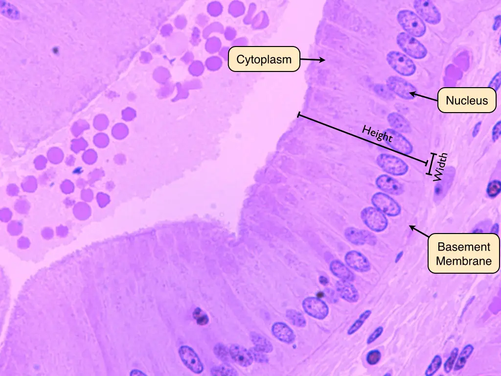
The single layer of cells in simple columnar epithelium are higher than they are wide. This form of epithelia borders the small intestine, absorbing nutrients from the lumen. The stomach also contains simple columnar epithelia, which secretes acid, digesting enzymes, and mucus.
Simple cuboidal epithelium under microscope
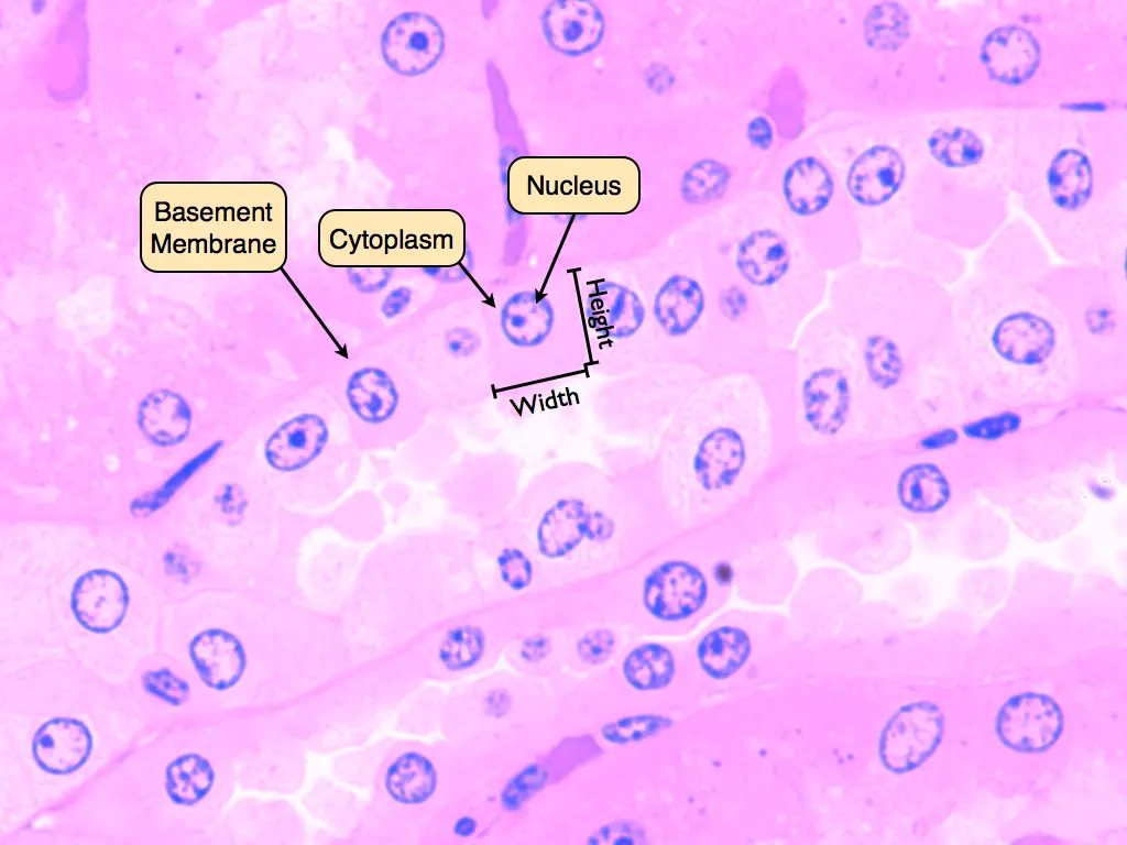
Simple cuboidal epithelium is made up of a single layer of cells that are roughly as tall as they are wide. This form of epithelium lines collecting ducts and tubes and is responsible for secreting or absorbing substances into the ducts or tubes.
FAQ of Simple Microscopes
- What is the definition of a simple microscopes?
- What is the uses of simple microscopes?
- What is simple microscope magnification?
- When was the simple microscope invented?
- What was the first microscope called?
- Who discovered microscope first time?
- What Are The Parts Of Simple Microscope?
- What is the working principle of a Simple Microscope?
the first scientist to describe living cells as seen through a simple microscope
Antonie van Leeuwenhoek was the first scientist to describe living cells as seen through a microscope. Van Leeuwenhoek was a Dutch scientist who lived in the late 17th and early 18th centuries, and he is considered to be the father of microbiology. He was the first person to observe and describe living cells, including bacteria, protozoa, and red blood cells, using a microscope.
Van Leeuwenhoek was not the inventor of the microscope, but he was the first person to use it to study living cells in detail. He designed and built his own microscopes, which were much more powerful than any that had been made before. His microscopes used a small, powerful lens to focus the image of a sample onto an eyepiece, and he was able to achieve magnifications of up to 275x with them.
Van Leeuwenhoek’s observations of living cells revolutionized our understanding of biology and laid the foundation for modern microscopy. His work laid the foundation for the field of microbiology and opened up new areas of research that have led to many of the scientific and medical advances we enjoy today.
Simple staining is often necessary to improve contrast in which microscope?
Simple staining is a technique that is often used to improve contrast in both compound and simple microscopes. Simple staining involves applying a dye or other coloring agent to a sample to highlight specific structures or features. It is often used to make it easier to see the details of a sample, particularly when the sample is transparent or has a similar color to the background.
In both compound and simple microscopes, simple staining can be used to improve contrast by making it easier to see the details of the sample. For example, simple staining can be used to highlight the cell walls of bacteria, the cytoplasm of cells, or the stomata of a leaf. It is a relatively simple technique that can be used to improve the clarity and resolution of the image being viewed.
Simple staining is typically used in combination with other techniques, such as brightfield microscopy, to enhance the contrast and resolution of the image. It is an important tool in microscopy and is widely used in scientific and medical research, as well as in education and other applications.
Who invented the simple microscope?
The simple microscope, also known as the monocular microscope, was developed by Dutch mathematician and astronomer Christiaan Huygens in the 17th century. Huygens designed a simple microscope that used a single lens to magnify the image of an object, similar to the design of a modern refracting simple microscope.
Huygens’ simple microscope was not widely used at the time because it had a relatively low magnification and was not capable of resolving small structures or organisms. However, it was an important precursor to the compound microscope, which was developed a few decades later and used multiple lenses to achieve higher magnifications and better resolution.
The compound microscope was invented by Dutch scientist Antonie van Leeuwenhoek in the late 17th century. Van Leeuwenhoek used a small, powerful lens to focus the image of a sample onto an eyepiece, and he was able to achieve magnifications of up to 275x with his microscopes. Van Leeuwenhoek’s microscopes were the first to reveal the existence of microorganisms, and they played a crucial role in the development of modern biology and medicine.
What is a simple microscope?
A simple microscope is a type of microscope that has only one lens, which is used to magnify the image of an object. Simple microscopes are also known as monocular microscopes, and they are typically less expensive and easier to use than compound microscopes, which have multiple lenses and can achieve higher magnifications. Simple microscopes are typically used for low-magnification applications and are not suitable for studying small structures or organisms in great detail.
Simple microscopes can be further divided into two types: refracting and reflecting. Refracting simple microscopes, also known as Galilean microscopes, use a converging lens to magnify the image of the sample. Reflecting simple microscopes, also known as Keplerian microscopes, use a concave mirror to focus the image onto an eyepiece. Both types of simple microscopes are limited in their magnification and resolution compared to compound microscopes, and they are not commonly used in scientific or medical research. However, they can be useful for certain educational or hobbyist applications, such as viewing small insects or examining the structure of leaves and flowers.
How many lenses does a simple microscope have?
A simple microscope has only one lens, which is used to magnify the image of an object. Simple microscopes are also known as monocular microscopes, and they are typically less expensive and easier to use than compound microscopes, which have multiple lenses and can achieve higher magnifications. Simple microscopes are typically used for low-magnification applications and are not suitable for studying small structures or organisms in great detail.
Simple microscopes can be further divided into two types: refracting and reflecting. Refracting simple microscopes, also known as Galilean microscopes, use a converging lens to magnify the image of the sample. Reflecting simple microscopes, also known as Keplerian microscopes, use a concave mirror to focus the image onto an eyepiece. Both types of simple microscopes have only one lens, which is used to magnify the image of the sample.
In contrast, compound microscopes have multiple lenses, including objective lenses and an eyepiece, which work together to magnify the image of the sample. The objective lenses of a compound microscope are located near the sample being viewed and are used to magnify the image, while the eyepiece is used to view the image and typically has a magnification of 10x or 15x. The total magnification of the microscope is the product of the magnification of the objective lens and the eyepiece. Compound microscopes are more powerful and capable of higher magnifications and better resolution than simple microscopes, and they are commonly used in scientific and medical research.
What is the difference between simple and light microscope?
The terms “simple microscope” and “light microscope” are often used interchangeably to refer to a type of microscope that uses visible light to magnify the image of an object. However, there are some subtle differences between these two types of microscopes.
A simple microscope is a type of microscope that has only one lens, which is used to magnify the image of an object. Simple microscopes are also known as monocular microscopes, and they are typically less expensive and easier to use than compound microscopes, which have multiple lenses and can achieve higher magnifications. Simple microscopes are typically used for low-magnification applications and are not suitable for studying small structures or organisms in great detail.
A light microscope, on the other hand, is a type of microscope that uses visible light to magnify the image of an object. Light microscopes can be either simple or compound microscopes, and they are typically used to study small structures or organisms in detail. Light microscopes are widely used in scientific and medical research, as well as in education and other applications, to study the structure and function of cells and other small structures.
In general, the term “simple microscope” refers to a type of microscope that has only one lens, while the term “light microscope” refers to a type of microscope that uses visible light to magnify the image of an object. Both simple and light microscopes can be either refracting or reflecting in design, and they are typically used for low-magnification applications. Compound microscopes, on the other hand, are more powerful and capable of higher magnifications and better resolution, and they are commonly used in scientific and medical research.
Who is the father of microscope?
Antonie van Leeuwenhoek is often referred to as the “father of the microscope.” Van Leeuwenhoek was a Dutch scientist who lived in the late 17th and early 18th centuries, and he is considered to be the first person to use a microscope to study living cells in detail.
Van Leeuwenhoek was not the inventor of the microscope, but he was the first person to design and build a microscope that was powerful enough to reveal the existence of microorganisms. He used a small, powerful lens to focus the image of a sample onto an eyepiece, and he was able to achieve magnifications of up to 275x with his microscopes.
Van Leeuwenhoek’s observations of living cells revolutionized our understanding of biology and laid the foundation for modern microscopy. His work laid the foundation for the field of microbiology and opened up new areas of research that have led to many of the scientific and medical advances we enjoy today. As such, he is often referred to as the “father of the microscope.”
Reference
- https://en.wikipedia.org/wiki/Optical_microscope
- https://www.slideshare.net/KirtiSharma87/microscope-ppt-63079222#close
- https://www.yourarticlelibrary.com/micro-biology/working-principle-and-parts-of-a-simple-microscope-with-diagrams/26490
- http://www.funscience.in/study-zone/Physics/OpticalInstruments/SimpleMicroscope.php#sthash.JyiHHclF.nWDg2919.dpbs
- https://laboratoryinfo.com/simple-microscope-parts/

