Table of Contents
Stroma in chloroplast
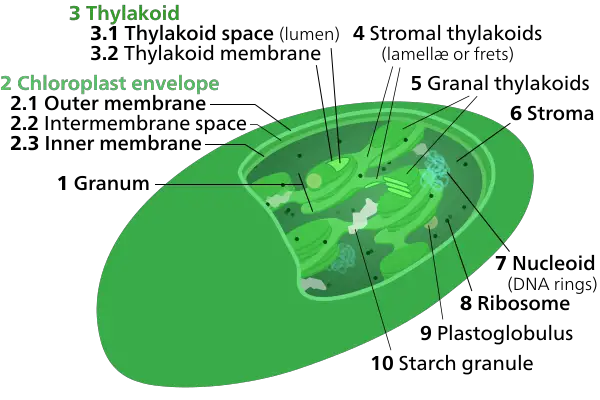
Stroma, as a term in botany, refers to the transparent fluid that surrounds the grana inside the chloroplast. In the stroma, there are the grana (stacks of the thylakoid) and sub-organelles, or daughter cells in which photosynthesis begins prior to the chemical reactions being completed within the stroma.
Photosynthesis takes place in two phases. In the initial stage that is light-dependent, reactions absorb all the light energy, and utilize it to create the energy storage compounds ATP as well as NADPH. In the second phase, the light-dependent reactions utilize these substances to remove carbon dioxide by capturing and decreasing carbon dioxide.
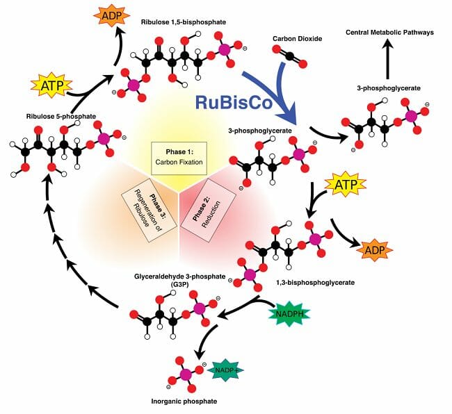
The biochemical redox reactions that take place within the stroma are collectively referred to as the Calvin cycle, also known as light-independent reactions. There are three stages: carbon fixation, reduction reactions and ribulose 1,5-bisphosphate (RuBP) renewal.
The stroma is also the location of chloroplast DNA and chloroplast ribosomes, and thus also the location of molecular processes including chloroplast DNA replication, and transcription/translation of some chloroplast proteins.
Definition of stroma in chloroplast
Stroma typically refers to the inner space that is filled with fluid of the chloroplasts that surround thylakoids as well as the grana. At first, the stroma was thought to be merely a the necessary support for the pigmented thylakoids. But it’s now clear that the stroma has starch as well as chloroplast DNA and the ribosomes, along with all the enzymes needed for photosynthesis-dependent light-dependent reactions commonly referred to by the Calvin cycle.
Originating from Greek word for the bed or layer Stroma may also mean other supporting structures like the connective tissues of organs, as well as the fungal tissues which transports the spores.
Structure of stroma in chloroplast
A microscopic inspection of the chloroplast will reveal obvious characteristics. It’s comprised of an outer membrane as well as an intricate web of inner membranes that form disc-like structures that are called Grana. Different grana are linked to one another via membranous extensions.
The membranes inside contain chlorophyll as well as other pigments that are that are involved in capturing light energy. Their obvious cytological look as well as the existence of pigments that are primary conferred importance on the membrane’s inner, grana, and their constituent thylakoids. But once the exterior membrane in the chloroplast was damaged and examined, it was discovered that reduction and carbon fixation ended. Also the transparent aqueous matrices or stroma, which seemed to only support the substructures that were pigmented, appeared to play a significant function in photosynthesis.
Chloroplasts evolved from prokaryotes that were free-living which formed an endosymbiotic connection with cells of the eukaryotic family. The stroma is still containing DNA and ribosomes that perform the synthesis of proteins. These proteins are crucial in the light-dependent reaction of photosynthesis and reactions that fix organic minerals like nitrates within organic molecules.

Function of stroma in chloroplast
The chloroplast is a unique organelle in that it is responsible for the main function that a plant cells has and is having the genome of its own. Numerous genes that are essential for their function have been included in its nuclear genome. This means that it must be capable of altering its metabolic function to support the functions of the cell. The stroma is crucial for this, as the stroma does not just house the necessary enzymes for carbon fixationbut but it also regulates the response of the chloroplast to stresses in the cell and also signals between organelles. It plays a crucial role in both light-dependent and photosynthesis reactions that are light-independent. In extreme stress the stroma is able to undergo autophagy in a selective manner without damaging the membranous structures that surround it as well as pigment molecules. Finger-like protrusions that extend from the stroma which do not contain any thylakoids, appear to be closely linked with the nucleus and the endoplasmic reticulum which contribute to the development of sophisticated regulation mechanisms.
Function of Chloroplast Stroma in Photosynthesis
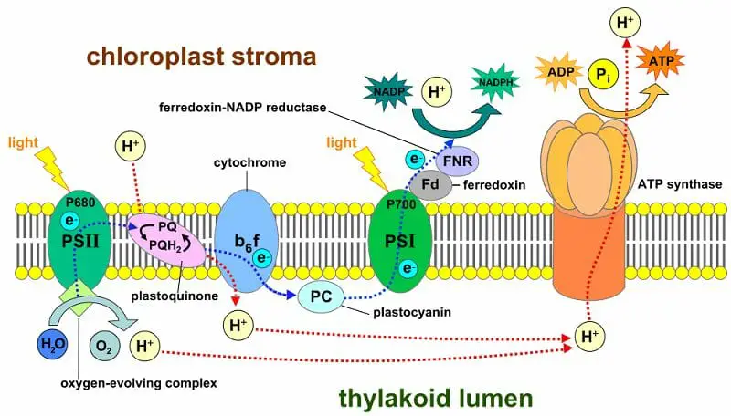
The stroma is first able to take part in photosynthesis as the energy of light taken up by pigment molecules is transformed into chemical energy by the electron transportation chain.

The photosystems I II (PSI and PSII) on the thylakoid membrane are composed of pigments that absorb light energy and utilize it to release high-energy electrons. These electrons pass through membrane-bound proteins like cytochrome B6F complex and plastoquinone (pq) in which simultaneous reduction and oxidation (redox) reactions take place. These reactions draw certain energy produced by the electron, in addition to pumping protons along their concentration gradients from the thylakoid to the thylakoid. If these protons are redirected back to the stroma, the actions of membrane-bound ATP Synthase drives the formation of ATP molecules. PSI can also be involved in production of the coenzyme reduced NADPH within the stroma through the action of ferrodoxin. The stroma is therefore home to the products that are the result of photochemical reactions that depend on light which include ATP and NADPH setting the scene for subsequent steps of photosynthesis.
The most important enzyme in the light-independent reactions, or the Calvin cycle, is RuBisCO or Ribulose-1,5-bisphosphate (RuBP) carboxylase. The enzyme is responsible for the initial stage of light-dependent reactions that involve carbon fixation. RuBisCO absorbs atmospheric carbon dioxide and diffuses into chloroplast stroma. It then fixates it in the form of organic molecules. Each CO2 molecule is combined with one RuBP molecule that has five carbon atoms. This will create two molecules of phosphoglycerate that are tri-carbon molecules.
The Calvin cycle involves two additional actions that occur within the stroma, namely the reduction of phosphoglycerate as well as the revival of RuBP. These steps require the usage in the form of ATP as well as NADPH. The light-dependent reactions require the two compounds in NADPH as well as three molecules ATP for the fixation of one CO2 molecule in the atmosphere.

Function of Chloroplast Stroma in Stress Response
Stresses induced by abiotic factors like salinity or temperature fluctuation or deficiency of nutrients could result in the development of protrusions shaped like fingers that arise from the chloroplast , referred to as Stromules. Stromules are observed in alpine plants that are exposed to greater temperatures. They are not a source of the thylakoids or grana and can be observed more frequently in the context of the deprivation of nutrients or the infection caused by pathogens.
Under extreme tension, the chloroplast tries to respond by focusing the stromal proteins for autophagy, transforming them into an evaporole. The autophagy induced by stress is controlled through the creation in an isolating membrane which blocks thylakoids and the grana. The membrane is then taken away from the chloroplast and subsequently targeted for destruction in the vacuole. This process is visible in microscopically by visible RuBisCO and other fluorescent proteins localized to stroma within the vacuole.
Function of Chloroplast Stroma in Intra-Organelle Signaling
Chloroplasts are semi-autonomous since they have their own genome. However, they also import many small molecules and proteins from the cell’s cytoplasm. Although they started out as autotrophs living in freedom, with the passage of time, some the genes they had were transferred to their nucleus of the host. These genes have been altered to ensure that they are targeted at the chloroplast and are believed to be in the hands of the combined control of the nucleus and the chloroplast. Signaling from the nucleus towards the plastid is known as anterograde signaling , while signals that travel towards the nucleus are referred to as retrograde signals. Both of these signals appear to be transmitted through the stromules that also play a part in the exchange of signals with two different plastids.
Calvin Cycle: Light-Independent Reactions
The stroma is the location for the three steps within the Calvin Cycle – carbon fixation Reduction, carbon fixation and regeneration.
Carbon fixation starts with the reaction of one CO2 molecule and one RuBP molecule. The six carbon atoms as well as two phosphate groups join to make two phosphoglycerate molecules which is a three-carbon molecule that contains the phosphate group. The reaction repeats three times and results in six phosphoglycerate molecules.
In the next step, phosphoglycerate accepts electrons to form glyceraldehyde-3-phosphate (G3P). The primary power for the reduction reaction results in the process of converting NADPH in NADP+ and the conversion of ATP to ADP. Therefore, ADP along with NADP+ are created for use in light-dependent reactions.
There is one more step to be completed – the process of regenerating RuBP. Out of the six molecules in G3P produced in the earlier stage, five are used to reform RuBP while the sixth is ejected from the chloroplast in order to create glucose.

Stroma in Animal Tissue
In mammals, stroma is referred to the tissues and cells that provide the essential functions in an organ. As an example, within the heart, muscles and neurons serve the major function, whereas the coronary circulation as well as immune system make up the stroma. Additionally, stroma comprises non-cellular components like collagen fibers, glycolipids, and glycoproteins which provide the structure for the organ and tissue.
Types of Stroma Tissue
- Loose connective tissue – It is found beneath the epithelial membranes as well as glandular epithelium. It connects the epithelia to various tissues. It is responsible for supporting blood vessels and nerves that are connected to epithelia. In addition, it is the primary site for inflammatory reaction in the body.
- Dense irregular connective tissue – The function of this kind is to bind at the highest tensile strength between tissues, converting tension from one point to another.
Examples of Animal Stroma
The stroma of every organ or tissue has certain generic functions, like transporting metabolites and fuels and the structural supports, within certain organs, they perform specific roles. The endocrine glands’ stroma aid in the production of hormones in the follicles as well as the lobules within the organ. In the Thymus, the stroma can influence the differentiation of T cells through either negative or positive selection. Organs that have to react quickly to the ever-changing requirements of the human body for example, the bone marrow or eye’s iris, also require special cells called stroma.
1. Stroma of the bone marrow
The stroma in the bone marrow isn’t directly involved in hematopoiesis however, it creates the microenvironment that boosts the activity of the cells that are involved in the creation of blood. The stroma is a source of growth factors, houses cells that are involved with bone metabolism and and contains fat cells, as well as macrophages. Macrophages are especially important since they play a role in the production of red blood cells. They also supply the iron required for hemoglobin production.
2. Stroma of the iris
The human iris starts to develop during the first trimester of gestation. It is among the few organs within the body that are easily observed. The iris is comprised of an epithelium that is pigmented as well as the muscles that are required to contract and dilate pupils. The cells serve as the primary role of the iris. They is supported by the vascular stroma. It is which is a loose and shattered connective tissue layer that contains collagen-forming cells and ligaments. It is because pigment filters the light that falls on the retina and lets some of it be absorbed by the pupil and create images on the retina. The pigmentation of the retina depends on the amount of pigment and melanin that is present in the stroma in people with brown eyes caused by heavy pigmentation and those with blue irises that produce very tiny amounts of pigment.
3. Stroma of cornea
The cornea’s stroma (or the substantia propria) is hard, fibrous and unbreakable, completely transparent and thickest corneal layer of the eye. It lies between Bowman’s membrane in the anterior and Descemet’s membrane laterally.
The corneal stroma of the human body is made of approximately 200 lamellae flattened (layers composed of collagen fibrils) that are that are superimposed on one on another. They measure 1.5-2.5 millimeters thick. The anterior lamellae are interwoven more than the posterior lamellae. The lamella’s fibrils are parallel to each other however they are at angles different to the lamellae adjacent to them. Lamellae are made by Keratocytes (corneal connective tissue cells) that occupy around 10 percent of the substantia propria.
Disorders of stroma in cornea
- Keratoconus can be described as a condition that is caused by disorganised lamellae. It leads to a conical cornea that is thinned.
- Macular corneal dystrophy is caused by the loss of the keratan sulfur
4. Stroma of ovary
The ovary’s stroma is a distinct type of connective tissue that is abounds with blood vessels. It is composed for the vast majority of spindle-shaped cells. They look like the fibroblasts. The stroma also includes normal connective tissue like collagen and reticular fibers. The ovarian stroma is distinct from other connective tissue due to the fact that it is a plethora of cells. The stoma cells are arranged in a manner so that it looks scattered. Stromal cells that are associated with maturing follicules can acquire endocrine function and release estrogens. Ovarian stroma extremely bloody.
On the outside of the organ, this tissue is very condensed and is an albuginea-like layers (tunica albuginea) comprised of connective tissue fibers, and fusiform cells in between.
The ovary’s stroma could contain interstitial cells that resemble the testis.
5. Lymph node stromal cell
Lymph node stromal cells are vital in the functioning and design of lymph nodes. Their roles include creating an intracellular tissue framework for to support hematopoietic cell growth; release of small-molecule chemical messengers that aid in interactions between hematopoietic cell lines; the facilitation of the movement of hematopoietic cell lines; the release of antigens into immune cells in the process of establishing in the immune adaptive system and the maintenance of the number of lymphocytes. Stromal cells are derived from mesenchymal stem cells that are multipotent.
6. Mesenchymal stem cell/mesenchymal stromal cells
Mesenchymal stem cell (MSCs) are also referred to as mesenchymal cells (also known as medicinal signaling cells) are stromal cells with multipotency that are able to develop into a variety cells, including osteoblasts (bone cells) as well as the chondrocytes (cartilage cells) and myocytes (muscle cells) and Adipocytes (fat cells that give rise to the adipose tissue of the marrow).
Mesenchymal stem cells are distinguished on morphological grounds by a compact cell body that has several cell processes that are lengthy and thin. The body of the cell has the nucleus, which is large and round, with a prominent nucleolus that is enclosed by finely scattered chromatin particle that give the nucleus an attractive appearance. The remaining cell’s body has a small portion of Golgi apparatus as well as the rough endoplasmic retina, polyribosomes, and mitochondria. These cells are large and thin, can be broadly dispersed. The extracellular matrix is filled with some reticular fibrils, but is not filled with the various collagen fibrils. These distinct morphological characteristics of mesenchymal stem cells may be seen in live imaging of cells.
