Table of Contents
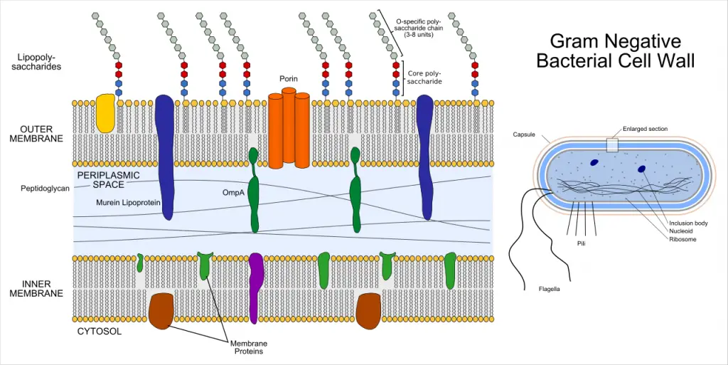
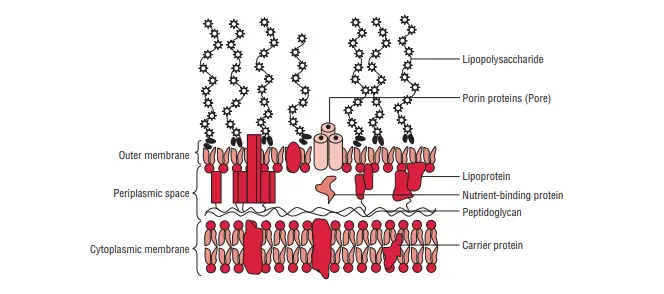
Cell wall of the Gram-negative is more complicated than the Gram-positive cell wall. The amount of peptidoglycan present in the Gram-negative cell wall is considerably lower than that of that of the cell’s Gram positive wall. There are only a few layers of peptidoglycan (2-8 millimeters) are visible in the cell membrane’s outermost. A Gram-negative wall that lies outside the peptidoglycan layer has three primary components–(a) the lipoprotein layer, (b) outer membrane as well as (c) Lipopolysaccharides.

Lipoprotein layer
The layer of lipoprotein is comprised of Braun’s lipoprotein. Braun’s lipoprotein can be described as a small lipoprotein which is covalently attached to the peptidoglycan underneath and is embedded within the membrane’s outer part through its hydrophobic ends. lipoprotein helps to stabilize the outer layer of the cell wall that is Gram-negative.

Outer membrane
The membrane’s outer layer has a two-layered design. its inner layer is similar the composition of the membrane of cells, and the outer membrane is made up of a distinct component known as lipopolysaccharide. The plasma and the outer membrane are directly in contact with many locations within the wall of Gram negative. The outer membrane contains many proteins, that include:
(a) Porins
The outer membrane is a special channels that are made of porins, which are protein molecules. Porins play a variety of roles:
- They allow the diffusion of low-molecular-weight hydrophilic substances, like amino acids, sugars and certain ions.
- They are hydrophobic molecules that are not included; and
- They are used to guard the cell.
(b) Outer membrane proteins (OMPs)
They include:
- Omp C D, F and PhoE and LamB are the four main protein components of the membrane’s outer which are responsible for the majority of the transmembrane transport of maltose and maltodextrins.
- Tsx The receptor for T6 bacteria is responsible for transmembrane transmission of nucleosides and certain amino acids.
- Omp A protein binds the membrane’s outer layer in the peptidoglycan layer. It also acts as the sex pilus receptor
F-mediated conjugation between bacteria. The outer membrane also has proteins that play a role in the transportation of certain molecules, like Vitamin B12 and iron-siderophore complexes it also houses a tiny number of minor proteins like phospholipases, enzymes and proteases.
Structure of a lipopolysaccharide
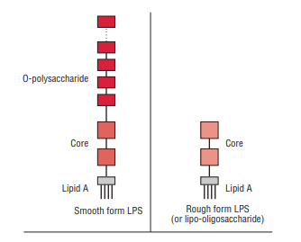
Lipopolysaccharides (LPS) are complex molecules that reside in the outer membrane of Gram-negative bacteria. The structurally, LPS comprise three major components: lipid A; the oligosaccharide that is the primary component, and the O polysaccharide also known as the O-antigen.

Lipid A
It is made up of phosphorylated disaccharide unit, to which several long-chain fatty acids have been attached. It also contains the hydroxymyristic acids, a distinctive fat that is associated with the endotoxic function that is a part of LPS. There is some variations in the structure lipid A in various species of Gram-negative bacteria. But it’s identical within bacteria from the same species.
Core oligosaccharide
The oligosaccharide’s core contains two sugars that are distinctive, ketodeoxyoctanoic acid (KDO) and a heptose that is joined by the lipid A. It is a genus-specific molecule and is common to every Gram-negative organism. Lipooligosaccharides (LOS) are glycolipids that are smaller in size. They possess very shorter multiantennary (i.e. branching) Glycans pre-selected by microorganisms (e.g., Neisseria meningitidis, N. gonorrhoeae, Haemophilus influenzae as well as Haemophilus ducreyi) which are found on mucosal surfaces. They show a wide antigenic and structural diversity , even within one strain. LOS is a major virus-causing factor. Epitopes that are present on LOS contain a N-acetyllactosamine (Gal()l-4-GlcNAc) residue that is immunochemically identical in structure to that of the precursor human erythrocyte I antigen. Sialylation of the N-acetyllactosamine molecule in vivo supplies this organism advantages of environmental of mimicry at molecular level of the antigens of the host, as well as the biological masking that is believed that is provided by sialic acid.
O polysaccharide or O-antigen
It is the area that extends beyond the core. It contains a range of unique sugars and differs in composition between strains of bacteria, giving specificity to a species-specific antigen. This is because it is exposed to the host immune system. Gram-negative bacteria could be able to thwart defenses of the host by changing the O side chains, thereby avoiding detection.
Characteristics gram-negative bacteria
Conventional Gram-negative (LPS-diderm) bacteria show these traits:
- A cell’s membrane inside is visible (cytoplasmic)
- A thin peptidoglycan layer can be found (this is more dense in Gram-positive bacteria)
- Its outer membrane is a source of lipopolysaccharides (LPS consisting of lipid A and core polysaccharide and the O antigen) in its leaflet exterior and phospholipids inside the leaflet’s inner layer.
- Porins reside within the outer membrane which function as pores for certain molecules
- Between the outer and cytoplasmic membrane , there is a gap that is filled with a gel-like substance known as periplasm.
- The S-layer is attached directly with the membrane’s outer surface and not that to the peptidoglycan
- If present, flagella are equipped with four rings of support instead of two.
- Teichoic acids and lipoteichoic acids are not present
- Lipoproteins are bonded by the polysaccharide backbone
- Some are made up of Braun’s lipoprotein that acts as a bridge between the membrane’s outer layer and the peptidoglycan chain through an oblique bond
- A majority, with some exceptions, don’t form spores.
