Table of Contents
The cell’s outer envelope is an organelle and physiological protection between the protoplasm (inside) of the cell and its external environment. The cell envelope guards bacteria from osmotic destruction and gives bacteria a rigid form. The cell envelope is composed of two elements that are a cell wall as well as a cytoplasmic or plasma membrane. It is a protective layer for the protoplasm composed of (i) the cytoplasm, (ii) inclusions in the cytoplasm (mesosomes, ribosomes and vacuoles, inclusion granules) and (iii) one circular DNA.
Following the time that Christian Gram developed the Gram stain in 1884, it quickly was evident that the majority of bacteria could be classified into two main categories based on how they react to the process of forming the Gram stain. Gram-positive bacteria colored purple, while Gram-negative bacteria stained either red or pink. The structural distinction between the two groups didn’t become apparent until the introduction that of transmission electron microscopy. In this article, we will discuss the long-held models of Gram positive and Gram-negative cell wall that were developed through these research studies. Recent studies on diverse types of bacteria have demonstrated that these models don’t apply to all bacteria. In light of ongoing discussions regarding these research, we will identify bacteria that are in line with the model as typical Gram-positive or Gram-negative bacteria.
Bacterial Cell Wall
- Prokaryotic cells are typically restricted by a rather robust and chemically complex structure found in between cell membranes and the the capsule/slime layer, also known as the cell wall.
- Peptidoglycan is the primary part of the cell’s wall. It is responsible for shaping and strength of the cell.
- Peptidoglycan is a disaccharide and contains two sugar derivatives–N-acetylglucosamine and N-acetylmuramic acid–joined together by short peptide chains.
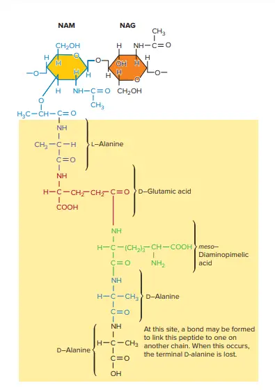
- N-acetylmuramic acid is the tetrapeptide side chain composed of Damino acids L and – (D-glutamic acid as well as L-alanine) together with mesodiaminopimelic acid (Gram-negative bacteria) or L-lysine (Gram-positive bacteria).
- Side chains of Tetrapeptide are linked via pentaglycine bridges.
- Most Gram-negative cell walls lack interpeptide bridge.
- Cell wall gives shape to cells and guards the bacteria from changes of inosmotic pressure. The pressure inside the cell of bacteria is 5-20 atmospheres.
- Bacterial cells are classified as Gram-positive and Gram-negative, based on the structural differences that exist between Gram-positive and Gram-negative cell walls.
- Cell walls of Gram-positive bacteria are more chemically simple structures than Gram-negative bacteria.
- The cells’ walls Bacillus subtilis, as well as a variety of other Gram-positive bacteria comprise of a single thick 20 to 80-nm uniform layer composed of peptidoglycan (murein) which is located just outside the plasma membrane.
Characteristics of Peptidoglycan
- The characteristic shared by the majority of bacterial cell walls are the presence of the peptidoglycan which creates a huge mesh-like structure, often referred to as the sacculus of peptidoglycan.
- Peptidoglycan is made up of several similar subunits.
- The sacculus is composed of two sugar derivatives: N-acetylglucosamine (NAG) and N-acetylmuramic Acid (NAM) as well as a variety of different amino acids.
- The amino acids are the peptide short, also known as the stem peptide comprising four alternate d- and lamino acids. The peptide is linked to the carboxyl section of NAM.
- Three amino acids aren’t found in proteins: d’glutamic acid mesodiaminopimelic acids.
- A presence of di-amino acid in the stem peptide helps protect against degrading by peptidases, which only recognize the amino acid l-isomers residues.
- The peptidoglycan sacculus develops by gluing the sugars of the peptidoglycan subunits in order to form a single strand. The strands are then linked to one another through covalent bonds created between the stem peptides which extend out from the strands.
- The backbone of every string is comprised of alternate NAM and NAG residues.
- It is an helical strand and the stem peptides stretch beyond the backbone in different directions.
- There are two kinds of cross-links: indirect and direct through peptide-based interbridges.
- Direct cross-links are defined by linking the carboxyl group that is an amino acid within one stem peptide with an amino amino acid found in an additional stem peptide.
- In particular some bacteria cross-link strands by linking the carboxyl portion of D-alanine at the 4th position in the stem peptide diaminopimelic acids (position 3,) of the peptidoglycan strand’s other stem peptide (the position 5 d’alanine is removed when the cross-link develops).
- Bacteria with indirect linkage utilize an interpeptide (also known as an interpeptide Bridge) which is which is a short chain of amino acids that connects the stem peptide from one peptidoglycan string to the other.
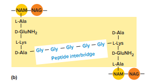
- Cross-linking produces a dense, interconnected system of peptidoglycan Strands.
- The peptidoglycan sacculus is a strong but elastically. It can expand and contract according to pressure from osmotic. It is because of the stiffness of the backbone and flexible cross-links.
- The sacculi of Peptidoglycans are also porous, allowing proteins with molecular weights of up to 50,000 to traverse, contingent upon whether or not the sacculus has been stretched or relaxed. Hence, only very large proteins are able to traverse the peptidoglycan.
- Different peptidoglycans are present in particular among Gram-positive bacteria. Some, for instance, substitute diamino acid Lysine for meso-diaminopimelic acid , and cross-link chains through interpeptide bridges.
- Interpeptide bridges are interpeptides that can vary significantly.
- Peptidoglycan also varies in the size of the chains and the degree of cross-linking. The bacteria that color Gram positive are likely to have more cross-linking and those which stain Gram negative are significantly less cross-linked.
Structure of Gram-Positive Cell Wall
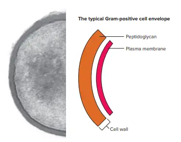
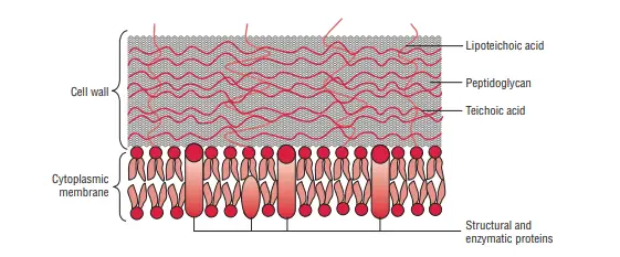
The cell’s Gram-positive wall can be described as thick (15-80 nm) and more homogeneous than the thin (2 millimeters) Gram-negative wall. The Gram-positive cell wall has a an extensive amount of peptidoglycan in multiple layers, which makes up around 40-80% of the its dry mass. The cell wall of the Gram-positive cells is composed mostly of teichoic and Teichuronic acids. These two substances make up as much as 50 percent from the total dry mass of the wall, and 10 percent in the weight dry of the entire cell.
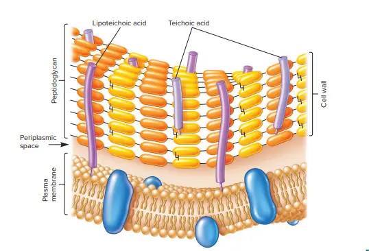
The periplasmic area is located between the plasma membrane as well as the cell wall . It is so small that it is rarely accessible to electron microscopy. The periplasm is home to a small amount of proteins. This is likely because the peptidoglycan sacculus porous, which means that a lot of proteins that move over the plasma membrane travel across the sacculus.
The secreted proteins of some are known as exoenzymes. Exoenzymes are often used to break down polymers, such as polysaccharides and proteins that are otherwise too big for transportation over the plasma membrane. the products of degradation, which are monomer-based building blocks, are then utilized by cells. The proteins that remain in the periplasmic area are typically linked with the plasma membrane. Some proteins that are secreted do not traverse the peptidoglycan sacculus but some of them are attached by it. These proteins play a role in cell interactions with the environment.
Teichoic acids
The majority of cultured bacteria that show Gram positivity are part of the family of phyla Firmicutes and Actinobacteria Most of them have cell walls that are thick and composed of peptidoglycan as well as large quantities of other polymers , such as teichoic acid.
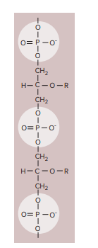
- Teichoic acids are polymers made of the glycerol or ribitol that are that are joined with phosphate group.
- Certain teichoic acids are tied to peptidoglycan and called wall teichoic acid.
- Other are covalently linked to the plasma membrane, they are known as lipoteichoic acids.
- Wall teichoic acids extend over the peptidoglycan’s surface. They are positively charged and to give the cell wall its negative charge.
- Teichoic acids aren’t present in other bacteria.
- Certain are not covalently bound to teichoic acid or different cell wall polymers and others are bound covalently with the peptidoglycan. Sortases, which are membrane-bound enzymes, catalyze the formation of covalent bonds which join these proteins to peptidoglycan.
- Teichoic acids come in two varieties: wall teichoic acids (WTA) and the lipoteichoic acid (LTA). They are connected to protein glycoglycan via a covalent link to the six hydroxyls of N-acetylmuramic acids in WTA and also to plasma membrane and lipids in LTA.
Functions of Teichoic acids
Teichoic acids have many functions:
- They are the major antigens on the surface of Grampositive species that have these. In Streptococcus pneumoniae the teichoic acids carry the antigenic determinants referred to as Forssman antigen. In Streptococcus Pyogenes, LTA is connected to the M protein which protrudes out of the cell membrane via the peptidoglycan layer. The M protein’s long molecules, along with LTA form microfibrils that aid in an attachment for S. Pyogenes to animal cells;
- They can also be used as antigens to classify serologically bacteria.
- They function as substrates for a variety of autolytic enzymes.
- These proteins are able to bind the magnesium ion, and could play a role in the supply of this ion to cells;
- They are involved in the normal function of the cell wall. They also serve as an external barrier against Gram-positive bacteria.
- Membrane teichoic acids serve to anchor the cell membrane.
- They assist in forming and maintaining an envelope of cells by anchoring it onto the plasma membrane.
- They are essential in cell division and protect cells from harmful substances found in environmental conditions (e.g. antibiotics, toxins and the host defense molecules).
- Additionally, they play a role in the uptake of ions and in the binding of pathogenic organisms in host tissue, starting the infection process.
2. Teichuronic acid
- Teichuronic acid is composed of sugar repeat units acids (such as D-glucuronic or N-acetylmannuronic acid).
- They are made in lieu of teichoic acids if the supply of phosphate to cells is restricted.
- The cell wall of Gram-positive cells also contains non-neutral sugars (such as arabinose, mannose, Rhamnose, and glucosamine) as well as acidsic sugars (such as mannuronic acid and glucuronic acid) and are subunits of polysaccharides found in cells’ wall.
References
- Chapot-Chartier, MP., Kulakauskas, S. Cell wall structure and function in lactic acid bacteria. Microb Cell Fact 13, S9 (2014). https://doi.org/10.1186/1475-2859-13-S1-S9
- https://en.wikipedia.org/wiki/Gram-positive_bacteria
- http://www.textbookofbacteriology.net/structure_9.html
- https://bio.libretexts.org/Bookshelves/Microbiology/Book%3A_Microbiology_(Kaiser)/Unit_1%3A_Introduction_to_Microbiology_and_Prokaryotic_Cell_Anatomy/2%3A_The_Prokaryotic_Cell_-_Bacteria/2.3%3A_The_Peptidoglycan_Cell_Wall/2.3A%3A_The_Gram-Positive_Cell_Wall#:~:text=The%20Gram%2Dpositive%20cell%20wall%20consists%20of%20many%20interconnected%20layers,interwoven%20through%20the%20peptidoglycan%20layers.
- https://open.oregonstate.education/generalmicrobiology/chapter/bacteria-cell-walls/
- https://www.slideshare.net/AshfaqAhmad52/bacterial-cell-wall-67925149
