Advertisements
Table of Contents
- These two organisms are phytoflagellates and are mainly found in freshwater pools and canals.
- The abundant Chlamydomonas and Volvox can produce green scum on ponds.
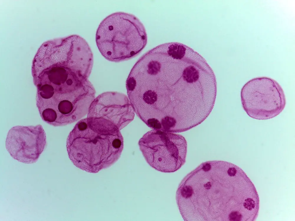
- Chlamydomonas are single-celled green flagellates while Volvox is a part of colonial phytoflagellate that develops a hollow spheroid with several thousand cells enclosed in its mucilaginous wall.
Volvox
- These are the polyphyletic genus of chlorophyte green algae.
- Volvox develops globular colonies which contain more than 50,000 cells.
- They are mostly found in freshwater.
- In 1700, a Dutch businessman and scientist Antonie van Leeuwenhoek first reported this organism.
- They reproduced by both sexual and asexual methods.
Chlamydomonas
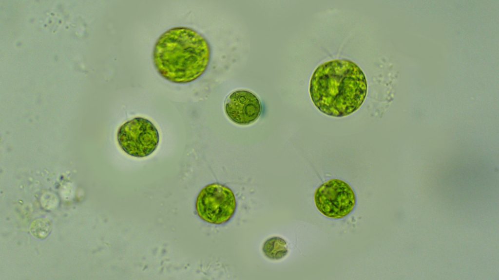
- It is a genus of green algae with 325 species.
- They are Motile unicellular algae.
- The cell wall of Chlamydomonas is composed of glycoprotein and non-cellulosic polysaccharides rather than cellulose.
- They can be found in dirty water and on moist soil, in freshwater, seawater, and also in snow as “snow algae”.
- In molecular biology, it is used as a model organism to study the motility and chloroplast dynamics, biogenesis, and genetics.
- They contain ion channels (channelrhodopsins). These ion channels are activated by lights.
- Chlamydomonas are spherical or slightly cylindrical shape.
- The papilla may be present or absent on them.
- They contain two anterior flagella and each as long as the other.
- They contain cup-shaped Chloroplasts.
- From the mode of nutrition, most of them are obligate phototrophs except C. reinhardtii and C. dysostosis, these two are facultative heterotrophs in nature.
Whole mount preparation for Chlamydomonas or Volvox
Aim
To prepare a temporary mount of Chlamydomonas or Volvox in order to examine the shape, structure, and arrangement.
Materials Required
- Clean microscopic slide.
- Clean cover slide.
- Culture or a drop of water containing the flagellates.
- 10% methylcellulose
- compound microscope
- Different Stains such as acetiocarmine, Noland’s solution, iodine solution
Procedure
- Place a drop of culture or a drop of water containing the flagellates on a clean slide.
- Then add a drop of 10% methylcellulose on this water drop or culture drop. It will restrict the movement of the organism and let you examine the organism.
- Now, gently place a clean coverslip over the drop and make sure no air bubbles are formed between the glass slide and coverslip.
- Observe the glass slide under a compound microscope. You will be able to observe the flagellar movement better by cutting down the light.
- You can see the other organs within the organism by applying different stains on them. For example, apply acetiocarmine to observe the nucleus, Noland’s solution to observe the flagella, and iodine solution to observe the starch grains.
Observation
- In case of Chlamydomonas, under high power magnification, you will observe two flagella, cellulose cell wall, two small contractile vacuoles, cytoplasm and cup-shaped chloroplast, pigment spot, and pyrenoid.
- In case of flagellate Volvox, you will find that the flagellar beat of the individual cells is correlated with each other so that the colony shifts in an oriented fashion.
Result
Based on the observation the supplied organism is Chlamydomonas or Volvox (write the name of the organism based on your observation).
Advertisements


