Table of Contents
In this article, we will learn how to prepare a temporary mount of Rhizopus.
Characteristics of Rhizopus
- It is a saprophytic fungi and found on plants.
- They are parasitic on animals.
- Rhizopus is multicellular fungi, with approximately 8 species. The eigh species are Rhizopus reflexus, Rhizopus schipperae, Rhizopus delemar, Rhizopus homothallicus, Rhizopus microsporus, Rhizopus arrhizus (Rhizopus oryzae), Rhizopus caespitosus, and Rhizopus stolonifer.
- Rhizopus is responsible for invasive mucormycosis in humans and animals, it is an opportunistic infection.
- There are few species of mould that are used in industries for their industrial importance for example Rhizopus stolonifer which caused bread mold.
- Rhizopus diseases may also be a difficulty of ketoacidosis.
- Rhizopus oryzae and Rhizopus microsporus has a medical importance.
- Rhizopus can be found on soil, decaying fruits, grown fruits and vegetables, jellies, tobacco, peanuts, leather, bread, and syrups.
- Rhizopus develop both asexual and sexual spores.
Study of Rhizopus by using Temporary Mount.
Aim
To prepare a temporary mount of Rhizopus in order to examine the shape, structure, and arrangement.
Requirements
The following instruments and reagents are required for the preparation of a temporary mount of Rhizopus.
- Glass slide
- Coverslip
- Water/Shear’s mounting medium/Melzer’s Solution/Lacto-fuchsin
- A pair of gloves
- Microscope
- Dropper
Prepare the Rhizopus
- Take a slice of soft bread and leave it in open air for about 60 min, place the bread within a plastic bag.
- Spray some water over the bread to have dampness then seal the bag. After that leave some air within the bag.
- Store this bag in a dark and warm place.
- Check the sample after 5 days, a black patch Or thread-like growth with masses of black, grey, and green fine dotted structures are formed on the bread.
Procedure
- Take a clean glass slide and place a drop of water on it by using a dropper (placed at the center of slide).
- Now place a very little portion of the mould on the drop of water by using a sterile toothpick.
- Get a clean coverslip and place it at an angle to the slide, so that one end of it, touches the water drop, then thoroughly drop it over the drop so that the coverslip shields the specimen without catching air bubbles beneath.
- Now make sure no excess water remains at the edges of the coverslip. If remains use tissue paper or blotting paper to blot up all the excess water.
- The slide is ready to observe under the compound microscope. Follow this step by step Instruction to operate a compound microscope.
Observation

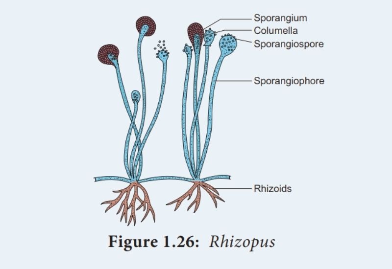
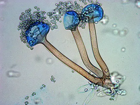
- The collected bread mould is made of fine thread-like projections termed hyphae and thin stems having knob-like structures known as Sporangia.
- There are hundreds of minute spores in Sporangia.
- When the sporangium ruptures, the small spores are scattered in air.
Result
Based on the observation the collected mold is Rhizopus sp.

Precautions
- If you have allergies and asthma, then stay away from mould.
- Avoid touching the mould with bear hands.
- Thoroughly wash your hands after touching the bread sample.

Great yes it is uceful for us
Thank You Baira
Great