Table of Contents
Toluidine blue is a basic thiazine metachromatic dye that stains nuclei blue, and can be used to differentiate different types of granules (e.g. within mast cells). It has a high affinity for acidic tissue components.
This basic stain binds with the acidic tissue. It has a high affinity for nucleic acids, hence it binds to nuclear material such as DNA and RNA. When it stains structures blue, it is termed orthochromatic.
Purpose
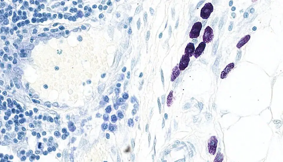
This stain is used to identify the mast cells. These cells are found widely distributed in the connective tissue. Their cytoplasm contains granules composed of heparin and histamine. These granules are metachromatic.
Cellular and Molecular Immunology, Roitt’s Essential Immunology, Kuby Immunology…

Principle of Toluidine blue stain
Mast cells should stain red-purple (metachromatic staining) and the background stain blue (orthochromatic staining). Metachromasia, tissue elements staining a different color from the dye solution, is due to the pH, dye concentration and temperature of the basic dye. Blue or violet dyes will show a red color shift, and red dyes will show a yellow color shift with metachromatic tissue elements.
Fixative: 10% formalin
Sections: paraffin sections at 5 um.
Solutions and Reagents
- Preparation of toluidine blue stain: Mix 1g of Toluidine blue O (Sigma) with 100 ml of 70% alcohol (Avoid contact and inhalation).
- Sodium Chloride (1%): Mix 0.5g Sodium chloride with 50 ml Distilled water. Adjust pH to 2.0~2.5 using glacial acetic acid or HCl.
- Toluidine Blue Working Solution (pH 2.0~2.5): Mix 5ml Toluidine blue stock solution with 45ml of 1% Sodium chloride. The pH should be around 2.3 and less than 2.5. Make this solution fresh and dump after use. pH higher than 2.5 will cause staining less contrast (Avoid contact and inhalation).
Steps in Toluidine Blue Staining
- Deparaffinize and hydrate to distilled water.
- Working Toluidene blue, 1-2 minutes.
- Rinse in distilled water, 3 changes.
- Dehydrate quickly through 95% and absolute alcohols.
- Clear in xylene, coverslip.
Results

- Mast cells: Stains violet
- Background: shades of blue
Other results
- Mastocytes: purple
- Cartilage: purple
- Mucins: purple/red
- Nuclei: blue
References
- Theory and Practice of Histotechnology by Barbara Hrapchak, D. Sheehan
- Manual of Histologic Staining Methods of the Armed Forces Institute of Pathology by Lee G Luna
- Hazardous Materials in the Histopathology Laboratory: Regulations, Risks, Handling, and Disposal 4th Edition by by Janet Crookham Dapson, Richard W. Dapson
