Table of Contents
What are Centrioles?
- Centrioles are integral cellular components, primarily found in the cells of most eukaryotic organisms. These structures, absent in higher plants such as conifers and flowering plants, are not present in prokaryotes, red algae, yeast, and certain non-flagellated protozoans. As cylindrical, barrel-shaped organelles, centrioles measure approximately 0.2 μm in diameter and 0.4 μm in length, and are devoid of membranes, DNA, and RNA.
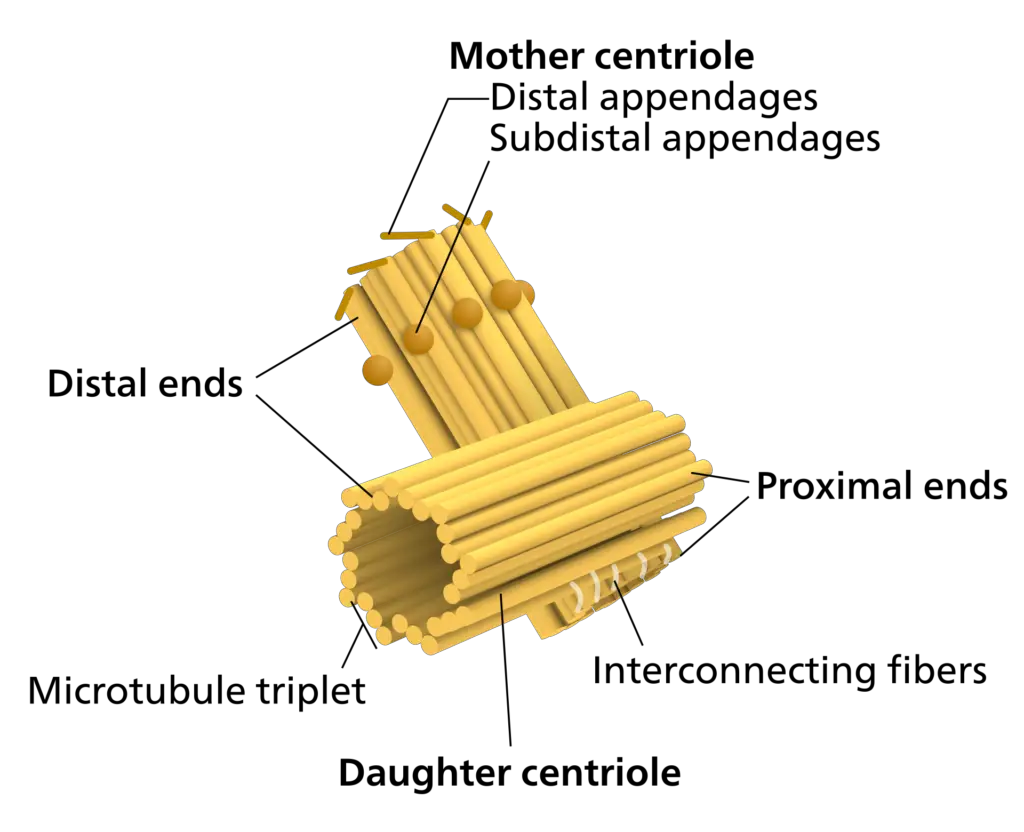
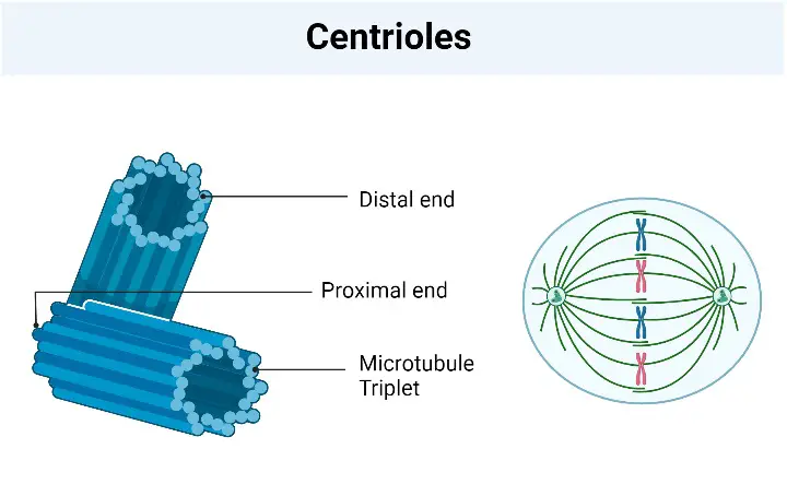
- Structurally, centrioles are composed of nine sets of microtubule triplets arranged in a cylinder. This standard composition can vary, as seen in organisms like crabs and Drosophila melanogaster embryos, which have nine doublets, and in Caenorhabditis elegans sperm cells and early embryos, with nine singlets. These structures are primarily made of the protein tubulin, along with additional proteins such as centrin, cenexin, and tektin.
- The primary function of centrioles is the organization and production of microtubules. During interphase, they are instrumental in forming cilia and flagella, which are essential for cell movement. In cell division, centrioles play a crucial role in producing the aster and the spindle, facilitating chromosome separation. They form a part of the centrosome, a larger structure that comprises a bound pair of centrioles surrounded by pericentriolar material (PCM), which serves as a nucleation site for microtubule growth.
- Historically, the discovery of the centrosome and centrioles can be traced back to the late 19th century. The centrosome was discovered independently by Walther Flemming in 1875 and Edouard Van Beneden in 1876, with Van Beneden making the first observation of centrosomes composed of two orthogonal centrioles in 1883. The terms “centrosome” and “centriole” were introduced by Theodor Boveri in 1888 and 1895, respectively. The basal body, a key component in this structure, was named by Theodor Wilhelm Engelmann in 1880. The pattern of centriole duplication was first elucidated by Étienne de Harven and Joseph G. Gall around 1950.
- In summary, centrioles are vital for the proper functioning of eukaryotic cells, facilitating processes such as cell division, movement, and the structural integrity of the cell. Their absence in certain organisms highlights a diverse range of cellular structures and mechanisms across different life forms.
Definition of Centrioles
Centrioles are cylindrical, microtubule-based structures found in most eukaryotic cells, playing a crucial role in cell division by aiding in the formation of the spindle apparatus and in cellular processes like the formation of cilia and flagella. They are typically composed of nine sets of microtubule triplets and lack a membrane, DNA, or RNA.
Occurrence of Centrioles
- Centrioles are cellular structures predominantly found in a wide array of eukaryotic organisms, exhibiting a specific pattern of occurrence across different species. In the realm of algae, centrioles are generally present, with the notable exception of red algae. Moving on to the plant kingdom, they are found in moss cells and certain fern cells. However, it is essential to note that centrioles are conspicuously absent in prokaryotes, which include bacteria and archaea, indicating a fundamental difference in cellular organization between prokaryotes and eukaryotes.
- In the diverse world of animal cells, centrioles are almost universally present. This prevalence underscores their critical role in cellular processes such as mitosis and meiosis, where they facilitate the segregation of chromosomes. However, the presence of centrioles is not uniform across all eukaryotes. In the specific case of plants, centrioles are absent in both cone-bearing plants (conifers) and flowering plants (angiosperms). This absence is significant as it highlights alternative mechanisms employed by these organisms for cell division and organization.
- Furthermore, in the context of protozoans, a heterogeneous group of eukaryotic microorganisms, the presence of centrioles is varied. In non-flagellated and non-ciliated protozoans, such as certain amoebae, centrioles are absent. Interestingly, some amoebae exhibit a dual-phase life cycle, encompassing both flagellated and amoeboid stages. In these organisms, centrioles develop during the flagellated stage but subsequently disappear in the amoeboid phase. This transient presence of centrioles during only a part of the life cycle of such amoebae is a remarkable example of the adaptability and diversity of eukaryotic cellular structures.
- Therefore, the occurrence of centrioles in eukaryotic cells is a subject marked by significant variability. Their presence in most algal cells (excluding red algae), certain plants, and almost all animal cells, juxtaposed with their absence in prokaryotes, yeast, conifers, angiosperms, and specific protozoans, illustrates a complex pattern of distribution. This distribution is reflective of the diverse strategies employed by different life forms in cell division and structure.
Structure of Centrioles

Centrioles are cylindrical structures, typically measuring 0.15–0.25 μm in diameter and 0.3–0.7 μm in length. However, their size can vary, with some as short as 0.16 μm and others extending up to 8 μm. These dimensions are discernible under an electron microscope, providing a detailed view of the centriole’s intricate structure.
- Cylinder Wall: Centrioles are cylindrical structures with a diameter ranging from 0.15 to 0.25 µm and a length between 0.3 to 0.7 µm, although some may be as short as 0.16 µm or as long as 8 µm. The cylinder wall of a centriole is formed by an array of nine triplet microtubules evenly spaced around the perimeter of an imaginary cylinder. These triplets are arranged like the blades of a pinwheel, tilted at a 45° angle inward towards the central axis. The tubules of each triplet twist helically from one end to the other, defining the centriole’s internal and external boundaries in the absence of an outer membrane.
- Triplets: The triplets consist of three subunit microtubules named A, B, and C, with A being the innermost. Each tubule has a diameter of about 200-260 Å. The A tubule is round with thirteen 40-45 Å globular subunits around its perimeter, and shares 3-4 subunits with the B tubule, which in turn shares several subunits with the C tubule. These triplets run parallel to each other and the long axis of the cylinder but turn in a long-pitched helix relative to the cylinder axis.
- Linkers: The tubule A of each triplet is connected to the tubule C of the adjacent triplet by protein linkers. These linkers maintain the radial orientation of the triplets and stabilize the cylindrical microtubule array.
- Cartwheel and Satellites or Pericentriolar Bodies: Centrioles lack central microtubules but sometimes exhibit protein spikes radiating from the core of each triplet, forming a cartwheel-like pattern. This configuration is crucial for determining the centriole’s structural and functional polarity, with growth occurring from the distal end where procentrioles form at a 90° angle. Surrounding the centrioles are electron-dense structures known as satellites or pericentriolar bodies, likely serving as nucleation sites for microtubules.

Chemical Composition
- Primary Composition of Microtubules: The microtubules constituting centrioles are primarily composed of the protein tubulin. Tubulin, a globular protein, forms the backbone of these microtubular structures, providing them with their characteristic rigidity and shape. This protein is essential in maintaining the structural integrity of centrioles.
- Presence of Lipid Molecules: Besides tubulin, lipid molecules are also present in the microtubules of centrioles and basal bodies. These lipid molecules play a role in maintaining the structural stability and functional dynamics of centrioles, contributing to their overall biochemical composition.
- Concentration of ATPase Enzyme: Centrioles and basal bodies exhibit a high concentration of ATPase enzyme. ATPase, an enzyme that catalyzes the decomposition of ATP (Adenosine Triphosphate) into ADP (Adenosine Diphosphate) and a free phosphate ion, is crucial for energy transfer within the cell. This enzyme’s presence in centrioles suggests its involvement in energy-dependent processes within these organelles.
- Debate Over Nucleic Acids: The presence of nucleic acids (DNA and RNA) in centrioles and basal bodies has been a topic of debate. While these organelles are generally not associated with containing their own DNA or RNA, some studies have questioned this assertion. Fulton (1971), for instance, raised doubts about the presence of nucleic acids in these structures. However, the consensus remains that centrioles and basal bodies are typically devoid of nucleic acids, distinguishing them from other cellular organelles like mitochondria and chloroplasts.
- Overall Chemical Composition: Therefore, the chemical composition of centrioles is primarily characterized by the presence of tubulin proteins and lipid molecules, supplemented by a significant concentration of ATPase enzyme. The absence or controversial presence of nucleic acids further defines their biochemical makeup, emphasizing their unique role in cellular functions devoid of genetic material.
Origin of Centrioles And Basal Bodies
The notion that new centrioles develop from the division of old centrioles is no longer widely held. Instead, it appears that new centrioles are either generated from scratch or synthesised using an existing centriole as a template (semi-autonomous replication).
1. Origin of centrioles by duplication of pre-existing centrioles
- In cultured fibroblasts, the beginning of DNA synthesis coincides with the beginning of centriole duplication (interphase).
- First, the two members of a pair of centrioles separate; next, a procentriole is created perpendicular to each original centriole, with the two organelles separated by 50 to 100 nm.
- An immature centriole has a ninefold symmetric array of single microtubules; each microtubule apparently serves as a template for the construction of adult centrioles’ triplet microtubules.
- In late prophase, each daughter centriole reaches its full size while retaining its tight proximity and orientation to the mother centriole. Consequently, when the interphase nuclei rejoin at the conclusion of nuclear division, a centrosome comprising two centrioles is present next to each nucleus.
- The development of the centriole (or basal body) has been examined in Paramecium, Tetrahymena, Xenopus, and chicken tracheal epithelium. The stages of development are basically same across the board.
- In an amorphous mass, the development of a basal body begins with the production of a single microtubule. Microtubules are introduced one by one until a ring of nine is formed with uniform spacing.
- As the microtubules appear, the amorphous mass disappears, as if eaten by the microtubule creation process.
- There is evidence that connectives exist between microtubules, which may function to set their spacing apart.
- Thus, a ring of nine full microtubules (i.e., A tubules) is generated, followed by the development of C-shaped B microtubules and C microtubules.
- The hub and the wheel are added centrally. The A-C linkages are not established until development is complete.
2. Origin of basal bodies
- In a ciliated vertabrate cell, which may include hundreds of cilia, the precursor cell’s centrioles give rise to the numerous basal bodies necessary to nucleate the cilia in the mature cell.
- During the development of ciliated epithelial cells that line the oviduct and trachea, for instance, the centriole pair migrates from its typical site near the nucleus to the apical area of the cell, where the cilia will form.
- In this case, instead of generating a single daughter centriole, each pair of centrioles creates several electrondense fibrogranular satellites.
- From these satellites, many basal bodies subsequently travel to the membrane to commence the development of cilia.
3. The de novo origin of centrioles and basal bodies
- In certain instances, centrioles appear to originate from scratch. Unfertilized eggs of many animals, for instance, lack functional centrioles and rely on the sperm centriole for the initial mitotic division (for cleavage); however, under certain experimental conditions, such as extreme ionic imbalance or electrical stimulation, the unfertilized egg can produce a variable number of centrioles.
- Each of these centrioles nucleates the creation of a small aster, one of which can be exploited by the egg for cleavage division, resulting in the parthenogenesis of a haploid organism.
- In fact, centriole precursors are maintained in the cytoplasm of unfertilized eggs and can be activated under certain conditions to produce a new centriole.
- Similar to the centrioles, the basal bodies are capable of self-assembly and abruptly appear in Naegleria as it transforms from an amoeboid to a normal ciliate.
- Centrioles have been hypothesised to be fully autonomous, self-replicating organelles due to their peculiar mechanism of duplication and their continuity over multiple generations.
- Although it is now known that this is not the case and that centrioles can develop spontaneously in the cytoplasm under specific conditions.
- Consequently, it is possible that some information required for centriole development is typically contained inside the centriole itself (just as the replication of mitochondria and chloroplasts depends on extrachromosomal genes carried in the organelles).
- In Chlamydomonas, for instance, a group of genes encoding proteins important in basal body shape and flagellar assembly are carried on a genetic element that segregates independently from the major chromosomes.
- This genetic element’s nature and placement have yet to be discovered.
Functions of Centrioles
- Formation of Basal Bodies and Cilia: Centrioles are instrumental in forming basal bodies, which are crucial for the development of cilia. These cilia play a significant role in various cellular processes, including movement and signal transduction.
- Role in Centrosome and Microtubule Organization: In most animal cells, centrioles act as focal points within the centrosome. The centrosome is vital for organizing the array of cytoplasmic microtubules during interphase and duplicates during mitosis to nucleate the two poles of the mitotic spindle. This function is essential for proper cell division.
- Dual Functionality in Chlamydomonas: In certain organisms like Chlamydomonas, centrioles can perform dual functions before each cell division. The basal bodies, which are modified centrioles, leave their position to act as mitotic poles after the absorption of flagella.
- Involvement in Spermatozoon Structure: Centrioles are responsible for the formation of the tail fiber or flagellum in spermatozoa, contributing to sperm motility and functionality.
- Role in Sensory Reception: Centrioles are involved in receiving optical, acoustic, and olfactory signals, serving as devices for locating the directions of these signal sources. This function highlights their role in sensory processes.
- Spindle Apparatus Formation: A critical function of centrioles is in the formation of the spindle apparatus during cell division. The spindle apparatus is essential for the correct segregation of chromosomes.
- Impact on Cell Division: The absence of centrioles can lead to errors and delays in the mitotic process, underscoring their importance in cell division.
- Anchor Point for Cilia and Flagella: A single centriole forms the basal body, anchoring each cilium or flagellum. This anchoring is crucial for the proper functioning of these cellular structures.
- Direction of Cilia and Flagella Formation: Basal bodies, derived from centrioles, direct the formation of cilia and flagella, influencing their orientation and functionality.
- Functioning as Pairs or Singles: While centrioles typically function as a pair in most animal cells, in the context of cilia and flagella, they function individually.
- Spatial Arrangement in Cells: Centrioles, as components of centrosomes, play a significant role in organizing microtubules in the cytoplasm. Their position determines the nucleus’s position and is crucial for the cell’s spatial arrangement.
- Importance in Sperm and Embryo Development: Sperm centrioles are vital for forming the sperm flagellum and for embryo development post-fertilization. The sperm’s centriole forms the centrosome and microtubule system of the zygote.
- Determination of Cilia and Flagella Position: The mother centriole determines the position of the flagellum or cilium, transitioning to the basal body in flagellates and ciliates. This function is crucial for the proper development and functioning of these structures.
- Link to Genetic and Developmental Diseases: Inability to utilize centrioles for functional flagella and cilia formation has been linked to various genetic and developmental diseases, including Meckel–Gruber syndrome, which is associated with improper centriole migration prior to ciliary assembly.
FAQ
What do centrioles do?
Centrioles are tiny cylindrical structures found within animal cells, typically located near the nucleus. They play an important role in cell division, specifically in the formation of the spindle fibers that pull the chromosomes apart during mitosis.
During cell division, the centrioles duplicate and move to opposite poles of the cell. From there, they organize the microtubules that make up the spindle fibers, which are responsible for separating the chromosomes into two identical sets. This process is crucial for the accurate distribution of genetic material to the daughter cells.
Additionally, centrioles are involved in the formation of cilia and flagella, which are hair-like structures that protrude from the cell surface and play roles in cellular movement and sensory perception. In this context, centrioles serve as the basal bodies that anchor the cilia and flagella to the cell membrane.
What are centrioles?
Centrioles are tiny cylindrical structures found in most animal cells. They are composed of microtubules and are typically located near the nucleus of the cell. Centrioles play an important role in organizing the microtubules that make up the cytoskeleton of the cell, which provides structural support and helps maintain the cell’s shape.
During cell division, the centrioles duplicate and move to opposite poles of the cell. From there, they play a critical role in the formation of the spindle fibers that pull the chromosomes apart during mitosis. This ensures that the genetic material is accurately distributed to the daughter cells.
Centrioles are also involved in the formation of cilia and flagella, which are hair-like structures that protrude from the cell surface and play roles in cellular movement and sensory perception. In this context, centrioles serve as the basal bodies that anchor the cilia and flagella to the cell membrane.
What is the role of the centrioles?
Centrioles play several important roles in animal cells. One of their primary functions is to organize the microtubules that make up the cytoskeleton of the cell, which provides structural support and helps maintain the cell’s shape.
During cell division, centrioles play a critical role in the formation of the spindle fibers that pull the chromosomes apart during mitosis. This ensures that the genetic material is accurately distributed to the daughter cells.
Centrioles are also involved in the formation of cilia and flagella, which are hair-like structures that protrude from the cell surface and play roles in cellular movement and sensory perception. In this context, centrioles serve as the basal bodies that anchor the cilia and flagella to the cell membrane.
Overall, centrioles are essential organelles that are involved in multiple cellular processes, including cell division and cell motility.
What is the function of centrioles?
The function of centrioles is to play a critical role in several cellular processes in animal cells.
One of their primary functions is to organize the microtubules that make up the cytoskeleton of the cell, providing structural support and helping maintain the cell’s shape.
During cell division, centrioles play a crucial role in the formation of spindle fibers, which pull the chromosomes apart during mitosis to ensure the accurate distribution of genetic material to the daughter cells.
Centrioles are also involved in the formation of cilia and flagella, which are hair-like structures that protrude from the cell surface and play roles in cellular movement and sensory perception. In this context, centrioles serve as the basal bodies that anchor the cilia and flagella to the cell membrane.
Overall, the function of centrioles is essential for maintaining the structural integrity of the cell and ensuring the accurate distribution of genetic material during cell division, as well as contributing to cellular movement and sensory perception.
What happens to the centrioles during mitosis?
During mitosis, the centrioles play a crucial role in the organization and separation of the genetic material.
Before mitosis begins, the centrioles duplicate, forming two pairs of centrioles that move towards opposite poles of the cell. These pairs of centrioles are known as the spindle poles.
As mitosis progresses, the spindle fibers begin to form between the two spindle poles. The centrioles play a critical role in organizing these spindle fibers, which are responsible for pulling the chromosomes apart during cell division.
As the spindle fibers organize, they attach to the chromosomes at specific points called kinetochores. The spindle fibers then contract, pulling the chromosomes towards the center of the cell.
The centrioles and spindle fibers continue to work together to align the chromosomes along the cell’s equator. Once the chromosomes are aligned, the spindle fibers pull them apart, separating the sister chromatids into two identical sets of chromosomes.
After the chromosomes have been separated, the spindle fibers and centrioles begin to disassemble, and the cell completes the process of cytokinesis, forming two identical daughter cells. The centrioles will then duplicate again in preparation for the next round of cell division.
What do centrioles look like?
Centrioles are small, cylindrical structures found in most animal cells. They are typically about 0.2 to 0.5 micrometers in diameter and 0.5 to 2 micrometers in length.
Each centriole consists of nine sets of microtubule triplets, arranged in a cylindrical shape. Each triplet is composed of three microtubules, arranged in a circular pattern. The triplets are arranged perpendicular to each other, forming a cylinder with a hollow center.
Under a microscope, centrioles appear as two small, darkly stained structures located near the nucleus of the cell. They are often visible during cell division, when they play a crucial role in the organization and separation of the genetic material.
Centrioles are found in pairs, and each pair is oriented at right angles to each other. This orientation is important for the formation and organization of the spindle fibers during cell division. Overall, the structure and arrangement of centrioles are essential for their function in multiple cellular processes.
at which phase are centrioles beginning to move apart in animal cells?
Centrioles begin to move apart from each other during the prophase stage of mitosis in animal cells.
In prophase, the duplicated pairs of centrioles move towards opposite poles of the cell, forming the two spindle poles. This movement is facilitated by microtubules, which begin to form between the centrioles and push them apart.
As mitosis progresses, the spindle fibers continue to form and attach to the chromosomes, allowing for their separation and accurate distribution to the daughter cells.
Once mitosis is complete, the two sets of centrioles will begin to duplicate again in preparation for the next round of cell division. The precise timing of centriole separation and duplication is tightly regulated to ensure the accurate distribution of genetic material and maintain the structural integrity of the cell.
Where are centrioles found?
Centrioles are found in most animal cells, including human cells. They are typically located near the nucleus of the cell, in a region called the centrosome.
The centrosome is an organelle that serves as the main microtubule organizing center (MTOC) of the cell. It is composed of two centrioles, which are arranged perpendicular to each other and surrounded by a matrix of proteins that help to organize the microtubules.
In addition to the centrosome, centrioles can also be found in specialized structures called basal bodies, which anchor cilia and flagella to the cell membrane. These structures are important for cellular movement and sensory perception.
Overall, centrioles are essential organelles found in animal cells that play crucial roles in cell division, cytoskeletal organization, and cellular movement.
What are centrioles made of
Centrioles are made up of a cylindrical arrangement of microtubules. Specifically, each centriole consists of nine sets of microtubule triplets, which are arranged in a cylinder with a hollow center.
Each triplet is composed of three microtubules, which are protein filaments made up of alpha and beta tubulin subunits. The triplets are arranged perpendicular to each other, forming a cylindrical structure that is important for organizing microtubules in the cell.
The microtubules that make up centrioles are surrounded by a matrix of proteins, which help to regulate their organization and function. The precise composition of these proteins can vary depending on the cell type and the specific function of the centrioles.
Overall, the structure and composition of centrioles are essential for their role in multiple cellular processes, including cell division and cytoskeletal organization.
Why are the centrioles important in the cell cycle?
Centrioles are important in the cell cycle for several reasons.
Firstly, during interphase, the centrioles play a role in organizing the microtubules of the cytoskeleton, which are important for maintaining the cell’s structure and shape. Additionally, the centrioles are involved in the formation of cilia and flagella, which are important for cell movement and sensory perception.
During mitosis, the centrioles play a crucial role in the organization and separation of the genetic material. Before mitosis begins, the centrioles duplicate, forming two pairs of centrioles that move towards opposite poles of the cell. These pairs of centrioles are known as the spindle poles.
As mitosis progresses, the spindle fibers begin to form between the two spindle poles. The centrioles play a critical role in organizing these spindle fibers, which are responsible for pulling the chromosomes apart during cell division.
Overall, the precise organization and duplication of centrioles are essential for the accurate distribution of genetic material during cell division and the maintenance of the cell’s structure and function. Any defects or abnormalities in centriole function can lead to genetic instability, developmental defects, and diseases such as cancer.
