Table of Contents
What is Euglena eyespot?
- The eyespot apparatus, commonly referred to as the stigma, is a specialized photoreceptive organelle predominantly observed in motile cells of green algae and certain unicellular photosynthetic entities, including euglenids.
- This organelle is integral for these organisms as it facilitates the detection of light intensity and direction. By doing so, it enables the cell to exhibit phototactic behaviors, either moving towards the light source (a phenomenon termed positive phototaxis) or steering away from it (known as negative phototaxis).
- Furthermore, when these cells encounter a sudden surge in light intensity, a distinct behavior called the “photoshock” or photophobic response is triggered.
- This entails the cell momentarily halting its movement, reversing its direction, and subsequently altering its swimming trajectory. Such light-mediated perceptions are crucial for these unicellular organisms to locate environments that offer optimal light conditions, which are conducive for photosynthesis.
- Structurally, eyespots are often described as nature’s rudimentary “eyes.” They are constituted by photoreceptors and are typically accompanied by vivid orange-red pigment granules. These granules are primarily composed of carotenoids, which bestow the eyespot with its characteristic hue. The interaction between the photoreceptors and these pigment granules results in modulating the flagellar beating patterns, culminating in a phototactic response.
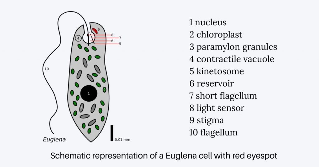
- In the context of Euglena, a protist renowned for its photosynthetic capabilities, the eyespot apparatus is a salient feature. Located anteriorly, near the paraflagellar body, the eyespot in Euglena is discernible as a bright red spot, a hue attributed to the carotenoid pigment granules it houses.
- Beyond its pigmentation, the eyespot is a complex assembly of photoreceptors, structural proteins, metabolic enzymes, and signaling molecules. The carotenoid-rich granules are strategically positioned around a reservoir, with a photoreceptor crystal overlaying them. These photoreceptor proteins are typically embedded in the plasma membrane that overlies the pigmented granules.
- Eyespots, in their functional capacity as light detectors, are not uniform across all organisms. They exhibit variations and are classified into five primary types, ranging from Type-A to Type-E. Each type is characterized by its unique protein composition. For instance, flavoproteins and retinylidene proteins are two predominant eyespot proteins.
- In Euglena, the eyespot is categorized as a Type-D, which predominantly contains flavoproteins. When subjected to staining procedures using osmium tetraoxide, the eyespot or stigma in Euglena yields a distinctive black precipitate.
- In summation, the eyespot apparatus is an evolutionary marvel that equips unicellular photosynthetic organisms with the ability to navigate their environment based on light cues, optimizing their photosynthetic efficiency.
Definition of Eyespot
An eyespot is a specialized, light-sensitive organelle found in certain unicellular organisms, enabling them to detect light direction and intensity, thereby facilitating phototactic behaviors.
Location of Eyespot Apparatus in Euglena
The Euglenophyta, a taxonomic group encompassing phytoflagellates, prominently feature the eyespot apparatus as a crucial organelle within their cellular architecture. Specifically, the eyespot in Euglenophyta is strategically positioned at the anterior end of the cell or in proximity to a specialized structure known as the reservoir. This characteristic placement ensures optimal light perception and phototactic responses in these photosynthetic organisms.
The eyespot itself is readily identifiable within the cell, manifesting as a discernible dark spot. Its location is particularly noteworthy, situated near the paraflagellar body (PFB) and in close proximity to the base of the large flagellum, a critical structural feature of Euglenophyta. This positioning is essential for efficient light detection and response mechanisms.
An integral component of the eyespot apparatus is the paraflagellar body, which serves as the connecting link between the eyespot and the flagellum. Importantly, the paraflagellar body houses light-sensitive photoreceptor proteins, which play a pivotal role in facilitating phototaxis – the organism’s ability to navigate in response to light stimuli. This complex interaction between the eyespot, paraflagellar body, and photoreceptor proteins underscores the precise localization and functional significance of the eyespot in Euglenophyta.
Concomitantly, the large flagellum, in addition to its structural role, imparts motility to Euglenophyta members. This motility is essential for their survival and ability to move towards or away from light sources, thereby optimizing their photosynthetic processes.
In summary, the eyespot apparatus in Euglenophyta is strategically positioned near the paraflagellar body and the base of the large flagellum, ensuring efficient light perception and enabling phototactic responses that are vital for these phytoflagellates’ adaptation and ecological success.
Eyespot Structure

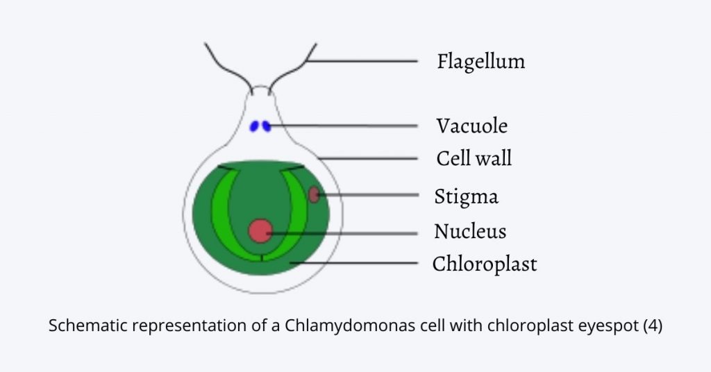
The structure of the eyespot, a vital photoreceptive organelle found in various photosynthetic microorganisms, exhibits remarkable complexity and functionality. This description provides a scientific overview of the eyespot structure, encompassing key details observed under both light and electron microscopy, with a focus on representative organisms such as Euglena and Chlamydomonas.
- Appearance under Light Microscopy: Under the light microscope, the eyespot presents itself as conspicuous dark, orange-reddish spots or stigmata. This distinctive coloration arises from the presence of carotenoid pigments contained within specialized organelles known as pigment granules. Significantly, these pigments contribute to the eyespot’s ability to detect and process light stimuli. Additionally, the photoreceptor components of the eyespot are situated within the plasma membrane overlaying these pigmented bodies.
- Eyespot Apparatus in Euglena: In the case of Euglena, the eyespot apparatus comprises an essential component known as the paraflagellar body. This structure serves as the connector linking the eyespot to the flagellum, thus playing a crucial role in the organism’s phototactic responses. When subjected to electron microscopy, the eyespot apparatus in Euglena reveals a highly ordered lamellar structure characterized by membranous rods arranged in a helical pattern. This intricate arrangement underpins the eyespot’s photoreceptive capabilities.
- Eyespot Structure in Chlamydomonas: In Chlamydomonas, the eyespot takes on a distinctive membranous sandwich structure, which is integral to its function. This structure is assembled from various chloroplast membranes, including the outer, inner, and thylakoid membranes, as well as carotenoid-filled granules. These granules are overlaid by the plasma membrane. Notably, the stacks of carotenoid-filled granules within the eyespot act as a quarter-wave plate, a specialized optical element that reflects incoming photons back toward the overlying photoreceptors. This arrangement effectively shields the photoreceptors from light originating from other directions, enhancing the eyespot’s sensitivity to light directionality.
- Dynamic Nature of Eyespot Structure: The eyespot structure exhibits dynamic properties, particularly during cell division. It disassembles as part of the cell division process and subsequently reforms in the daughter cells. This reformation occurs in an asymmetric fashion relative to the cytoskeleton. This asymmetry is crucial for ensuring proper phototactic responses in the organism, as it influences the eyespot’s orientation within the cell.
In summary, the eyespot structure is a highly organized and functionally specialized organelle, crucial for photoreception and phototactic behaviors in photosynthetic microorganisms such as Euglena and Chlamydomonas. Its intricate arrangement of pigmented granules, chloroplast membranes, and photoreceptor components, coupled with its dynamic properties during cell division, highlights its essential role in guiding these microorganisms in response to light stimuli and optimizing their photosynthetic activities.

Different Eyespot Proteins
Eyespots, the photoreceptive organelles prevalent in various unicellular organisms, employ a diverse array of proteins to facilitate their light-sensing functions. These proteins play pivotal roles in the detection and processing of light stimuli, thereby contributing to the organisms’ phototactic responses. This discussion delves into the different types of proteins found within eyespots, shedding light on their significance in the realm of photoreception.
- Photoreceptor Proteins: At the core of eyespot-mediated light perception are photoreceptor proteins. These proteins are primarily responsible for sensing and transducing light signals. In the context of unicellular organisms, photoreceptor proteins can be categorized into two main groups:
- Flavoproteins: Flavoproteins are characterized by the presence of flavin molecules as chromophores. These chromophores enable them to absorb and respond to specific wavelengths of light, thus initiating the phototransduction process. In Euglena, it is likely that the photoreceptor protein falls into the flavoprotein category.
- Retinylidene Proteins (Rhodopsins): Rhodopsins are another group of photoreceptor proteins found in unicellular organisms. They contain retinal as their chromophore. Archaeal-type rhodopsins, in particular, play a crucial role in phototaxis in organisms like Chlamydomonas. These proteins are instrumental in capturing and transducing light signals for navigation.
- Structural, Metabolic, and Signaling Proteins: Beyond photoreceptor proteins, eyespots encompass a diverse array of structural, metabolic, and signaling proteins. These proteins contribute to the overall functionality and maintenance of the eyespot apparatus. In Chlamydomonas, for instance, the eyespot proteome comprises an impressive collection of approximately 200 different proteins. While the photoreceptor proteins are central to light perception, these other proteins play essential roles in supporting the organelle’s structure and associated biochemical processes.
In summary, eyespots utilize a multifaceted ensemble of proteins to detect and respond to light stimuli. Photoreceptor proteins, including flavoproteins and retinylidene proteins, serve as the primary agents for light perception, each with their distinctive chromophores. Additionally, a myriad of structural, metabolic, and signaling proteins collectively contribute to the eyespot’s overall functionality. This intricate protein complement underscores the sophistication of the eyespot apparatus and its critical role in guiding unicellular organisms in response to light cues.
The Photoreception and signal transduction
Photoreception and signal transduction are intricate processes that underpin the ability of certain microorganisms to perceive and respond to light stimuli. This discussion delves into the specific mechanisms employed by two distinct organisms, Euglena and Chlamydomonas, to achieve photoreception and subsequent signal transduction.
Photoreception and Signal Transduction in Euglena:
Euglena, a unicellular organism, utilizes a blue-light-activated photoreceptor known as adenylyl cyclase to sense and respond to light. When these photoreceptor proteins are excited by blue light, they initiate a series of biochemical events. One significant outcome of this process is the production of cyclic adenosine monophosphate (cAMP), which functions as a second messenger within the cell.
The presence of cAMP as a second messenger sets in motion chemical signal transduction pathways. These pathways, in turn, have a profound impact on the flagellar beat patterns within Euglena. Alterations in flagellar beating patterns result in coordinated cell movement. Therefore, the photoreception and signal transduction mechanism in Euglena culminate in the modulation of flagellar movements, enabling the organism to respond to light stimuli with directed motion.
Photoreception and Signal Transduction in Chlamydomonas:
Chlamydomonas, another unicellular microorganism, employs a distinct set of photoreceptor proteins called archaeal-type rhodopsins for light perception. These rhodopsins carry an all-trans retinylidene chromophore, which undergoes photoisomerization to a 13-cis isomer in response to light stimulation.
The photoisomerization of the chromophore triggers the activation of photoreceptor channels within Chlamydomonas. This activation, in turn, leads to changes in the membrane potential and cellular calcium ion concentration. The alteration in membrane potential and calcium ion levels constitutes a photoelectric signal transduction cascade.
Importantly, the changes induced by photoelectric signal transduction extend to the flagellar strokes of Chlamydomonas. The modulation of flagellar strokes is the cellular response to the light signal, resulting in the directed movement of the organism.
In both Euglena and Chlamydomonas, photoreception is the initial step in perceiving light stimuli, and the subsequent signal transduction processes involve intricate biochemical and physiological mechanisms. These processes ultimately culminate in the adjustment of flagellar movements, enabling these microorganisms to navigate their environments in response to light cues.
Types of Eyespot
The classification of eyespots encompasses five distinct types, each characterized by specific structural and organizational features. These types provide valuable insights into the diversity and complexity of eyespot structures across various microorganisms. The following is an overview of the five types of eyespots:
- Type A: Type A eyespots are integrated into the chloroplast structure but are not directly associated with flagella. This type is exemplified by microorganisms belonging to the Chlorophyceae and Cryptophyceae classes. In these organisms, the eyespot plays a role in photoreception and light-guided behavior, despite its lack of direct connection to flagellar components.
- Type B: Type B eyespots are also situated within the chloroplast but are not linked to swollen flagella. This category includes microorganisms from the Chrysophyceae, Xanthophyceae, and Phaeophyceae classes. Similar to Type A, Type B eyespots contribute to photoreception and light-directed movement, despite their distinct structural arrangement within the chloroplast.
- Type C: Type C eyespots represent independent clusters of osmophilic granules. Organisms from the Euglenophyceae class fall into this category. These granule clusters are a crucial part of the eyespot apparatus in Euglenophyceae, aiding in the perception of light stimuli and subsequent phototactic responses.
- Type D: Type D eyespots consist of osmophilic granules and also include membranous structures. This type is observed in microorganisms from the Dinophyceae class. The presence of membranous structures within the eyespot apparatus adds a layer of complexity to the photoreception mechanism in these organisms, enhancing their ability to respond to light cues.
- Type E: Type E eyespots represent the largest category among the five types, although the description concludes abruptly. It is worth noting that additional information regarding Type E eyespots is needed to provide a comprehensive understanding of their characteristics and the microorganisms in which they are found.
In summary, the classification of eyespots into five distinct types underscores the structural and organizational diversity of these photoreceptive organelles in various microorganisms. Each type exhibits unique features that contribute to the organism’s ability to sense and respond to light stimuli, ultimately playing a critical role in phototactic behaviors and environmental adaptation.
Mechanism of Eyespot-Mediated Light Perception
The mechanism of eyespot-mediated light perception in Euglena is a finely orchestrated process involving several distinct stages, each characterized by precise molecular events and cellular responses. This mechanism serves as the foundation for the organism’s phototactic behaviors, which enable it to navigate its environment based on light cues.
- Activation of Photosensory Pigments: The process commences with the activation of photosensory pigments, specifically in response to blue light. Within the Euglena cell, this activation stimulates the activity of photosensitive adenylate cyclase, a key enzyme involved in signal transduction pathways.
- Conversion of Light Signal into a Chemical Signal: Adenylate cyclase, once activated by the incident blue light, initiates a cascade of biochemical reactions. One notable outcome of this cascade is the generation of cyclic adenosine monophosphate (cAMP), a secondary messenger molecule. This conversion of a light signal into a chemical signal is a pivotal step in the process.
- Transfer of Information to the Flagella: The newly synthesized cAMP serves as the messenger that carries information regarding the light stimulus. This information is subsequently relayed to the flagella, the motile appendages of Euglena. The cAMP-triggered chemical transduction events lead to alterations in the flagellar beat patterns.
- Phototactic Movement: Ultimately, the changes in flagellar beat patterns result in a coordinated cell movement known as phototaxis. Euglena exhibits phototaxis in response to the direction and intensity of light. This phenomenon can manifest as either positive or negative phototaxis, depending on whether the organism moves towards or away from increasing light intensity.
- Positive Phototaxis: In the case of positive phototaxis, Euglena exhibits a directional movement towards areas of increasing light intensity. This behavior is fundamental for Euglena’s ability to seek out optimal light conditions for photosynthesis.
Euglena utilizes its large flagellum for steering during phototaxis. This is intricately coupled with the light-sensitive “Eyespot apparatus” or “Stigma.” The eyespot, a prominent feature within Euglena, contains numerous lipid globules enriched with carotenoid pigments. These pigments are integral to the phototactic response.
The photoreceptors present in Euglena contain flavin molecules as chromophores, and they, in conjunction with the pigmented shading provided by the eyespot apparatus, play a pivotal role in controlling the phototactic movement. The process of phototaxis in Euglena can be divided into two stages:
- Orientation: During this stage, Euglena rotates about its long axis, and the flagellum moves around its base or anterior portion, resulting in the organism moving backward.
- Directed Movement: When light falls on one side of the photoreceptor, the eyespot periodically shades the photoreceptors. This shading initiates a succession of photophobic responses that propel Euglena towards the increasing light source.
It is important to note that once the organism achieves its orientation, continuous shading of the photoreceptor occurs, and no further photophobic responses are elicited. This finely tuned mechanism allows Euglena to navigate its environment efficiently in response to light stimuli, optimizing its positioning for photosynthetic activity.
Functions of Eyespot Apparatus
The eyespot apparatus is a pivotal and intricate component found in various photoreceptive microorganisms, including Euglena. It serves a multitude of functions that are central to the organism’s ability to perceive and respond to light stimuli. In the realm of scientific literature, the eyespot apparatus is recognized for its photoreceptive capabilities and its structural intricacies.
- Photoreception: At its core, the eyespot apparatus is chiefly responsible for photoreception. Within its confines, it houses carotenoid-rich globules that function as screening pigments or reflector devices. These pigments play a fundamental role in capturing and processing incoming light, allowing the organism to perceive the direction and intensity of light in its environment.
- Photoreceptor Proteins: The photoreceptor proteins present in Euglena, notably flavoproteins, are integral components of the eyespot apparatus. These proteins possess photo-behavioral functions, enabling them to detect and respond to variations in light conditions. Through these proteins, Euglena can gauge the presence and characteristics of light, which, in turn, influence its behavioral responses.
- Pigmented Apparatus with Stacked Membrane: Structurally, the eyespot is characterized as a pigmented apparatus with a stacked membrane configuration. This structural organization plays a crucial role in the efficient capture and processing of light. The stacked membrane architecture enhances the sensitivity of the eyespot to changes in light conditions, further augmenting the organism’s photoreceptive capabilities.
- Light-Guided Behavior and Signaling: The eyespot’s primary function is to facilitate light-guided behavior in Euglena. It serves as the nexus for signaling between the photoreceptors (sensors) and effector organelles, such as the flagella. This intricate signaling network enables Euglena to exhibit phototaxis, responding to light stimuli by moving either towards or away from the light source.
- Optimal Light Conditions for Photosynthesis: Euglena’s ability to sense light stimuli through the pigmented stigma within the eyespot is instrumental in its quest to locate environments with optimal light conditions. This capability is of paramount importance as it allows the organism to position itself in areas where sunlight is most abundant, thereby optimizing the photosynthetic process.
- Influence on Flagellar Movement: Beyond its role in light perception, the eyespot apparatus also exerts influence over flagellar movement in Euglena. Changes in light conditions trigger alterations in flagellar beating patterns, which are essential for the organism’s motility and navigation in response to light cues.
In conclusion, the eyespot apparatus in Euglena is a multifaceted organelle with critical functions in photoreception, light-guided behavior, and the optimization of photosynthesis. Its intricate interplay of pigments, photoreceptor proteins, and structural features underscores its significance in the organism’s ecological adaptation and survival.
Quiz
What is the primary function of the Eyespot Apparatus in microorganisms?
a) To capture prey
b) To sense and respond to light
c) To assist in reproduction
d) To store nutrients
Which organelle within the Eyespot Apparatus contains carotenoid pigments responsible for its color?
a) Mitochondria
b) Chloroplast
c) Nucleus
d) Golgi apparatus
In Euglena, where is the Eyespot typically located within the cell?
a) Near the nucleus
b) At the posterior end
c) Near the paraflagellar body
d) In the cytoplasm
Which photoreceptor proteins are commonly found in the Eyespot Apparatus of microorganisms?
a) Hemoglobin
b) Flavoproteins and retinylidene proteins
c) Ribosomes
d) Collagen
What is the role of cAMP in the Eyespot-mediated response to light?
a) It reflects light
b) It functions as a secondary messenger
c) It anchors the Eyespot to the cell membrane
d) It stores energy
Which type of phototaxis involves the movement of microorganisms toward areas of increasing light intensity?
a) Negative phototaxis
b) Positive phototaxis
c) Neutral phototaxis
d) Random movement
In Chlamydomonas, what type of rhodopsins are responsible for phototaxis?
a) Bacterial rhodopsins
b) Animal rhodopsins
c) Archaeal-type rhodopsins
d) Plant rhodopsins
What is the significance of the membranous structures within Type D Eyespots?
a) They produce light
b) They store nutrients
c) They enhance photoreception
d) They provide structural support
Which class of microorganisms typically exhibits Type C Eyespots?
a) Chlorophyceae
b) Chrysophyceae
c) Euglenophyceae
d) Cryptophyceae
In which class of microorganisms is Type E Eyespot found, and what additional information is needed to classify it?
a) Dinophyceae; Structural details
b) Diatomophyceae; Functional properties
c) Euglenophyceae; Color characteristics
d) Cryptophyceae; Organizational features
FAQ
What is the Eyespot Apparatus in microorganisms?
The Eyespot Apparatus is a photoreceptive organelle found in various microorganisms, primarily unicellular ones. It enables these organisms to sense and respond to light stimuli.
What is the main function of the Eyespot Apparatus?
The primary function of the Eyespot Apparatus is to detect the direction and intensity of light, allowing microorganisms to perform phototaxis (movement in response to light) for optimal positioning.
Where is the Eyespot typically located within a cell?
The precise location of the Eyespot varies among different microorganisms. In Euglena, for example, it is often found near the paraflagellar body.
What pigments are responsible for the color of the Eyespot?
Carotenoid pigments are typically responsible for the orange-red coloration of the Eyespot. These pigments aid in light absorption.
What are the major types of photoreceptor proteins found in the Eyespot Apparatus?
The major types of photoreceptor proteins in the Eyespot Apparatus include flavoproteins and retinylidene proteins (rhodopsins).
How do photoreceptor proteins in the Eyespot function?
Photoreceptor proteins absorb specific wavelengths of light and initiate signal transduction pathways. This leads to changes in flagellar beat patterns, resulting in directed movement in response to light.
What is the difference between positive and negative phototaxis?
Positive phototaxis is a directional movement of microorganisms toward increasing light intensity, while negative phototaxis involves movement away from increasing light intensity.
Which class of microorganisms exhibits archaeal-type rhodopsins for phototaxis?
Archaeal-type rhodopsins are commonly found in microorganisms like Chlamydomonas, where they play a role in phototactic responses.
What is the significance of membranous structures in certain Eyespot types, such as Type D?
Membranous structures within Eyespots, like Type D, enhance photoreception and contribute to the organism’s ability to respond to light cues.
How many different types of Eyespots are there, and what are they based on?
There are five main types of Eyespots (Type A, Type B, Type C, Type D, and Type E). They are classified based on structural, organizational, and functional characteristics observed in various microorganisms.

What organisms have eyespots?
Eyespots, or ocelli, are eye-like markings found in a diversity of organisms including lepidopterans (butterflies, moths, and skippers), reptiles, fish, birds, and cats.