Table of Contents
What is Anabaena?
- Anabaena, a fascinating genus of filamentous cyanobacteria, holds a crucial place in the realm of microbial life. These organisms exhibit remarkable adaptability and versatility, forming colonies that consist of both solitary cells and interconnected groups of filamentous cells. Through the process of photosynthesis, Anabaena cells harness light energy to produce organic compounds and, as an intriguing byproduct, release oxygen gas into their surroundings, contributing to the oxygenation of aquatic environments.
- One of the most intriguing aspects of Anabaena is its dual role as both a beneficial agent and a potential threat in various ecological contexts. Notably, several species of Anabaena have demonstrated their prowess as natural fertilizers when utilized in rice paddies, fostering healthier crop growth through the enrichment of nitrogen content in the soil. This symbiotic relationship between Anabaena and rice plants showcases the potential of these cyanobacteria to contribute positively to agricultural practices.
- However, Anabaena also possesses a more enigmatic side, as some of its species produce neurotoxins that can have harmful effects on domestic animals, farm animals, pets, and local fauna. These neurotoxins are believed to play a role in Anabaena’s intricate symbiotic interactions, acting as a defense mechanism against grazing stress and predators, thereby aiding in the protection of the cyanobacterial colonies.
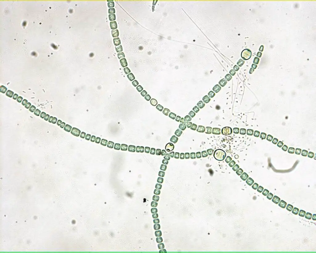
- In terms of its structural characteristics, Anabaena exhibits a filamentous morphology that shares similarities with another cyanobacterial genus, Nostoc. The filaments of Anabaena are composed of a chain of bead-like cells, and intercalary heterocysts are interspersed along the trichome. These heterocysts display morphological traits resembling vegetative cells, further contributing to the complexity of Anabaena’s cellular organization. While the filaments of Nostoc and Anabaena can be challenging to distinguish from one another, a key distinction lies in the presence of mucilage that envelops Nostoc filaments, allowing them to form cohesive colonies, whereas Anabaena lacks this mucilaginous covering.
- Anabaena’s ecological influence extends to its role as planktonic organisms, inhabiting diverse aquatic environments. A notable trait of Anabaena is its ability to fix nitrogen, a process essential for nutrient cycling and the sustenance of various ecosystems. This capacity for nitrogen fixation has led to symbiotic relationships with certain plant species, such as the mosquito fern. Moreover, specific Anabaena species have been identified as endophytes within the roots of Cycas and Azolla, underscoring the intricate connections between these cyanobacteria and higher plants.
- Although Anabaena’s presence in water ecosystems is widespread, nutrient-rich water bodies can support extensive growth or blooms of these cyanobacteria. These blooms, while visually striking, can have adverse consequences. As Anabaena cells decay, they discolor the water and release unpleasant odors, potentially disrupting the ecological balance of aquatic habitats.
- In conclusion, Anabaena stands as a remarkable example of cyanobacterial diversity and ecological significance. Its ability to fix nitrogen, form symbiotic relationships with plants, and serve as a natural fertilizer highlights its potential for positive contributions to agriculture and ecosystem dynamics. Simultaneously, its production of neurotoxins and role in forming disruptive blooms underline the complex interplay between organisms in various environments. Further research into Anabaena’s biology and interactions promises to unveil more about the intricate web of life within aquatic ecosystems.
Requirement for Observation of Anabaena Under Microscope
- Light Compound Microscope: A versatile instrument capable of illuminating the microscopic universe, the light compound microscope is an indispensable tool for this endeavor. With magnification options of 10×, 40×, and 100×, it enables the gradual uncovering of Anabaena’s fine details and intricacies. This apparatus serves as the gateway to the microcosm, allowing us to witness the wonders of Anabaena’s cellular realm.
- Lens Paper: Precise clarity is the hallmark of a successful microscopic examination. Lens paper, a delicate yet efficient companion, facilitates the maintenance of clean microscope lenses. Ensuring the elimination of dust, smudges, and obstructions, lens paper is a vital ally in obtaining crisp and accurate observations of Anabaena’s minute structures.
- Prepared Slide of Anabaena or Nostoc: A meticulously crafted slide harboring a specimen of Anabaena or its close relative, Nostoc, lies at the heart of this undertaking. This prepared slide serves as a canvas on which the vibrant tableau of Anabaena’s existence is displayed. With careful placement and adjustment, it unveils the cyanobacterial world in its full glory, inviting scrutiny and exploration.
- Images of Anabaena or Nostoc: In the absence of physical specimens, the inclusion of high-resolution images of Anabaena or Nostoc acts as a viable alternative. These visual representations offer a window into the morphology and structure of these cyanobacteria, allowing for informed observation and analysis. The power of visual aids transforms the microscopic study into an engaging visual journey.
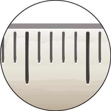
- Special Slide with Micron Ruler or Clear Millimeter Ruler: Precision is the hallmark of scientific inquiry, and a specialized slide equipped with a micron ruler or a clear millimeter ruler serves as the compass for measurement and scale. This calibrated guide ensures accurate assessment of Anabaena’s dimensions, fostering meticulous documentation and analysis.
Procedure for observation of Anabaena Under Microscope
This procedure outlines the steps for creating a wet mount slide and observing the prokaryotic alga Anabaena under a microscope.
- Materials: Gather the required materials from your lab room, typically located near the sinks. You will need a clean microscope slide, a plastic pipette, Anabaena culture, coverslips, a dissection needle (if necessary), a compound microscope, and paper towels.
- Prepare Microscope Slide: Place a clean microscope slide on a flat surface.
- Collect Anabaena: Use a plastic pipette to carefully collect a few drops of Anabaena culture. Avoid overfilling the pipette. Gently shake the culture if cells have settled.
- Apply Anabaena to Slide: Dispense one or two drops of Anabaena onto the center of the microscope slide.
- Add Coverslip: Hold a coverslip by its edges at a 45-degree angle. Touch the coverslip’s bottom edge to the slide at the edge of the Anabaena drop. Slowly lower the outer edge of the coverslip onto the sample. Use a dissection needle if needed.
- Eliminate Air Bubbles: Ensure there are no visible air bubbles under the coverslip. If large air bubbles are present, restart the process. If excess water causes the coverslip to float, absorb the water with a paper towel.
- Microscope Observation: Place the prepared slide on the compound microscope’s stage. Begin with the low-power objective to find a focus point. Adjust the iris diaphragm for proper lighting.
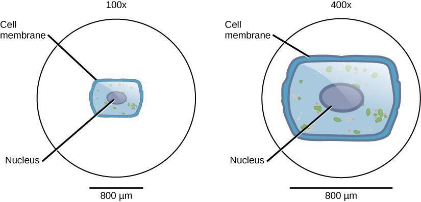
- Switch to Higher Magnification: Change to the four-power objective lens for a closer examination after observing at low power.
- Disposal and Cleaning: Safely dispose of the coverslip in a sharps disposal bin. Rinse the slide, wash with soap, rinse again, and dry with a paper towel before returning it to the slide box.
- Conclusion: This procedure provides a basic method for creating a wet mount slide and observing Anabaena under a microscope. Follow safety guidelines and specific instructions from your lab instructor.
Structured Inquiry
Step 1: Estimating Field of View
Begin by placing a clear millimeter ruler on the microscope stage, a simple act that lays the foundation for accurate observations. Estimate the size of the field of view across different magnifications, ensuring a calibrated perspective for subsequent analyses. Convert these estimates from millimeters to micrometers, a vital conversion that paves the way for precise measurements of cell dimensions during the inquiry.

Step 2: Hypothesizing and Predicting
With the groundwork laid, transition to the realm of hypothesis and prediction. In your notebook, vividly depict what you anticipate encountering under the microscope. Craft a visual representation of your expectations, envisioning the size, features, and potential structures of Anabaena cells. Delve into questions about organelles, cell walls, and any anticipated colorations, weaving your insights into a comprehensive prediction.

Step 3: Detailed Drawing and Exploration
Translate your hypotheses into reality as you embark on a detailed drawing of Anabaena’s structure. Utilize a sharp pencil to meticulously capture the essence of these cyanobacteria, paying heed to cell sizes, shapes, and organization. Integrate color into your drawings where applicable, noting the hues employed and labeling any discernible structures. This step lays the groundwork for visual documentation that forms the cornerstone of your inquiry.
Step 4: Critical Analysis and Reflection
With your detailed drawing as a reference point, embark on a critical analysis of your observations. Evaluate the alignment between your predictions and actual findings, pondering over the colors and characteristics you encountered. Contemplate the significance of the observed photosynthetic nature of the cyanobacteria and its implications on coloration. Reflect on the presence or absence of organelles, cell walls, and heterocysts, inviting a deeper exploration of these microorganisms’ functional adaptations.
Collaborating with your partner, engage in thoughtful discussions to dissect the implications of your observations. Document your insights and revelations in your notebook, forming a comprehensive record of your structured inquiry into Anabaena’s microscopic domain. Through this methodical approach, you not only enhance your understanding of these fascinating cyanobacteria but also cultivate critical thinking skills that are indispensable to scientific exploration.

Anabaena Under Microscope Video
FAQ
What is Anabaena, and why is it of interest for microscopic observation?
Anabaena is a genus of filamentous cyanobacteria. Its microscopic observation provides insights into its cellular structure, arrangement, and adaptations, shedding light on its role in ecosystems and potential applications.
What magnification levels are recommended for observing Anabaena?
Typically, magnification levels of 10×, 40×, and 100× objectives are recommended for observing Anabaena under a light compound microscope.
How can I estimate the size of Anabaena cells using a microscope?
Place a clear millimeter ruler on the microscope stage and estimate the size of the field of view at different magnifications. Convert these estimates to micrometers for accurate measurements of cell sizes.
What are some common structures observed in Anabaena under the microscope?
Common structures include bead-like cells forming filamentous chains, intercalary heterocysts (specialized cells), and potentially visible sheaths.
What coloration should I expect when observing Anabaena?
Anabaena is photosynthetic, so you might observe green or blue-green coloration due to the presence of chlorophyll and other pigments.
Can I observe organelles in Anabaena cells under the microscope?
While you may observe structures resembling organelles, such as heterocysts, the lack of membrane-bound organelles like those found in eukaryotic cells makes detailed organelle observation challenging.While you may observe structures resembling organelles, such as heterocysts, the lack of membrane-bound organelles like those found in eukaryotic cells makes detailed organelle observation challenging.
How can I distinguish Anabaena from other similar cyanobacteria under the microscope?
The absence of mucilage covering the filaments can help distinguish Anabaena from closely related cyanobacteria like Nostoc. Intercalary heterocysts and other structural features also aid in differentiation.
What is the significance of heterocysts in Anabaena?
Heterocysts are specialized cells involved in nitrogen fixation, a crucial process that converts atmospheric nitrogen into a usable form for plants and other organisms.
How do I document my observations of Anabaena under the microscope?
Record detailed drawings of the observed structures, noting cell sizes, arrangements, and any unique features. Use your notebook to document your predictions, observations, analysis, and reflections.
What insights can microscopic observation of Anabaena provide for research and education?
Microscopic observation of Anabaena contributes to understanding its ecological role, adaptations, and potential applications in fields like agriculture and environmental science. It also provides a valuable educational tool to teach about microbial diversity and interactions.


