Table of Contents
What is Codon?
- A codon, in the realm of molecular biology, is a sequence of three nucleotides within DNA or RNA that corresponds to a specific amino acid or serves as a regulatory signal in protein synthesis. The very essence of life’s continuity across generations hinges on the processes of replication, transcription, and translation of the genetic code harbored within DNA and RNA.
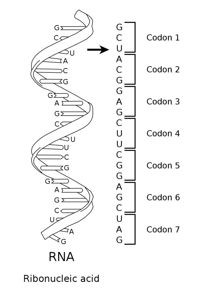
- DNA, the fundamental molecule of life, is composed of four nucleotide bases: adenine (A), guanine (G), cytosine (C), and thymine (T). This genetic code is transcribed into RNA, which then serves as a blueprint for synthesizing proteins. Unlike DNA, RNA comprises adenine, guanine, cytosine, and uracil (U). These nucleotide sequences provide the instructions for arranging amino acids in a specific order to construct proteins.
- The reading of the genetic code occurs in triplets of nucleotides, termed as codons. Each distinct codon either designates a particular amino acid or functions as an indicator to initiate or terminate protein synthesis. The groundbreaking discovery of the first codon is attributed to Marshall Nirenberg and J. Heinrich Matthaei. Their pioneering poly-U experiment unveiled that a triplet of uracil units (UUU) in RNA encoded for the amino acid phenylalanine. This monumental revelation paved the way for subsequent research, spearheaded by Nirenberg and his team, which identified all 64 codons and their associated amino acids. For their invaluable contributions to the understanding of the genetic code, Nirenberg, Robert W. Holley, and Har Gobind Khorana were conferred the Nobel Prize in Physiology or Medicine in 1968.
- In essence, the genetic code serves as a set of guidelines employed by living cells to decode the information encapsulated within DNA or RNA sequences into proteins. This intricate process of translation is executed by the ribosome, which meticulously arranges proteinogenic amino acids as directed by messenger RNA (mRNA). This is achieved with the assistance of transfer RNA (tRNA) molecules, which not only transport amino acids but also interpret the mRNA in sets of three nucleotides. Remarkably, this genetic code exhibits a high degree of similarity across diverse organisms and can be succinctly represented in a table encompassing 64 entries.
- To elucidate further, codons play a pivotal role in determining the subsequent amino acid to be incorporated during the intricate process of protein biosynthesis. Generally, a triad of nucleotides in a nucleic acid sequence corresponds to a singular amino acid. While the majority of genes adhere to this canonical genetic code, certain exceptions and variant codes, such as those observed in mitochondria, do exist.
What is a Codon Chart?
A Codon Chart, often referred to as a codon table, is an essential scientific reference tool that provides a systematic representation of the relationship between sequences of three nucleotides, known as codons, and the amino acids they encode. This chart serves as a key to decode the genetic information stored in DNA or RNA, facilitating the accurate translation of nucleotide sequences into the corresponding amino acid chains that form proteins.
The chart encompasses 64 distinct codons. Among these, 61 codons are designated to encode the 20 standard amino acids, while the remaining three function as stop codons, indicating the termination of protein synthesis. A noteworthy feature of the genetic code, as depicted in the codon chart, is its redundancy or degeneracy. This means that while each codon specifies a unique amino acid, a single amino acid can be represented by multiple codons. An illustrative example of this phenomenon is the amino acid proline, which is encoded by four different codons: CCU, CCC, CCA, and CCG.
To utilize the codon chart effectively, one identifies the sequence of nucleotides in a given codon and then cross-references it with the chart to determine the corresponding amino acid. For instance, the codon sequence UAC, when referenced in the chart, corresponds to the amino acid tyrosine.
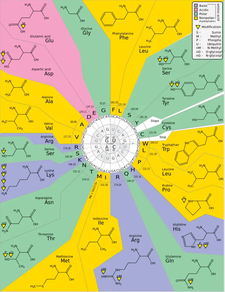
For clarity, the codon chart also typically includes a list of the 20 standard amino acids, accompanied by both their three-letter and one-letter abbreviations. A brief overview is as follows:
- Alanine (Ala, A)
- Arginine (Arg, R)
- Asparagine (Asn, N)
- Aspartic acid (Asp, D)
- Cysteine (Cys, C)
- Glutamic acid (Glu, E)
- Glutamine (Gln, Q)
- Glycine (Gly, G)
- Histidine (His, H)
- Isoleucine (Ile, I)
- Leucine (Leu, L)
- Lysine (Lys, K)
- Methionine (Met, M)
- Phenylalanine (Phe, F)
- Proline (Pro, P)
- Serine (Ser, S)
- Threonine (Thr, T)
- Tryptophan (Trp, W)
- Tyrosine (Tyr, Y)
- Valine (Val, V)
| Amino Acid | Abbreviation | One-Letter Abbreviation |
| Alanine | Ala | A |
| Arginine | Arg | R |
| Asparagine | Asn | N |
| Aspartic acid | Asp | D |
| Cysteine | Cys | C |
| Glutamic acid | Glu | E |
| Glutamine | Gln | Q |
| Glycine | Gly | G |
| Histidine | His | H |
| Isoleucine | Ile | I |
| Leucine | Leu | L |
| Lysine | Lys | K |
| Methionine | Met | M |
| Phenylalanine | Phe | F |
| Proline | Pro | P |
| Serine | Ser | S |
| Threonine | Thr | T |
| Tryptophan | Trp | W |
| Tyrosine | Tyr | Y |
| Valine | Val | V |
Codon Chart
| First Base | Second Base | U | C | A | G |
|---|---|---|---|---|---|
| U | U | UUU Phe | UCU Ser | UAU Tyr | UGU Cys |
| UUC | UCC | UAC | UGC | ||
| C | UUA Leu | UCA Ser | UAA STOP | UGA STOP | |
| UUG | UCG | UAG STOP | UGG Trp | ||
| C | U | CUU Leu | CCU Pro | CAU His | CGU Arg |
| CUC | CCC | CAC | CGC | ||
| C | CUA Leu | CCA Pro | CAA Gln | CGA Arg | |
| CUG | CCG | CAG | CGG | ||
| A | U | AUU Ile | ACU Thr | AAU Asn | AGU Ser |
| AUC | ACC | AAC | AGC | ||
| C | AUA Ile | ACA Thr | AAA Lys | AGA Arg | |
| AUG Met | ACG | AAG | AGG | ||
| G | U | GUU Val | GCU Ala | GAU Asp | GGU Gly |
| GUC | GCC | GAC | GGC | ||
| C | GUA Val | GCA Ala | GAA Glu | GGA Gly | |
| GUG | GCG | GAG | GGG |


How do you read a codon table?
Reading a codon table is essential for determining the amino acid that corresponds to a specific three-nucleotide sequence (codon) in mRNA. Here’s a step-by-step guide on how to read a codon table:
- Understand the Structure: A typical codon table is a 4×4 grid, organized by the four nucleotide bases: U (Uracil), C (Cytosine), A (Adenine), and G (Guanine). The mRNA sequence is read 5′ to 3′.
- First Base: The first letter of the codon (reading left to right) determines which row you’ll be looking at in the codon table. This is usually the leftmost column of the chart.
- Second Base: The second letter of the codon determines the column. This is usually the top row of the chart.
- Third Base: Once you’ve located the correct row and column based on the first two letters of the codon, you’ll see a set of codons in that cell. The third letter of your codon will direct you to the specific amino acid (or Start/Stop indication) within that cell.
- Determine the Amino Acid: Once you’ve used all three letters of your codon to navigate the chart, you’ll be left with a specific amino acid abbreviation (like Phe for Phenylalanine, Met for Methionine, etc.) or a “Start” or “Stop” indication.
- Special Codons:
- Start Codon: AUG is the start codon, and it codes for Methionine (Met). It signals the beginning of translation.
- Stop Codons: There are three stop codons (UAA, UAG, UGA) that do not code for an amino acid. Instead, they signal the end of translation.
- Practice: Try reading a few sample codons using the table. For instance, if you have the codon GCU, you’d start with G (row), move to C (column), and then find U within that cell. The result would be the amino acid Alanine (Ala).
Remember, the codon table is based on mRNA sequences. If you’re working with DNA, replace all the Thymine (T) bases with Uracil (U) to use the codon table.
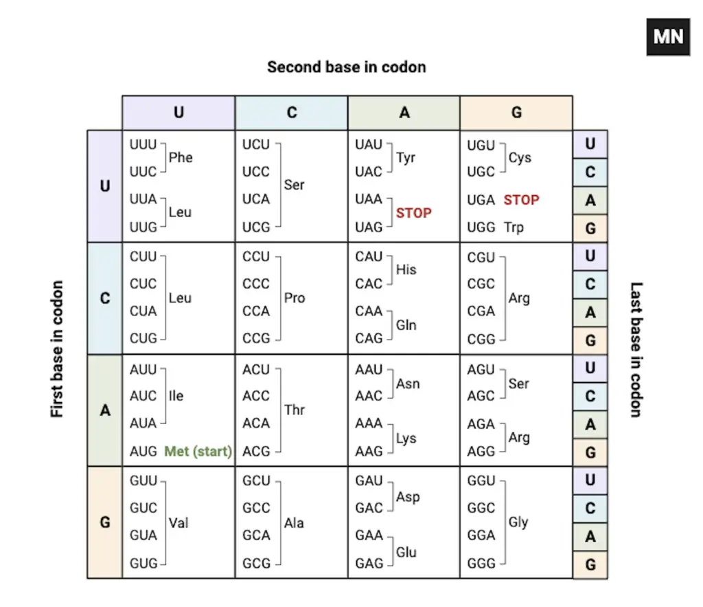
Here is an example – How do you read a codon table?

Reading this codon chart involves determining the amino acid that corresponds to a specific three-nucleotide sequence (codon) in mRNA. The chart helps translate the mRNA sequence into the respective amino acid. Here’s how you can read this codon chart:
- Understand the Axes:
- First Base in Codon: The leftmost column provides the first base of the codon.
- Second Base in Codon: The top row represents the second base of the codon.
- Last Base in Codon: Each cell in the chart will be subdivided based on the third base of the codon.
- Find the Cell:
- First Base: Look at the first nucleotide/base of your codon and go to the corresponding row labeled on the left side.
- Second Base: Look at the second nucleotide/base and move to the corresponding column labeled at the top.
- Determine the Amino Acid:
- Once you’ve found the right cell, the third base of your codon will pinpoint the exact amino acid. Within the cell, you’ll see codons with their corresponding amino acids. Match the third base of your codon to one of these to find the amino acid.
- Special Codons:
- Start Codon: AUG is labeled as the start codon and it codes for Methionine (Met). It’s where the translation begins in the protein synthesis process.
- Stop Codons: UAA, UAG, and UGA are stop codons. These don’t code for an amino acid but signal the end of translation.
- Example:
- For the codon UGC, start with U (row), move to G (column), and within that cell, find C. The resulting amino acid is Cys (Cysteine).
Remember, this codon chart is specific to mRNA sequences. If you’re working with DNA, you’d replace all the Thymine (T) bases with Uracil (U) to use this codon chart.
What are Start Codons?
- In the intricate realm of molecular biology, the term “Start Codon” refers to specific codons within an mRNA sequence that demarcate the commencement of protein translation. The codon AUG is predominantly recognized as the principal start codon, acting as a beacon for the initiation of the translation process.
- In the diverse cellular environments of eukaryotes, the AUG codon encodes the amino acid methionine (Met). Contrastingly, within prokaryotic organisms, the same AUG codon is designated to code for formyl methionine (fMet). The distinction in the amino acid coded by AUG in these two cellular domains underscores the evolutionary nuances and complexities of the translation process.
- The initiation of translation is not a solitary act of the start codon. It is a coordinated endeavor, involving initiation factors and specific tRNA molecules. These molecular entities collaborate to accurately recognize the AUG start codon, thereby setting the stage for the subsequent translation process.
- While AUG stands as the primary beacon for translation initiation, the world of genetics is replete with exceptions. In eukaryotic systems, there exist alternative codons that, albeit infrequently, can also function as start sites. These non-AUG start codons are relatively scarce, emphasizing the predominant role of AUG in protein synthesis.
- Delving into prokaryotic organisms, such as the bacterium E. coli, one observes a broader repertoire of start codons. Apart from the canonical AUG, codons like GUG and UUG also partake in signaling the onset of translation.
- In summation, the start codon, predominantly represented by AUG, plays a pivotal role in the molecular orchestration of protein synthesis, marking the genesis of the translation process across diverse cellular domains.

What are Stop Codons?
- In the intricate tapestry of molecular biology, stop codons occupy a pivotal role within the genetic code. Among the 64 codons that constitute this code, three distinct codons stand out, not for their association with amino acids, but rather for their lack thereof. These unique codons are termed as “stop codons.”
- Stop codons, alternatively referred to as termination or nonsense codons, function as molecular sentinels, signaling the cessation of protein synthesis during the translation process. These codons do not correspond to any amino acid, and their primary role is to indicate the termination point for the ribosome, ensuring the accurate completion of protein synthesis.
- The triad of stop codons comprises UAA, UAG, and UGA. Their discovery can be traced back to 1965, credited to the groundbreaking experiments conducted by Sydney Brenner on the T4 bacteriophage. Among these, UAG holds the distinction of being the inaugural stop codon identified and is colloquially termed the “amber” codon. The other two codons, UGA and UAA, are respectively dubbed as the “opal” and “ochre” codons.
- In essence, stop codons serve as molecular checkpoints, ensuring the precise and timely termination of protein synthesis, a critical aspect of cellular function and genetic expression.

What is Codon Translation?
- Codon translation is an integral component of the protein synthesis pathway, a fundamental biological process that orchestrates the assembly of proteins, the quintessential macromolecules vital for cellular function and structure. This process is a meticulous translation of the genetic code, ensuring the accurate construction of protein molecules.
- The journey of protein synthesis commences not with the direct utilization of the genetic blueprint stored in DNA, but with an intermediary step known as transcription. During transcription, the genetic directives of DNA are transcribed into RNA through the action of the enzyme RNA polymerase. This results in the formation of messenger RNA (mRNA), a single-stranded RNA molecule that carries the transcribed genetic information.
- Subsequent to transcription, the process of translation is initiated in the cellular cytoplasm. Here, the mRNA, in conjunction with cellular machinery comprising ribosomes and transfer RNA (tRNA), deciphers the encoded genetic instructions, translating them into polypeptide chains composed of amino acids. The translation process is characterized by the sequential reading of mRNA codons, with each codon corresponding to a specific amino acid. This amino acid is then added to the growing polypeptide chain by tRNA molecules. The culmination of this translation process is signaled by the presence of a stop codon within the mRNA sequence.
- The precision and specificity of codon translation are underpinned by the unique structure and function of tRNA molecules. Resembling a cloverleaf, the tRNA molecule boasts a distinct architecture comprising four double-helical stems interspersed with three single-stranded loops. Of these loops, the central one, termed the anticodon loop, harbors a triad of nucleotides known as the anticodon. This anticodon sequence is complementary to the mRNA codon, ensuring an accurate and specific interaction between the two, thereby facilitating the correct placement of amino acids during protein synthesis.
- Post the completion of transcription and translation, the nascent amino acid chain undergoes a series of modifications, culminating in the formation of a fully functional protein, ready to partake in the myriad cellular activities and processes. In essence, codon translation is the bridge that connects genetic information to functional proteins, underscoring the intricate and harmonized nature of cellular biology.
