Table of Contents
Cell division is a crucial process that allows organisms to grow, repair, and reproduce. One important aspect of cell division is cytokinesis, the process of separating one cell into two daughter cells. While cytokinesis is a universal process, it can vary greatly between different types of cells. In this article, we will compare and contrast cytokinesis in plant and animal cells and discuss the key differences between the two processes.
What is Cytokinesis?
Cytokinesis is the process of separating one cell into two daughter cells during cell division. It is a critical step in the cell division cycle, as it ensures that each daughter cell receives a complete and accurate copy of the genetic material.
The process of cytokinesis is similar in both plant and animal cells, but there are several key differences between the two. Understanding these differences is important for researchers, as it can provide insights into the unique characteristics of different types of cells.

What Happens During Cytokinesis?
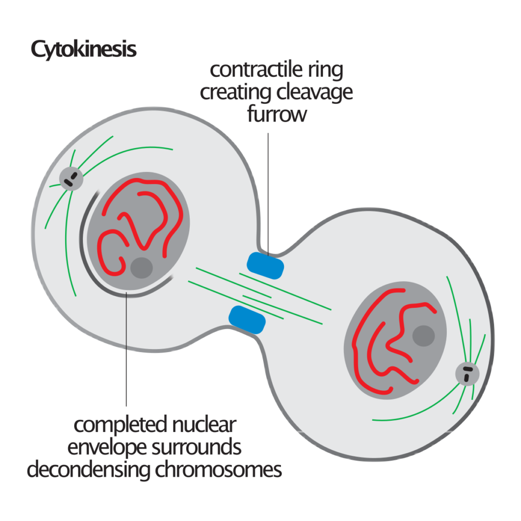
Cytokinesis is the final stage of cell division in eukaryotic cells, where the cytoplasm divides and forms two separate daughter cells. During cytokinesis, the cell membrane invaginates to form a cleavage furrow, which eventually pinches inwards to separate the cytoplasm into two distinct cells. The furrow is guided by a contractile ring of actin and myosin filaments, which pulls the plasma membrane inward. Once the cleavage furrow reaches the center of the cell, a new cell membrane forms to separate the two daughter cells. This process ultimately results in the division of the cytoplasm and the formation of two genetically identical daughter cells.
Cytokinesis in Plant Cells
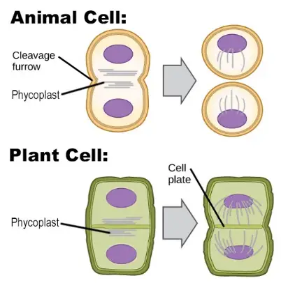
Plant cells undergo cytokinesis through a unique process known as cell plate formation. During this process, a cell plate forms at the center of the cell and gradually expands, eventually dividing the cell into two daughter cells. The cell plate is composed of vesicles that transport materials needed for cytokinesis, such as cell wall components.
Process of Cell Plate Formation
The process of cell plate formation is a crucial aspect of cell division and growth in plants. This process ensures the separation of two daughter cells, leading to cell expansion and organogenesis. In this article, we will explore the five steps involved in the formation of the cell plate.
- Phragmoplast Formation: The first step in the cell plate formation process is the formation of the phragmoplast. A phragmoplast is a microtubule array that supports and guides the formation of the cell plate. These microtubules are remnants of the spindle and play a crucial role in the formation of the phragmoplast.
- Trafficking of Vesicles and Fusion with Microtubules: Once the phragmoplast is formed, vesicles containing essential components such as proteins, lipids, and carbohydrates are trafficked into the mid-zone by the microtubules. These vesicles are sourced from the Golgi apparatus and are required for the formation of the cell plate.
- Formation of Membrane Sheets through Widened Microtubules Fusion: The next step involves the lateral fusion of the widened microtubules to form a planar sheet, known as the cell plate. Other cell wall components, including cellulose, deposit on the cell plate, leading to its maturation.
- Recycling of Cell Membrane: Materials After the formation of the cell plate, unwanted membrane materials are removed from it through clathrin-mediated endocytosis. This step is crucial for the proper functioning of the cell plate.
- Fusion of the Cell Plate with the Existing Cell Wall: The final step in the cell plate formation process is the fusion of the cell plate with the existing cell membrane. This separation leads to the formation of two daughter cells, and the edges of the cell plate are fused with the parental cell membrane. Most of the time, this fusion occurs in an asymmetrical manner. Additionally, strands of the endoplasmic reticulum passing through the newly formed cell plate act as precursors to plasmodesmata, a type of cell junction found in plant cells.
In conclusion, the cell plate formation process is a crucial aspect of cell division and growth in plants. The five steps involved in this process, including phragmoplast formation, trafficking of vesicles and fusion with microtubules, formation of membrane sheets through widened microtubules fusion, recycling of cell membrane materials, and fusion of the cell plate with the existing cell wall, work together to ensure the proper functioning of the cell plate. Different cell wall components, such as cellulose, hemicellulose, pectins, and arabinogalactan proteins, are deposited on the cell plate, leading to its maturation and proper functioning.
Cytokinesis in Animal Cells
In animal cells, cytokinesis is achieved through the formation of a contractile ring. This ring is composed of actin and myosin filaments that contract and squeeze the cell, dividing it into two daughter cells. The contractile ring is responsible for separating the cytoplasm and pulling the cell membrane inward, resulting in the formation of two separate cells.
Process of Cytokinesis in Animal Cell
1. Anaphase Spindle Recognition
The formation of the central spindle is essential for efficient cytokinesis in animals and C. elegans. During anaphase, the decline in CDK1 activity leads to the recognition of the spindle. The stabilization of microtubules forms the central spindle, also known as the spindle midzone, while non-kinetochore microtubules form bundles between the opposite poles of the parent cell.
The Chromosomal Passenger Complex (CPC) is dephosphorylated due to the decline in CDK1 activity, leading to its translocation to the central spindle. The CPC regulates the phosphorylation of central spindle component proteins, including PRC1 and MKLP1. The phosphorylated PRC1 forms a homodimer that binds to the antiparallel microtubules, facilitating the spatial arrangement of microtubules on the central spindle.
The GTPase-activating protein CYK-4 and phosphorylated MKLP1 form the centralspindlin complex, a higher-order cluster bound to the central spindle. Multiple central spindle components are phosphorylated to initiate the self-assembly of the central spindle, which controls the position of the cleavage furrow, maintains membrane vesicle delivery to the cleavage furrow, and controls midbody formation at the end of cytokinesis.
The Role of CPC in Regulating Central Spindle Formation
2. Division Plane Specification
The specification of the division plane is governed by three hypotheses – the astral stimulation hypothesis, the central spindle hypothesis, and the astral relaxation hypothesis. The cleavage furrow is positioned to the cell cortex by two redundant signals from the spindle, one from the central spindle and the other from the spindle aster.
“The Three Hypotheses of Division Plane Specification”
3. Actin-Myosin Ring Assembly and Contraction
The contractile ring, formed by actin and the motor protein myosin-II, drives the cleavage process. The Rho protein family regulates the formation and contraction of the contractile ring in the middle of the cell cortex. The RhoA protein promotes the formation of the contractile ring, which also contains scaffolding proteins such as anillin, linking the equatorial cortex and the central spindle.
“The Power Behind the Contractile Ring: The Role of Actin and Myosin-II”
4. Abscission
The cleavage furrow ingresses to form the midbody structure, with a diameter of 1-2 μm. The midbody is completely cleaved in a process known as abscission, during which intercellular bridges are filled with antiparallel microtubules, the cell cortex is constricted, and the plasma membrane is fashioned.
“The Final Step: Understanding Abscission in Animal Cell Cytokinesis”
Animal cell cytokinesis is a highly regulated process powered by Type II Myosin ATPase, which generates contractile forces. The four distinct steps
Differences between Plant and Animal Cell Cytokinesis

- The key difference between plant and animal cell cytokinesis is the mechanism by which the cells are divided. In plant cells, cytokinesis is achieved through the formation of a cell plate, while in animal cells, it is achieved through the contraction of a contractile ring.
- Another difference is that plant cells have a cell wall that is laid down during cytokinesis, while animal cells do not. The cell wall provides additional structural support to the plant cell and helps to maintain its shape.
- In addition, plant cells have a unique structure called the tonoplast, which is a specialized organelle involved in the regulation of cellular water content. Tonoplasts play a role in cytokinesis by facilitating the formation of the cell plate.
- Finally, plant cells often undergo cytokinesis without the assistance of centrosomes, while animal cells require centrosomes to divide properly. Centrosomes play a role in the formation of the contractile ring and help to maintain the proper orientation of the cell during division.
| Feature | Plant Cells | Animal Cells |
|---|---|---|
| Method of Division | Cell plate formation | Cleavage furrow formation |
| Structure involved | Cell wall | Contractile ring (microfilaments) |
| Result | New cell wall is formed between the two daughter cells | Cleavage furrow deepens until it pinches the cell in two |
| Regulating Mechanism | Cell plate deposition regulated by vesicles containing cell wall materials | Contractile ring contraction is regulated by actomyosin filaments |
Note: Cytokinesis is the final stage of cell division where the cell is physically divided into two daughter cells.
Conclusion
In conclusion, while both plant and animal cells undergo cytokinesis during cell division, there are several key differences between the two processes. These differences stem from the unique structures and mechanisms present in each type of cell. Understanding these differences is important for researchers studying cell biology and can provide insights into the distinct characteristics of different types of cells.
In summary, plant cells undergo cytokinesis through cell plate formation, while animal cells divide through the contraction of a contractile ring. Additionally, plant cells have a cell wall and tonoplast, while animal cells have centrosomes, which play a role in cytokinesis.
