Table of Contents
What is Metaphase?
- Metaphase, a pivotal stage in eukaryotic cell division, is characterized by the alignment of chromosomes along an imaginary central plane termed the metaphase plate. This alignment is a consequence of the intricate interplay between chromosomes and the microtubule network, which ensures precise chromosome segregation.
- In the preceding stages, namely prophase and prometaphase, chromosomes undergo condensation, the nuclear envelope disintegrates, and spindle fibers emerge from centrosomes. These spindle fibers, composed of microtubules, are dynamic structures.
- While they exhibit stability proximal to the centrosomes, their distal ends display a remarkable dynamism, characterized by a pattern of growth and shrinkage. This “three steps forward, two steps back” progression enables the microtubules to explore the cellular space and eventually establish connections with kinetochores, specialized protein structures located at the centromeres of chromosomes.
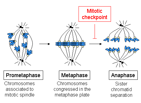
- The spindle assembly checkpoint is a critical surveillance mechanism operational during metaphase. It ensures that all chromosomes are correctly attached to the spindle fibers and are poised for equal segregation.
- This checkpoint is of paramount importance as any aberration in chromosome attachment or alignment can lead to aneuploidy, a state where cells possess an abnormal number of chromosomes. Such anomalies during meiosis can culminate in non-viable offspring or birth defects, while during mitosis, they can predispose cells to oncogenic transformation.
- The metaphase stage is not merely a passive alignment of chromosomes. It is a dynamic equilibrium maintained by opposing forces exerted by spindle fibers attached to kinetochores on either side of the chromosome.
- This tug-of-war ensures that chromosomes remain centrally positioned until the onset of anaphase, where sister chromatids are pulled apart to opposite poles, paving the way for the culmination of cell division.
- In summary, metaphase is a critical juncture in cell division, ensuring the fidelity of chromosome segregation. The intricate interplay between chromosomes and the microtubule network, coupled with rigorous checkpoint mechanisms, underscores the precision and intricacy of cellular processes that sustain life.
Definition of Metaphase
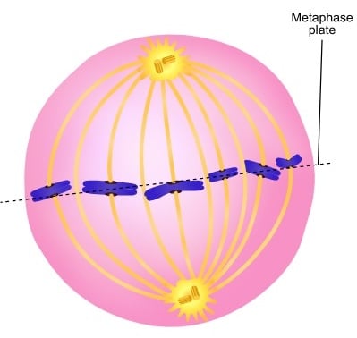
Metaphase is a stage in eukaryotic cell division where chromosomes align at the central plane, known as the metaphase plate, facilitated by spindle fibers, ensuring precise chromosome segregation in subsequent phases.

Key features of Metaphase
- Chromosomal Condensation: During metaphase, chromosomes achieve their highest level of condensation, rendering them distinctly visible under a microscope.
- Nuclear Envelope Disintegration: The nuclear envelope fully dissolves, allowing chromosomes to reside within the cytoplasm.
- Cohesin Complex: Sister chromatids remain conjoined at the centromere due to a protein complex known as cohesin.
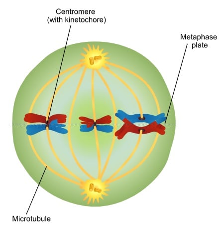
- Kinetochore Attachment: A multiprotein structure, the kinetochore, is affixed to each centromere. Microtubules establish a connection with these kinetochores, facilitating chromosome movement.
- Bipolar Attachment: Each sister chromatid is tethered to kinetochore microtubules originating from diametrically opposite poles.
- Chromosomal Alignment: The tug-of-war exerted by microtubules ensures that sister chromatids oscillate until they achieve a central alignment along the metaphase or equatorial plate.
- Equal Segregation Assurance: Metaphase plays a pivotal role in guaranteeing that subsequent cell division results in an equitable distribution of chromosomes to the progeny cells.
- Karyotyping: This stage is optimal for conducting karyotyping, a technique employed to detect chromosomal anomalies and for research purposes.
- Colchicine’s Role: Derived from Colchicum autumnale, colchicine is an alkaloid that halts cell division at metaphase by disrupting spindle fiber formation.
- Transition to Anaphase: Concluding metaphase, the cell progresses to anaphase, marked by the partitioning of sister chromatids, which then migrate to opposing cell poles.
In essence, metaphase is a meticulously orchestrated stage in cell division, ensuring the fidelity of chromosomal distribution and laying the foundation for subsequent phases.
What happens in metaphase?
In metaphase, a critical phase of cell division, the nucleus of the parent cell prepares to distribute its duplicated genetic material equally into two daughter cells. This phase is characterized by several meticulously orchestrated events:
- Chromosomal Alignment: The chromosomes, each consisting of two sister chromatids connected at the centromere, align themselves at the cell’s center. This alignment is a result of dynamic forces, akin to a cellular “tug of war,” ensuring precise positioning.
- Kinetochore Formation: Preceding metaphase, during prophase, specialized structures called kinetochores form around the centrosome. These kinetochores play a pivotal role in chromosome movement.
- Microtubule Attachment: Extending from the poles at either end of the cell are long protein filament microtubules, termed kinetochore microtubules. These microtubules establish connections with the kinetochores.
- Chromosomal Oscillation: The kinetochore microtubules exert forces on the sister chromatids, pulling them in opposing directions. This results in a back-and-forth movement until the chromosomes achieve a central alignment, forming an equatorial plane.
- Spindle Assembly Checkpoint: A crucial surveillance mechanism, the spindle assembly checkpoint, operates during metaphase. It assesses the proper alignment of chromosomes and the correct attachment of kinetochores to microtubules. This checkpoint ensures the cell’s readiness for the subsequent phase of division.
- Transition to Anaphase: Once the spindle assembly checkpoint confirms the correct alignment and attachment, the cell progresses to the next phase, anaphase, where the sister chromatids are separated and moved to opposite poles.
It’s worth noting that while mitosis and meiosis differ in their chromosomal alignment patterns, the spindle assembly checkpoint is imperative in both cell division processes to ensure genomic integrity.
In essence, metaphase is a phase marked by meticulous chromosomal alignment and rigorous checkpoint mechanisms, setting the stage for the subsequent phases of cell division.
Metaphase of Mitosis

Metaphase is a central phase in the mitotic cell division cycle, characterized by the alignment of chromosomes within the cellular environment. This alignment is crucial for ensuring the accurate distribution of genetic material into the daughter cells. Here are the key events and features of metaphase in mitosis:
- Chromosomal Condensation: Prior to metaphase, during prophase, chromosomes undergo a condensation process. This is vital to safeguard the integrity of the chromosomes from potential damage during the dynamic movements of metaphase.
- Nuclear Membrane Dissolution: As metaphase commences, the nuclear membrane disintegrates, leading to the dispersion of chromosomes within the cytoplasm.
- Microtubule Attachment: Radial microtubules, formed during the preceding phases, connect to each chromosome. Specifically, microtubules from opposing centrosomes establish connections with each chromosome, which comprises two identical sister chromatids.
- Role of Cohesins: These sister chromatids remain bound together at their centromeres by protein complexes termed cohesins. This connection ensures that the chromatids move in unison during metaphase.
- Dynamic Movement: The kinetochore microtubules play a pivotal role in the dynamic movement of chromosomes. They exert forces, both pulling and pushing, causing the chromatids to oscillate until they align at the cell’s equatorial region, forming the metaphase plate.
- Spindle Assembly Checkpoint: A critical surveillance mechanism, the spindle assembly checkpoint, operates during metaphase. It assesses the proper alignment of chromosomes and the correct attachment of kinetochores to microtubules. Only upon satisfying this checkpoint does the cell progress to the subsequent phase.
- Transition to Anaphase: Once the spindle assembly checkpoint is met, chromosomes emit a signal that activates the anaphase-promoting complex. This marks the culmination of metaphase and heralds the onset of anaphase, where sister chromatids are separated and move to opposite poles of the cell.
In essence, metaphase in mitosis is a meticulously orchestrated phase ensuring the precise and equal distribution of genetic material. The alignment of chromosomes at the metaphase plate is a testament to the cell’s commitment to genetic fidelity, paving the way for the creation of two genetically identical daughter cells.
Metaphase of Meiosis
Metaphase I in Meiosis

Meiosis is a specialized form of cell division that results in the production of haploid cells from a diploid precursor. Within this process, Metaphase I stands as a pivotal stage, distinguished by its unique chromosomal arrangements and subsequent segregation. Here are the salient features of Metaphase I in meiosis:
- Diploid to Haploid Transition: Meiosis I facilitates the transition of a cell from diploid, housing two sets of chromosomes, to haploid, containing a single set. This reduction is essential for sexual reproduction, ensuring offspring inherit a complete set of chromosomes when gametes fuse.
- Homologous Chromosome Pairs: Prior to Metaphase I, each chromosome, having undergone DNA replication, exists as sister chromatids. These chromatids pair up with their homologous counterparts, representing the same genomic regions but potentially different genetic variants or alleles.
- Alignment at the Metaphase Plate: Unlike mitosis, where individual chromosomes line up at the metaphase plate, Metaphase I in meiosis witnesses the alignment of these homologous pairs. This arrangement is crucial for the subsequent segregation of these pairs into separate cells.
- Kinetochore Microtubule Attachment: For each homologous pair, kinetochore microtubules emanating from opposite poles of the cell attach to the kinetochores of each chromosome. This attachment is vital for the ensuing separation of these pairs.
- Meiotic Spindle Checkpoint: A surveillance mechanism, the meiotic spindle checkpoint, ensures that all homologous pairs are correctly aligned and attached to microtubules from both poles. Only upon satisfying this checkpoint does the cell progress to the subsequent phase, Anaphase I.
- Significance for Genetic Diversity: The alignment and subsequent separation of homologous pairs during Metaphase I play a pivotal role in generating genetic diversity. The potential for different combinations of maternal and paternal chromosomes in the resulting gametes lays the foundation for varied genetic makeup in offspring.
- Consequences of Errors: Proper alignment and separation during Metaphase I are paramount. Any aberrations can lead to gametes with an incorrect chromosome number, potentially resulting in non-viable offspring or those with genetic disorders.
In summary, Metaphase I in meiosis is a meticulously orchestrated stage, ensuring the accurate segregation of homologous chromosome pairs. Its successful execution is vital for producing genetically diverse and viable gametes, underpinning the continuity of life across generations.
Metaphase II in Meiosis
Metaphase II stands as a pivotal juncture in the meiotic process, marking the second phase of meiosis II. Herein, we delve into the intricacies and significance of this stage:
- Initiation of Metaphase II: Following the brief interlude termed ‘interkinesis’, the cells re-enter the divisional phase. Notably, this resumption of division occurs without any preceding DNA replication, ensuring that each cell retains two copies of a singular allele for every gene.
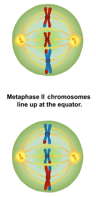
- Chromosomal Arrangement: As Metaphase II commences, the nuclear envelope undergoes dissolution. In contrast to Metaphase I, where homologous pairs were the focal point, Metaphase II centers on the sister chromatids. These chromatids, devoid of their homologous counterparts, align themselves on the metaphase plate.
- Kinetochore and Spindle Fibers: A distinguishing feature of Metaphase II is the interaction between kinetochores and spindle fibers. Specifically, the two kinetochores of each centromere bind to spindle fibers emanating from opposing poles. This arrangement is preparatory for the subsequent separation of sister chromatids.
- Meiotic Spindle Checkpoint: As with other phases of cell division, Metaphase II is governed by regulatory checkpoints. The meiotic spindle checkpoint ensures the correct alignment and attachment of chromosomes before permitting progression to the subsequent phase.
- Outcome of Metaphase II: Upon successful completion of Metaphase II, the stage is set for the separation of sister chromatids. This separation, which occurs in the ensuing anaphase, culminates in the formation of four distinct cells, each harboring a unique allele for every gene.
In essence, Metaphase II in meiosis serves as a bridge, transitioning from the initial separation of homologous chromosomes in Meiosis I to the final segregation of sister chromatids. This phase, underpinned by precise molecular interactions and regulatory checkpoints, ensures the generation of genetically diverse gametes, a cornerstone of sexual reproduction.

Metaphase in cytogenetics and cancer studies
Metaphase, a pivotal stage in cell division, holds paramount significance in the realms of cytogenetics and oncology. The unique attributes of chromosomes during this phase render them ideal for meticulous examination, facilitating profound insights into genetic structures and anomalies.
- Optimal Visualization in Metaphase: Chromosomes undergo pronounced thickening and coiling during metaphase, reaching an optimal state for visual scrutiny. This condensed state forms the archetype of chromosomes, commonly depicted in karyotypes.
- Classical Cytogenetic Analyses: Cells, when cultured for short durations, are arrested in the metaphase phase using mitotic inhibitors. Subsequently, these cells are employed for slide preparation, followed by chromosome banding or staining. Techniques such as Giemsa (G banding) or Quinacrine staining produce intricate patterns, revealing up to several hundred bands on chromosomes. Such detailed banding patterns are instrumental in discerning chromosomal structures and enumerations, forming the bedrock of karyotyping.
- Advanced Techniques and Metaphase: Normal metaphase spreads are integral to advanced methodologies like Fluorescence In Situ Hybridization (FISH) and serve as a foundational matrix for Comparative Genomic Hybridization (CGH) experiments.
- Metaphase in Cancer Studies: Malignant cells, whether sourced from solid tumors or leukemia samples, can be harnessed to produce metaphase preparations. A meticulous examination of these stained metaphase chromosomes unveils both numerical and structural aberrations within the tumor cell genome. Such anomalies can manifest as chromosomal segment losses or even translocations. A quintessential example is the emergence of chimeric oncogenes, such as bcr-abl, observed in chronic myelogenous leukemia. These genomic alterations, discernible through metaphase chromosome inspection, are pivotal in understanding the genetic underpinnings of various cancers.
- Applications in Clinical Diagnostics: Agents like Colcemid, when introduced during the metaphase, are instrumental in lymphocyte karyotyping, facilitating comprehensive chromosomal analyses. This is also applicable for amniotic fluid cell chromosome analysis, underscoring the clinical relevance of metaphase studies.
In summation, the metaphase stage, with its distinctive chromosomal attributes, serves as a linchpin in cytogenetic and cancer research. Through meticulous analyses of metaphase chromosomes, researchers can unravel the intricate tapestry of genetic information, paving the way for advanced diagnostic and therapeutic interventions in oncology.
Applications of Metaphase
Metaphase, a critical phase in cell division, offers a unique window into the intricacies of the cellular machinery and has profound implications in the realm of genetic analysis. Herein, we explore the pivotal applications of this stage:
- Karyotyping for Genetic Abnormalities: Metaphase serves as an opportune moment for the technique of karyotyping, a method employed to visualize and analyze the number, size, and shape of an individual’s chromosomes. Given that chromosomes are most condensed and distinctly visible during metaphase, it becomes feasible to detect structural and numerical chromosomal aberrations, aiding in the diagnosis of genetic disorders.
- Insight into Chromosomal Crossovers: During meiosis, metaphase is the juncture where chromosomal crossovers, or genetic recombination, transpire. This process is fundamental for generating genetic diversity within offspring. By studying these crossover events during metaphase, researchers can gain insights into the mechanisms of genetic variation and inheritance patterns.
- Understanding Chromosomal Damage: The dynamic forces exerted by kinetochore microtubules on chromatids during metaphase can, at times, lead to chromosomal damage, especially if the cell’s regulatory checkpoints, namely the mitotic and meiotic spindle checkpoints, are bypassed or malfunction. By examining cells in metaphase, scientists can identify and understand the consequences of such damages, which might have implications in conditions like cancer or birth defects.
In summary, the metaphase stage of cell division, beyond its fundamental role in cellular reproduction, serves as a crucial platform for genetic investigations. By harnessing the insights gleaned from this phase, researchers and clinicians can advance our understanding of genetics, paving the way for improved diagnostic and therapeutic strategies.
Metaphase in Mitosis and Meiosis Video
Difference between Metaphase 1 and Metaphase 2
| Topic | Metaphase 1 | Metaphase 2 |
| Origin | Metaphase 1 is associated with meiosis 1. | Metaphase 2 is associated with meiosis 2. |
| Arrangement of Chromosomes | Tetrads are arranged at the metaphase equator. | Single chromosomes are arranged at the metaphase equator. |
| Attachment of Chromosomes | Microtubules of one pole are attached to kinetochores of one of the two chromosomes facing to the same pole. | Microtubules are attached to the kinetochores of the centromere on either side of a single chromosome |
| Result | Single chromosomes move towards the opposing poles at anaphase 1. | One pair of sister chromatids move towards the opposing poles at anaphase 2. |
| Metaphase Plate | The metaphase plate is arranged in equidistant to the opposing poles. | The metaphase plate rotates 90 degrees compared to metaphase 1. |
Difference between metaphase of mitosis and metaphase 1 of meiosis
| Topic | Metaphase of mitosis | Metaphase I of meiosis |
| Definition | In mitosis, there is only one division and it produces two daughter cells. Involves one cell division and consequences in two daughter cells. | In meiosis there are two successive divisions, ultimately producing four daughter cells. Involves two successive cell divisions and consequences in four daughter cells |
| Chromosomes division | In the phase, centromeres of the chromosomes undergo division. | Centromeres of chromosome remain undivided. |
| Centromeres arranged | Centromeres are arranged along the equational plane and Chromosomes do not form any loop. | Centromeres remain a little bit any from the equational plane and Chromosomes form loops. |
| Crossing over | The nature of chromosomes remains unchanged as crossing over does not occur. | Crossing over occurs in the substage pachytene and the nature of chromosome remain unchanged |
| Steps | It has only one step. In mitosis, homologous chromosomes pair with each other (i.e., they form tetrads) and crossing-over never occur. | Metaphase of meiosis has two steps such as metaphase-1 and metaphase-2. In meiosis, homologous chromosomes pair with each other (i.e., they form tetrads) and crossing-over occur. |
| Chromosomes Number | Single chromosomes with two chromatids each, line up on the metaphase plate. | Paired chromosomes (bivalents) with four chromatids in each pair line up. |
| Chromosomes align | In metaphase of mitosis, individual chromosomes align there. | In metaphase I of meiosis, tetrads align on the metaphase plate. |
| Daughter cells | Results in diploid daughter cells (chromosome number remains the same as parent cell) and daughter cells are genetically identical. | Results in haploid daughter cells (chromosome number is halved from the parent cell) and daughter cells are genetically different. |
| Occurs | Occurs in all organisms except viruses and creates all body cells (somatic) apart from the germ cells (eggs and sperm) | Occurs only in animals, plants, and fungi and creates germ cells (eggs and sperm) only |
| Duration of Prophase | Prophase is much shorter and no recombination/crossing over occurs in prophase. | Prophase I takes much longer and involves recombination/crossing over of chromosomes in prophase I |
| line up of chromosomes | In metaphase individual chromosomes (pairs of chromatids) line up along the equator. | In metaphase, I pairs of chromosomes line up along the equator. |
| Sister chromatids | During anaphase, the sister chromatids are separated to opposite poles. | During anaphase I the sister chromatids move together to the same pole. |
Quiz
During which phase of cell division are chromosomes most condensed and aligned at the center of the cell?
a) Prophase
b) Metaphase
c) Anaphase
d) Telophase
In which phase of meiosis do homologous pairs of chromosomes align at the metaphase plate?
a) Metaphase I
b) Metaphase II
c) Anaphase I
d) Anaphase II
Which protein complex holds sister chromatids together at the centromere during metaphase?
a) Actin
b) Tubulin
c) Cohesin
d) Dynein
What is the name of the plate where chromosomes align during metaphase?
a) Centrosome Plate
b) Chromosomal Plate
c) Metaphase Plate
d) Kinetochore Plate
Which complex is responsible for the attachment of microtubules to chromosomes during metaphase?
a) Centrosome
b) Centriole
c) Kinetochore
d) Spindle
Which drug is known to arrest cell division at the metaphase stage by interfering with spindle fiber formation?
a) Aspirin
b) Colchicine
c) Penicillin
d) Ibuprofen
During metaphase II of meiosis, what aligns at the metaphase plate?
a) Homologous pairs of chromosomes
b) Sister chromatids
c) Non-sister chromatids
d) Centrosomes
Which checkpoint ensures that all chromosomes are properly attached to the spindle microtubules during metaphase?
a) G1 checkpoint
b) S checkpoint
c) G2 checkpoint
d) Spindle assembly checkpoint
Which phase immediately follows metaphase in mitosis?
a) Prophase
b) Telophase
c) Anaphase
d) Interphase
In which phase of the cell cycle do chromosomes decondense after metaphase?
a) Prophase
b) Telophase
c) Anaphase
d) Interphase
FAQ
What is metaphase?
Metaphase is a stage in cell division, both in mitosis and meiosis, where chromosomes align at the center of the cell before they are separated into daughter cells.
Why is metaphase important?
Metaphase ensures that chromosomes are properly aligned and attached to spindle fibers, which is crucial for the equal distribution of genetic material to the daughter cells.
How is metaphase different in mitosis and meiosis?
In mitosis, individual chromosomes align at the metaphase plate. In meiosis I, homologous pairs of chromosomes align, while in meiosis II, individual chromosomes align similarly to mitosis.
What follows after metaphase in the cell cycle?
After metaphase, the next phase is anaphase, where the chromosomes or chromatids are pulled apart to opposite ends of the cell.
How can one identify a cell in metaphase under a microscope?
A cell in metaphase will show chromosomes aligned at the center of the cell, appearing as a line or plate of distinct structures.
What role do kinetochores play during metaphase?
Kinetochores are protein complexes located at the centromere of each chromosome. They serve as attachment points for spindle fibers, ensuring proper alignment and separation of chromosomes.
Why is the spindle assembly checkpoint crucial during metaphase?
The spindle assembly checkpoint ensures that all chromosomes are properly attached to spindle fibers. This prevents premature separation and ensures equal distribution of chromosomes to daughter cells.
What happens if a cell fails the spindle assembly checkpoint during metaphase?
If a cell fails this checkpoint, it may not proceed to anaphase. This can lead to cell cycle arrest, cell death, or errors in chromosome separation, which can result in diseases or disorders.
How does metaphase contribute to genetic diversity during meiosis?
During metaphase I of meiosis, homologous chromosomes align at the metaphase plate. This alignment is random, leading to different combinations of maternal and paternal chromosomes in gametes, contributing to genetic diversity.
Are there any medical applications of studying cells in metaphase?
Yes, karyotyping is a technique where chromosomes in metaphase are stained and examined to detect chromosomal abnormalities. It’s used in diagnosing genetic disorders and certain cancers.
References
- https://www.genome.gov/genetics-glossary/Metaphase
- https://www.sparknotes.com/biology/cellreproduction/mitosis/section2/
- https://www.britannica.com/science/metaphase
- https://www.sciencedirect.com/topics/biochemistry-genetics-and-molecular-biology/metaphase
- https://www.nature.com/scitable/definition/metaphase-249/
- https://www.biologyonline.com/dictionary/metaphase-ii
- http://www.macroevolution.net/metaphase-ii.html
- https://pediaa.com/difference-between-metaphase-1-and-2/
- https://www.vedantu.com/question-answer/distinguish-between-metaphase-of-mitosis-and-class-12-biology-cbse-5f70e8fec8f93c434adb7ada
- https://qsstudy.com/biology/difference-metaphase-mitosis-metaphase-1-meioses

