Table of Contents
What is Muscular Tissue?
- Muscular tissue, also known as muscle tissue, is a specialized type of tissue found in animals that plays a vital role in generating force and facilitating movement within the body. It is composed of thin and elongated cells called muscle fibers, which possess the unique ability to contract and relax. This contractile property allows muscular tissue to exert forces on different parts of the body, enabling various body movements and functions.
- Muscle fibers contain cytoplasm, which is referred to as sarcoplasm. Within the sarcoplasm, there is a network of membranes called the sarcoplasmic reticulum. This structure is responsible for storing and releasing calcium ions, which are essential for muscle contraction. Surrounding the muscle fibers is a membrane called the sarcolemma, which helps maintain the integrity of the muscle cell and regulates the exchange of substances between the muscle fiber and its surrounding environment.
- Muscular tissue is a soft tissue and is categorized into three main types: skeletal or striated muscle, smooth muscle, and cardiac muscle. Skeletal muscle tissue consists of elongated muscle cells known as muscle fibers. These muscle fibers are responsible for the voluntary movements of the body, such as walking, running, and lifting objects. Skeletal muscle tissue is also associated with tendons and the perimysium, a connective tissue sheath that surrounds and supports the muscle fibers.
- Smooth muscle, also known as non-striated muscle, is found in the walls of hollow organs, blood vessels, and other structures within the body. Unlike skeletal muscle, smooth muscle contracts involuntarily, without conscious control. It is activated through various mechanisms, including the interaction of the central nervous system, peripheral plexus, or hormonal stimulation. Smooth muscle plays a crucial role in the movement and control of substances within the body, such as food through the digestive system or blood through blood vessels.
- Cardiac muscle is a specialized type of muscle tissue that forms the walls of the heart. It possesses characteristics of both skeletal and smooth muscle, combining the striated appearance of skeletal muscle with the involuntary contraction of smooth muscle. Cardiac muscle is responsible for the rhythmic contractions of the heart, which pump blood throughout the body.
- Muscle tissue contains specific contractile proteins called actin and myosin. These proteins interact to generate the force necessary for muscle contraction. Additionally, muscle tissue includes regulatory proteins like troponin and tropomyosin, which regulate the interaction between actin and myosin, controlling the initiation and termination of muscle contractions.
- The response of muscle tissue to various neurotransmitters and hormones, such as acetylcholine, noradrenaline, adrenaline, and nitric oxide, depends on the specific muscle type and its location within the body. Different muscle types have varying sensitivities and responses to these chemical signals, which contribute to their functional diversity.
- Muscle tissue can be further categorized based on factors like myoglobin content, presence of mitochondria, and myosin ATPase activity. These factors influence the muscle’s oxidative capacity, energy production, and contractile properties.
- In summary, muscular tissue is a specialized type of tissue found in animals that allows for movement and the application of forces within the body. It consists of muscle fibers, which contract and relax to generate the necessary forces. Muscular tissue is categorized into skeletal, smooth, and cardiac muscle, each serving distinct functions in the body. Actin, myosin, and regulatory proteins control muscle contraction, while neurotransmitters and hormones play a role in muscle activation.
Definition of Muscular Tissue
Muscular tissue is a specialized type of tissue found in animals that consists of muscle fibers capable of contracting and generating force, allowing for movement and control of body functions.
Properties/Characteristics of Muscular Tissue
Muscular tissue possesses several distinct properties that enable its functions and contribute to efficient movement and physiological processes.
- Contractibility: Muscular tissue has the remarkable ability to contract, or shorten forcefully. This property allows muscles to generate the necessary force for movement and perform various functions within the body. When muscle fibers receive a signal from a motor neuron or hormone, they undergo a contraction, resulting in the shortening of the muscle.
- Excitability: Muscular tissue exhibits excitability, which means it can respond to stimuli. When stimulated by a motor neuron or certain hormones, the muscle tissue initiates a response, leading to muscle contraction. This responsiveness to external signals is crucial for coordinated movement and proper functioning of the muscular system.
- Extensibility: Muscles also possess the property of extensibility, allowing them to be stretched without damage. This feature is essential for flexibility and range of motion. For example, when performing exercises or stretching routines, muscles can be extended, enabling movement across joints and facilitating the maintenance of muscle tone and overall muscle health.
- Elasticity: Muscular tissue demonstrates elasticity, which refers to its ability to recoil or return to its original length and shape after being stretched. This elasticity enables muscles to resume their resting position efficiently. It ensures that muscles can contract and relax effectively, contributing to smooth and coordinated movements.
- Highly vascularized: Muscular tissue is richly supplied with blood vessels, ensuring a robust blood flow to the muscles. This vascularization allows for the delivery of oxygen and nutrients required for muscle contraction and metabolism. Additionally, it facilitates the removal of waste products, such as carbon dioxide and lactic acid, which are byproducts of muscle activity.
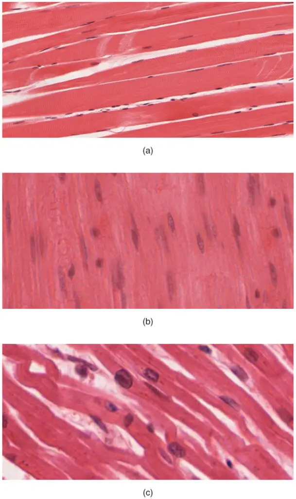
- Striated or smooth appearance: Under a microscope, muscular tissue can exhibit a striated or smooth appearance, depending on its type. Skeletal and cardiac muscle tissues are striated due to the arrangement of contractile proteins within their fibers, while smooth muscle tissue lacks this striated pattern. The presence or absence of striations is a characteristic feature used to distinguish between different types of muscular tissue.
The properties of muscular tissue collectively enable its role in movement, stability, posture, organ function, and other physiological processes. By contracting and relaxing, muscles generate forces required for locomotion and provide support to the skeletal system. They also contribute to the maintenance of body temperature and play a vital role in organ function, including the pumping action of the heart and the movement of substances through the digestive and urinary systems.
Structure of Muscular Tissue
The structure of muscular tissue is organized in a way that allows for its specialized functions and the generation of force for movement. Here are some key aspects of the structure of muscular tissue:
- Epimysium: Muscular tissue is bundled together and surrounded by a tough connective tissue called the epimysium. This layer provides structural support and protection to the muscle.
- Fascicles: Within the epimysium, the muscle fibers are grouped into bundles called fascicles. These fascicles consist of long fibers that run parallel to each other and contribute to the overall muscle structure and function.
- Perimysium: Each fascicle is surrounded by a layer of connective tissue known as the perimysium. This layer contains blood vessels and nerves that supply the individual muscle fibers within the fascicle.
- Endomysium: The muscle fibers within a fascicle are further surrounded and supported by a protective layer called the endomysium. It provides a suitable environment for the muscle fibers and allows for the exchange of nutrients and waste products.
- Sliding Filament Mechanism: The individual muscle fibers within the fascicles contain contractile units called sarcomeres. These sarcomeres are responsible for muscle contraction and give the muscle its striated appearance. During contraction, the actin and myosin filaments within the sarcomeres slide past each other, resulting in the shortening of the muscle fiber.
- Tendons: The epimysium extends beyond the muscle fibers and connects to tendons. Tendons are dense connective tissues that attach muscles to bones or other structures. They play a crucial role in transmitting the forces generated by the muscle to produce movement.
- Types of Muscular Tissue: In vertebrates, there are three main types of muscular tissue: skeletal, cardiac, and smooth. Skeletal muscle is typically attached to bones and is under voluntary control. Cardiac muscle is found in the walls of the heart and is responsible for its rhythmic contractions. Smooth muscle is found in the walls of various organs and structures and is involved in involuntary movements, such as those of the digestive system or blood vessels.
The structure of muscular tissue is intricately organized to allow for effective muscle contraction, transmission of forces, and the performance of specific functions depending on the type of muscle tissue. These structural adaptations contribute to the diverse range of movements and physiological processes that muscular tissue facilitates in the body.
Types of muscle tissue
The muscular tissue is of three types:
- Skeletal Muscle Tissue
- Smooth Muscle Tissue
- Cardiac Muscle Tissue

1. Skeletal Muscle Tissue
Skeletal muscle tissue, also known as voluntary muscle tissue, plays a vital role in the movement of our body. Here are some key characteristics and features of skeletal muscle tissue:
- Attachment to the Skeleton: Skeletal muscles are connected to the skeleton through tendons and help facilitate movement by pulling on the bones.
- Striated Appearance: Skeletal muscles have a distinct striped or striated appearance due to the arrangement of alternating light and dark bands within the muscle fibers. These bands are known as sarcomeres and contribute to the muscle’s contractile properties.
- Actin and Myosin Organization: Sarcomeres within skeletal muscle tissue contain highly organized structures of proteins, including actin and myosin. These proteins are responsible for muscle contraction and contribute to the muscle’s ability to generate force.
- Voluntary Control: Skeletal muscles are under voluntary control, meaning we have conscious control over their contraction and relaxation. This allows us to perform specific movements and adjust muscle activity as needed.
- Multinucleated Cells: Skeletal muscle fibers are multinucleated, meaning they contain multiple nuclei within a single muscle cell. This helps support the high energy demands and protein synthesis required for muscle function.
- Abundance and Mass: Skeletal muscles make up a significant portion of our body mass, accounting for approximately 40% of our total body weight. This highlights the importance of skeletal muscle in overall body function and movement.
- Oxygen Supply: Skeletal muscle tissue is well-supplied with blood vessels, ensuring a steady delivery of oxygen and nutrients to the muscle fibers. This enables sustained muscle activity during aerobic exercise.
- Fiber Types: Skeletal muscle is classified into different fiber types, such as Type I (slow-twitch) and Type II (fast-twitch) muscles. These fiber types exhibit variations in contraction speed, endurance, and energy sources, allowing for a diverse range of muscle functions.
Skeletal muscle tissue is essential for various movements, ranging from simple everyday activities to complex athletic performances. Its voluntary nature and striated appearance reflect its unique properties and role in our body’s biomechanics. The ability to consciously control skeletal muscles enables precise movements and coordination, contributing to our overall physical capabilities.
Structure of Skeletal Muscle Tissue
The structure of skeletal muscle tissue is highly organized and consists of various components that contribute to its function. Here are some key features of the structure of skeletal muscle tissue:
- Muscle Fibers: Skeletal muscle tissue is composed of long, cylindrical muscle fibers. These fibers can vary in length and diameter but are generally around 1.2 inches long and 0.004 inches in diameter. Each muscle fiber contains multiple nuclei, which are necessary for protein synthesis and maintaining cellular function.
- Myofibrils: Within each muscle fiber, there are numerous myofibrils. Myofibrils are thread-like structures that run parallel to each other and extend the entire length of the muscle fiber. They are responsible for the muscle’s contractile properties.
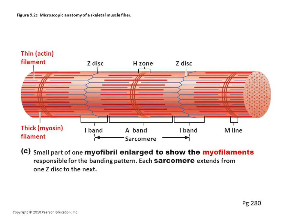
- Thick and Thin Filaments: Myofibrils are composed of thick and thin filaments. The thick filament is made up of larger proteins called myosin, while the thin filament consists of smaller proteins called actin. These filaments overlap with each other, creating the distinct pattern of alternating light and dark bands observed under a light microscope.
- A Bands and I Bands: The overlapping of thick myosin and thin actin filaments gives rise to the dark A bands (anisotropic bands) in the skeletal muscle tissue. These A bands do not allow light to pass through. The regions where only thin actin filaments are present create the light I bands (isotropic bands), which allow light to pass through.
- Z Lines and H Zone: Cutting across each I band is a dark Z line. Z lines act as anchor points for the actin filaments. Within the A band, there is a somewhat light H zone (Hensen’s disc), which consists only of myosin filaments. The H zone becomes narrower during muscle contraction.
- Sarcomeres: The area between two Z lines is known as a sarcomere. Sarcomeres are considered the fundamental contractile units of myofibrils. They represent the functional units responsible for muscle contraction. When muscles contract, sarcomeres shorten, leading to overall muscle fiber contraction.

The highly organized structure of skeletal muscle tissue allows for efficient force generation and coordinated muscle contractions. The arrangement of myosin and actin filaments, along with the presence of sarcomeres, provides the foundation for muscle contraction and the ability to generate movement in response to nerve signals.
Functions of Skeletal Muscle Tissue
Skeletal muscle tissue plays crucial roles in the movement and locomotion of the body. Here are some key functions of skeletal muscle tissue:
- Voluntary Movement: Skeletal muscles are under conscious control, allowing us to perform voluntary movements. They are responsible for various activities such as walking, running, lifting objects, and manipulating our environment with our hands. Skeletal muscles contract and relax in response to signals from the central nervous system (CNS) and peripheral nervous system (PNS).
- Locomotion: Skeletal muscles enable the movement of the body as a whole. By contracting and relaxing in a coordinated manner, skeletal muscles produce movements that allow us to change positions, maintain posture, and engage in physical activities.
- Fatigue and Recovery: Skeletal muscles are capable of powerful and rapid contractions, but they can also experience fatigue when subjected to prolonged or intense activity. Skeletal muscles have a limited endurance capacity and require rest periods to recover and replenish energy stores. Fatigue is a temporary decline in muscle performance due to the depletion of energy sources and the accumulation of metabolic byproducts.
- Energy Requirements: Skeletal muscles have high energy demands to support their contraction and movement. They are supplied with an extensive network of blood vessels that deliver oxygen and nutrients, including glucose, which is essential for energy production. Skeletal muscles also contain numerous elongated mitochondria, known as the powerhouses of the cell, where aerobic respiration takes place to generate ATP, the main energy source for muscle contraction. Additionally, skeletal muscles store glycogen, a form of glucose, which can be rapidly broken down to provide fuel during intense exercise.
- Distribution in the Body: Skeletal muscles are found throughout the body, attached to various structures. They are present in the head, trunk, and limbs, as well as in the body wall, tongue, pharynx, and esophagus. These muscles work together to provide the necessary force and movement required for different bodily functions.
- Nervous System Control: Skeletal muscle contractions are stimulated by electrical impulses transmitted by motor nerves originating from the CNS. When a signal is received from the nervous system, skeletal muscle fibers contract, resulting in movement. The neurotransmitter acetylcholine plays a vital role in facilitating skeletal muscle contractions.
Overall, skeletal muscle tissue is responsible for voluntary movement, locomotion, and maintaining posture. It relies on the coordination of the nervous system, a continuous supply of energy, and appropriate recovery periods to perform its functions effectively.
2. Cardiac Muscle Tissue
Cardiac muscle tissue, also known as myocardium, is a specialized type of muscle tissue that is unique to the heart. Here are some key features and functions of cardiac muscle tissue:
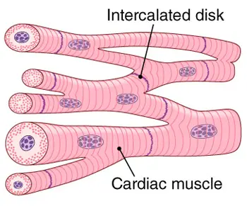
- Location and Structure: Cardiac muscle tissue is exclusively found in the walls of the heart, specifically in the myocardium. It forms a thick middle layer between the outer epicardium layer and the inner endocardium layer. Cardiac muscle cells, known as cardiomyocytes, are elongated and branched in structure, allowing them to interconnect with each other.
- Involuntary and Striated: Like skeletal muscle, cardiac muscle tissue is striated, meaning it exhibits alternating light and dark bands under a microscope. However, unlike skeletal muscle, cardiac muscle is involuntary, meaning it functions without conscious control. The contraction of cardiac muscle is regulated by specialized cardiac conducting cells and the autonomic nervous system.
- Unique Features of Cardiomyocytes: Cardiomyocytes have a single centrally located nucleus, distinguishing them from the multinucleated skeletal muscle fibers. These cells are interconnected through intercalated discs, which contain both anchoring junctions (desmosomes) and gap junctions. Desmosomes help maintain the structural integrity of the cardiac muscle, while gap junctions facilitate the rapid transmission of electrical impulses between cells, allowing for coordinated contraction of the heart.
- Contractions and Pumping Action: The main function of cardiac muscle tissue is to generate contractions that propel blood throughout the body. Coordinated contractions of the cardiac muscle cells in the atria and ventricles ensure the efficient pumping of blood into the systemic and pulmonary circulatory systems. This contraction and relaxation process is known as systole and diastole, respectively, and is responsible for maintaining the circulation of oxygenated and deoxygenated blood.
- Oxygen and Nutrient Supply: Cardiac muscle cells have high energy demands and rely on a continuous supply of oxygen and nutrients to function effectively. The coronary arteries, which branch off the aorta, provide the necessary blood supply to the myocardium. These arteries deliver oxygen and nutrients while removing waste products, such as carbon dioxide, from the cardiac muscle cells.
- Syncytium and Coordinated Contraction: Cardiac muscle cells form a functional syncytium, meaning they behave as a single unit. The intercalated discs allow the cells to synchronize their actions, enabling a rapid and coordinated contraction of the heart. This synchronization ensures efficient pumping of blood and maintains the rhythmicity of the heartbeat.
In summary, cardiac muscle tissue is uniquely structured and specialized for its role in the involuntary contraction of the heart. The interconnected cardiomyocytes, intercalated discs, and coordinated contractions contribute to the efficient pumping of blood throughout the body, providing the necessary oxygen and nutrients to support overall cardiovascular function.
Structure of Cardiac Muscle Tissue

Cardiac muscle tissue possesses a distinct structure that enables its unique function within the heart. Here are some key features of the structure of cardiac muscle tissue:
- Striations: When viewed under a microscope, cardiac muscle tissue exhibits striations similar to skeletal muscle tissue. The striations are caused by the arrangement of contractile proteins within the cells.
- Cardiomyocytes: The cells of cardiac muscle tissue are known as cardiomyocytes. These cells are closely packed together, forming a network within the myocardium. Unlike skeletal muscle fibers, each cardiomyocyte contains a single nucleus. However, populations of cardiomyocytes with two to four nuclei can also be found.
- Intercalated Discs: Cardiac muscle cells are joined end to end by specialized cell junctions called intercalated discs. These discs are crucial for the structural and functional integrity of cardiac muscle tissue. Intercalated discs contain three important components:
- Desmosomes: Desmosomes are anchoring junctions that connect neighboring cells. They provide mechanical strength and stability to the tissue.
- Gap Junctions: Gap junctions are small channels that allow for direct communication between adjacent cells. They facilitate the passage of ions and small molecules, enabling the synchronization of contractions among cardiomyocytes.
- Fascia Adherens: Fascia adherens are adhesion belts that link the actin filaments of adjacent cells. They contribute to the structural integrity of intercalated discs.
- I and A Bands: Similar to skeletal muscle, cardiac muscle cells exhibit light I bands and dark A bands. The intercalated discs are always located at the Z-line, which divides the sarcomeres. The alternating arrangement of the actin and myosin filaments within the sarcomeres contributes to the striations observed in cardiac muscle tissue.
- Nervous Supply: Cardiac muscle tissue is supplied by both the central nervous system (CNS) and the autonomous nervous system (ANS). The CNS provides innervation through motor nerves, while the ANS regulates the rate and force of contractions through sympathetic and parasympathetic pathways.
- Rhythmic Contractions: Cardiac muscle tissue exhibits an inherent rhythm of contraction. Unlike skeletal muscle, which requires external stimulation, cardiac muscle cells can initiate contractions on their own. This intrinsic property allows the heart to maintain a coordinated and rhythmic pumping action.
- Fatigue Resistance: Cardiac muscle tissue is highly resistant to fatigue. It continuously contracts and relaxes throughout a person’s lifetime without becoming exhausted. This fatigue resistance is essential for the heart’s continuous pumping action and ensures the delivery of oxygenated blood to the body’s tissues.
In summary, the structure of cardiac muscle tissue is characterized by closely packed cardiomyocytes that are interconnected by intercalated discs. These discs play a crucial role in synchronizing contractions and maintaining the structural integrity of the tissue. The presence of striations, rhythmic contractions, and fatigue resistance are key features that distinguish cardiac muscle from other types of muscle tissue in the body.
Functions of Cardiac Muscle Tissue
Cardiac muscle tissue serves vital functions in the heart, ensuring its continuous pumping action and maintaining proper circulation throughout the body. Here are some key functions of cardiac muscle tissue:
- Pumping Blood: The primary function of cardiac muscle tissue is to generate forceful contractions that propel blood throughout the circulatory system. As the heart contracts, the cardiac muscle tissue contracts in a coordinated manner, squeezing the chambers (atria and ventricles) and forcing blood to be pumped out of the heart.
- Rhythmic Contractions: Cardiac muscle tissue possesses the unique ability to initiate and sustain its own rhythmic contractions. This self-contracting property, also known as automaticity, allows the heart to beat in a regular and synchronized fashion without requiring external stimulation. The rhythmic contractions of cardiac muscle tissue maintain the heart’s pumping action, ensuring the continuous circulation of blood.
- Autonomic Regulation: While cardiac muscle tissue can contract spontaneously, its contractions are also influenced and regulated by the autonomic nervous system (ANS). The ANS consists of the sympathetic and parasympathetic divisions, which exert control over the rate and force of cardiac contractions. Sympathetic stimulation increases heart rate and contractility, while parasympathetic stimulation decreases heart rate. The autonomic regulation of cardiac muscle tissue helps maintain appropriate cardiovascular responses to various physiological demands.
- Endurance and Reliability: Cardiac muscle tissue is designed to contract repeatedly and rhythmically throughout an individual’s lifetime. Unlike other types of muscle tissue, cardiac muscle does not experience fatigue or become exhausted easily. This endurance and reliability ensure that the heart can sustain its pumping function without interruption, providing a continuous supply of oxygenated blood to the body’s tissues and organs.
- Synchronization: The intercalated discs present in cardiac muscle tissue play a crucial role in synchronizing the contractions of individual cardiac muscle cells. These specialized cell junctions allow for the rapid transmission of electrical impulses and ions between neighboring cells, facilitating coordinated contractions throughout the heart. This synchronization ensures that the heart chambers contract in a well-coordinated manner, optimizing the efficiency of blood ejection.
- Maintenance of Circulation: The continuous rhythmic contractions of cardiac muscle tissue help maintain blood circulation throughout the body. By contracting and relaxing in a coordinated pattern, cardiac muscle ensures that blood flows through the heart’s chambers, into the blood vessels, and reaches various tissues and organs. This constant circulation supplies oxygen, nutrients, and hormones while removing waste products such as carbon dioxide.
In summary, cardiac muscle tissue functions as the muscle of the heart, enabling it to pump blood and maintain circulation throughout the body. Its self-contracting nature, autonomic regulation, endurance, synchronization, and role in sustaining circulation are essential for the heart’s rhythmic contractions and its vital role in delivering oxygen and nutrients to tissues and organs.
3. Smooth Muscle Tissue
Smooth muscle tissue, also known as non-striated muscle, is an important type of muscle found in various organs and structures of the body. Here are some key points about smooth muscle tissue:
- Involuntary and Controlled by the Autonomous Nervous System: Smooth muscle tissue is under involuntary control, meaning it functions without conscious effort. It is regulated by the autonomic nervous system, specifically the sympathetic and parasympathetic divisions, which coordinate and modulate its contractions. The smooth muscle tissue plays a crucial role in the contractility of organs such as the digestive system, urinary system, reproductive system, blood vessels, and airways.
- Lack of Striations: Unlike skeletal and cardiac muscle tissue, smooth muscle tissue does not exhibit striations under a microscope. This is due to the arrangement of actin and myosin filaments, which are thin and randomly distributed within the cells. The absence of striations is one of the distinguishing characteristics of smooth muscle tissue.
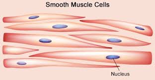
- Spindle-Shaped Cells with Single Nucleus: Smooth muscle cells, also called smooth muscle fibers or myocytes, have a distinct spindle shape. They are elongated with tapered ends, resembling a spindle or fusiform. Each smooth muscle cell contains a single centrally located nucleus, in contrast to skeletal muscle cells, which are multinucleated. The spindle shape of smooth muscle cells allows them to contract and relax in a coordinated manner.
- Types of Smooth Muscle: Smooth muscle can be classified into two main types: single-unit (unitary) and multiunit smooth muscle. In single-unit smooth muscle, the cells are connected by gap junctions, allowing them to function as a syncytium. This means that the entire bundle or sheet of smooth muscle contracts as a coordinated unit. Multiunit smooth muscle, on the other hand, consists of individual cells that are innervated separately. This arrangement provides fine control and allows for gradual responses in specific regions.
- Locations in the Body: Smooth muscle tissue is found in various locations throughout the body. It forms the walls of blood vessels (vascular smooth muscle) such as arteries, arterioles, and veins. Smooth muscle is also present in organs and structures such as the digestive tract, urinary bladder, uterus, male and female reproductive tracts, respiratory tract, arrector pili muscles in the skin, ciliary muscle in the eye, and the iris. The structure and function of smooth muscle cells remain similar across different organs, but the specific stimuli that induce contractions can vary, producing distinct effects in the body.
- Involuntary Movements and Contractions: Smooth muscle tissue plays a crucial role in the involuntary movements of internal organs. It provides the contractile component of organs involved in processes such as digestion, urination, reproduction, blood flow regulation, and airway constriction. The contractions of smooth muscle tissue are slow and sustained, allowing for the steady movement of substances and the regulation of organ function.
In summary, smooth muscle tissue is a non-striated, involuntary muscle type that is controlled by the autonomic nervous system. Its contractility is responsible for the movement and function of various organs and structures in the body, allowing for vital physiological processes to occur. The spindle-shaped cells, absence of striations, and diverse distribution within the body distinguish smooth muscle tissue from other muscle types.
Structure of Smooth Muscle Tissue

Smooth muscle tissue has a unique structure that sets it apart from other types of muscle tissue. Here are some key features of the structure of smooth muscle tissue:
- Muscle Fiber Shape: Smooth muscle fibers are elongated and spindle-shaped, but not as long as skeletal muscle fibers. They are slender and tapered at the ends. This shape allows smooth muscle tissue to fit into various organs and structures of the body.
- Single Nucleus: Each smooth muscle fiber contains a single nucleus, which is located at the center of the fiber at its broadest part. This is in contrast to skeletal muscle fibers, which are multinucleated.
- Sarcolemma and Myofibrils: Smooth muscle fibers are enclosed by a membrane called the sarcolemma, which surrounds the entire cell. Within the cell, numerous longitudinal myofibrils are present. These myofibrils are responsible for the contraction of the muscle fiber.
- Arrangement of Actin and Myosin: The actin and myosin myofilaments within the myofibrils of smooth muscle tissue are thinner compared to those in skeletal muscle tissue. Furthermore, their arrangement is more random, which results in the absence of visible stripes or striations. This is one of the distinguishing features of smooth muscle tissue.
- Characteristics of Contraction: Smooth muscle tissue exhibits two main characteristics during contraction. First, the contraction and relaxation periods are slower compared to skeletal muscle tissue. This slower contraction allows for sustained contractions, which are important for functions such as maintaining organ tone. Second, the action of smooth muscle tissue is rhythmic. Contraction can last for 30 seconds or more without fatigue. This sustained contraction ability, coupled with the ability to stretch, makes smooth muscle tissue suitable for controlling the movement and function of organs such as the stomach, intestines, urinary bladder, and uterus.
In summary, smooth muscle tissue has a distinctive structure characterized by spindle-shaped muscle fibers with a single nucleus. The arrangement of actin and myosin myofilaments is more random, resulting in the absence of striations. Smooth muscle tissue has slower and sustained contractions, making it well-suited for the controlled movement and function of organs involved in processes such as digestion, urination, and reproductive functions.
Functions of Smooth Muscle Tissue
Smooth muscle tissue serves several important functions in the body due to its presence in various organ systems. Here are some key functions of smooth muscle tissue:
- Regulation of Hollow Organ Contraction: Smooth muscle is found in the walls of hollow organs, including the stomach, bladder, uterus, and intestines. It plays a crucial role in regulating the contraction and relaxation of these organs, allowing for processes such as digestion, urine storage and release, and childbirth. The coordinated contractions of smooth muscle in these organs help propel substances, such as food or urine, through their respective passages.
- Control of Tubular Structures: Smooth muscle is present in tubular structures such as blood vessels, lymph vessels, and bile ducts. It contributes to the regulation of blood flow and pressure by adjusting the diameter of blood vessels, influencing circulation throughout the body. Smooth muscle in the walls of lymph vessels aids in the movement of lymph fluid. Additionally, smooth muscle in the bile ducts helps regulate the flow of bile from the liver to the intestine for digestion.
- Function in Sphincters: Sphincters are circular muscles that act as valves, controlling the flow of substances through openings in the body. Smooth muscle is essential for the function of various sphincters, such as the uterine cervix, which helps seal off the uterus during pregnancy, and the sphincter muscles in the eyes that regulate pupil size.
- Duct Regulation: Smooth muscle is involved in the regulation of ducts in exocrine glands. It helps control the release and flow of substances, such as saliva or pancreatic enzymes, from these glands into ducts for transportation or secretion.
- Slower and Sustained Contractions: Smooth muscle cells contract at a slower rate compared to skeletal muscle cells, but they have more prolonged contractions that can be sustained for extended periods. This sustained contraction is beneficial for functions such as maintaining organ tone and controlling the movement of substances through various passageways.
- Involuntary Control: Smooth muscle tissue is involuntary, meaning it is not under conscious control. Contractions of smooth muscle are regulated by the autonomic nervous system, allowing for automatic and involuntary responses without conscious effort.
In summary, smooth muscle tissue performs vital functions in the regulation of hollow organs, tubular structures, sphincters, and exocrine glands. It facilitates contractions and relaxations that control the movement of substances and contribute to the normal functioning of organ systems throughout the body.
Comparison of Structure and Properties of Muscle Tissue Types
| Muscle Type | Structural Elements | Function | Location |
|---|---|---|---|
| Skeletal | Long cylindrical fiber, striated, many peripherally located nuclei | Voluntary movement, produces heat, protects organs | Attached to bones and around entry & exit sites of body (e.g., mouth, anus) |
| Cardiac | Short, branched, striated, single central nucleus | Contracts to pump blood | Heart |
| Smooth | Short, spindle-shaped, no evident striation, single nucleus in each fiber | Involuntary movement, moves food, involuntary control of respiration, moves secretions, regulates flow of blood in arteries by contraction | Walls of major organs and passageways |
Functions of muscle tissue
Muscle tissue plays crucial roles in various physiological functions in the body. Here are some key functions of muscle tissue:
- Movement: Muscle tissue is responsible for generating the forces necessary for body movements. Skeletal muscles, attached to bones, contract and pull on them, allowing us to walk, run, lift objects, and perform various activities.
- Maintenance of Posture: Even when we are at rest, our muscles are actively engaged in maintaining posture and keeping our body upright. They provide the necessary support to keep us standing or sitting.
- Respiration: Muscles involved in respiration, such as the diaphragm and intercostal muscles, contract and relax to drive the movement of air into and out of our lungs.
- Heat Generation: Muscle contraction generates heat, which is essential for maintaining body temperature. When we are cold, our muscles may contract involuntarily, causing shivering, to generate heat and warm up the body.
- Communication: Muscles in the face, tongue, and throat allow us to communicate through speech, facial expressions, and gestures. They enable us to convey our thoughts, emotions, and intentions.
- Constriction of Organs and Blood Vessels: Smooth muscle tissue, found in the walls of organs and blood vessels, contracts and relaxes to facilitate various processes. It helps propel food through the digestive tract, expel urine from the bladder, move substances through ducts, and regulate blood pressure by constricting or dilating blood vessels.
- Pumping Blood: Cardiac muscle, specifically found in the heart, contracts rhythmically to pump blood throughout the body. It ensures the circulation of oxygen, nutrients, and waste products to and from the tissues.
- Protection of Organs and Body Cavities: Muscles play a crucial role in protecting delicate internal organs by enclosing and surrounding them. They provide structural support and help maintain the integrity of body cavities, preventing herniation or displacement of organs.
The functions of muscle tissue are diverse and essential for our everyday activities, maintaining homeostasis, and enabling proper organ and system functioning. The coordinated actions of different types of muscle tissue allow us to perform a wide range of movements and contribute to overall health and well-being.
Differences Between smooth muscle, cardiac muscle, skeletal muscle
Smooth Muscle | Cardiac Muscle | Skeletal Muscle | |
|---|---|---|---|
| Anatomy | |||
| Neuromuscular Junction | None | Present | Present |
| Fibers | Fusiform, short (<0.4 mm) | Branching | Cylindrical, long (<15 cm) |
| Mitochondria | Numerous | Many to few (by type) | Numerous |
| Nuclei | 1 | 1 | >1 |
| Sarcomeres | None | Present, max. length 2.6 µm | Present, max. length 3.7 µm |
| Syncytium | None (independent cells) | None (but functional as such) | Present |
| Sarcoplasmic Reticulum | Little elaborated | Moderately elaborated | Highly elaborated |
| ATPase | Little | Moderate | Abundant |
| Physiology | |||
| Self-regulation | Spontaneous action (slow) | Yes (rapid) | None (requires nerve stimulus) |
| Response to Stimulus | Unresponsive | “All-or-nothing” | “All-or-nothing” |
| Action Potential | Yes | Yes | Yes |
| Workspace | Force/length curve is variable | The increase in the force/length curve | At the peak of the force/length curve |
FAQ
What is muscular tissue?
Muscular tissue is a type of tissue in the body that is responsible for movement and contraction. It is composed of specialized cells called muscle fibers that have the ability to contract and relax.
How many types of muscular tissue are there?
There are three types of muscular tissue: skeletal muscle tissue, cardiac muscle tissue, and smooth muscle tissue.
What is the difference between skeletal, cardiac, and smooth muscle tissue?
Skeletal muscle tissue is attached to the bones and is responsible for voluntary movements. Cardiac muscle tissue is found in the heart and is responsible for involuntary contractions. Smooth muscle tissue is found in the walls of internal organs and blood vessels and is also involuntarily controlled.
What is the function of muscular tissue?
The main function of muscular tissue is to generate force and produce movement. It allows for locomotion, maintains posture, supports body structures, and enables the movement of substances within the body.
How do muscles contract?
Muscles contract through the interaction of actin and myosin, two types of proteins. When stimulated, myosin heads bind to actin filaments, causing them to slide past each other. This shortens the length of the muscle fiber and results in contraction.
Can muscle tissue regenerate?
Skeletal muscle tissue has a limited ability to regenerate and repair itself. Cardiac muscle tissue has a very limited regenerative capacity, while smooth muscle tissue has a higher regenerative potential.
How is muscular tissue supplied with oxygen and nutrients?
Muscular tissue is supplied with oxygen and nutrients through an extensive network of blood vessels. Capillaries deliver oxygen and nutrients to the muscle fibers and remove waste products such as carbon dioxide.
Can muscular tissue become fatigued?
Yes, muscular tissue can become fatigued, especially skeletal muscle tissue. Prolonged or intense muscle activity can deplete energy stores and result in muscle fatigue. However, smooth and cardiac muscle tissues have different fatigue properties.
What is the role of the nervous system in muscle function?
The nervous system plays a crucial role in muscle function. Motor neurons transmit electrical signals from the central nervous system to muscle fibers, initiating muscle contractions. The nervous system also regulates the coordination, strength, and timing of muscle contractions.
Can muscular tissue be affected by diseases or disorders?
Yes, muscular tissue can be affected by various diseases and disorders. Examples include muscular dystrophy, myasthenia gravis, muscle strains, and cardiac arrhythmias. These conditions can impair muscle function and lead to weakness, fatigue, or abnormal muscle contractions.
References
- https://www.visiblebody.com/learn/muscular/muscle-types
- https://open.oregonstate.education/aandp/chapter/4-4-muscle-tissue/
- https://content.byui.edu/file/a236934c-3c60-4fe9-90aa-d343b3e3a640/1/module7/readings/function_muscle_tissue.html
- https://www.kenhub.com/en/library/anatomy/muscles
- https://biologydictionary.net/muscle-tissue/
- http://histologyguide.com/slidebox/04-muscle-tissue.html
- https://www.lecturio.com/concepts/types-of-muscle-tissue/
- https://courses.lumenlearning.com/suny-dutchess-ap1/chapter/introduction-to-muscle-tissue/
- https://www.biologyonline.com/dictionary/muscle-tissue
- https://www.geeksforgeeks.org/muscular-tissue-structure-functions-types-and-characteristics/
- https://openstax.org/books/anatomy-and-physiology-2e/pages/10-1-overview-of-muscle-tissues
- https://www.onlinebiologynotes.com/muscular-tissue-skeletal-smooth-cardiac-muscle/
- https://www.toppr.com/guides/biology/structural-organisation-in-animals/muscular-tissue/


