Table of Contents
The earthworm, a common annelid found in gardens and soil worldwide, possesses a simple yet effective nervous system that serves as a model for understanding basic neural functions in more complex organisms. Unlike vertebrates with centralized brains, the earthworm’s nervous system is segmented and decentralized, reflecting its elongated, cylindrical body structure.
At the anterior end, just behind the mouth, lies the cerebral ganglion, often referred to as the “brain” of the earthworm. This structure sends nerve fibers down the length of the worm, forming the ventral nerve cord. Along this cord, at each body segment, there are ganglia or clusters of nerve cells that process sensory information and control motor functions specific to that segment. This segmented arrangement allows for coordinated movement and responsiveness to environmental stimuli.
The earthworm’s nervous system is also equipped with various sensory organs that detect changes in light, temperature, moisture, and vibrations. These sensory inputs enable the earthworm to navigate its subterranean environment, avoid predators, and seek out food sources.
In summary, the earthworm’s nervous system, while simple in comparison to higher organisms, provides essential functions that allow it to thrive in its habitat. Its study offers valuable insights into the evolutionary development of nervous systems across species.

Nervous system of Earthworm
The earthworm’s nervous system can be taxonomically delineated into three distinct components:
- Central Nervous System (CNS): The CNS is the principal command center of the earthworm. It comprises the cerebral ganglion, situated anteriorly just behind the mouth, which is often analogized to the “brain” in higher organisms. From this ganglion, a ventral nerve cord extends longitudinally, running the length of the worm. This cord is punctuated by segmental ganglia in each body segment, facilitating localized processing and response mechanisms.
- Peripheral Nervous System (PNS): The PNS acts as the communication conduit between the CNS and the rest of the earthworm’s body. It consists of a network of nerves branching out from the central nerve cord and the segmental ganglia. These nerves innervate various body parts, transmitting sensory information to the CNS and carrying motor commands to the muscles and other effector organs.
- Sympathetic Nervous System: While the term “sympathetic” might evoke associations with the mammalian autonomic nervous system, in earthworms, it refers to a set of nerves that primarily innervate the worm’s alimentary canal. This system plays a crucial role in regulating the digestive processes of the earthworm, ensuring efficient nutrient absorption and waste elimination.
1. Central nervous system of earthworm
The earthworm, a representative of the annelid phylum, possesses a central nervous system (CNS) that, while seemingly rudimentary, is intricate and specialized for its ecological niche. The CNS of the earthworm is primarily composed of two main components: the nerve ring and the ventral nerve cord.
a. Nerve Ring
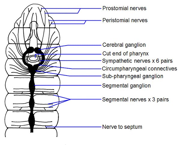
The nerve ring, also referred to as the brain ring, encircles the pharynx, spanning the 3rd and 4th segments of the earthworm. This oblique structure serves as the primary neural hub. The dorsal section of the nerve ring consists of the cerebral or supra-pharyngeal ganglia, which are often equated to the “brain” of the earthworm. These paired, fused ganglia are situated dorsally in the groove between the buccal cavity and the pharynx. On the ventral side, the nerve ring comprises the sub-pharyngeal ganglia, which are also fused. The dorsal and ventral ganglia are interconnected by circum-pharyngeal or peri-pharyngeal connectives, completing the ring-like structure.
b. Ventral Nerve Cord
Originating from the sub-pharyngeal ganglia, the ventral nerve cord extends posteriorly, running along the mid-ventral line to the terminal end of the earthworm. While it appears singular, it is essentially a fusion of two longitudinal cords. From the 5th segment onwards, each segment exhibits a ganglionic swelling, termed the segmental ganglion. Histologically, this double nerve cord is solid, comprising nerve cells and fibers. It is encapsulated by the epineurium, a protective sheath of connective tissue. External to the epineurium lies a layer of longitudinal muscle fibers, further enveloped by the visceral peritoneum. The nerve cord houses both ordinary fibers and specialized giant fibers or neurocords. These giant fibers, four in number (one median, one sub-median, and two laterals), are positioned dorsally and are instrumental in the swift conduction of neural impulses along the nerve cord. Their orientation and function vary, with lateral fibers conducting impulses antero-posteriorly, while the median and sub-median fibers facilitate postero-anterior conduction.
In essence, the central nervous system of the earthworm, with its nerve ring and ventral nerve cord, exemplifies the evolutionary adaptability of neural structures. Its design ensures efficient sensory integration and motor coordination, enabling the earthworm to thrive in its environment.
2. Peripheral nervous system (PNS) of earthworm
The earthworm, a vital member of the annelid group, boasts a meticulously organized peripheral nervous system (PNS) that interfaces with its central nervous system (CNS) to ensure optimal sensory and motor functions. The PNS is characterized by a network of nerves that emanate from the CNS and extend to various regions of the earthworm’s body.
- Origin and Distribution of Nerves: The cerebral ganglia, often referred to as the “brain” of the earthworm, give rise to 8-10 pairs of nerves. These nerves cater to the prostomium, buccal cavity, and pharynx. Additionally, the circum-pharyngeal or peri-pharyngeal connectives, which are pivotal components of the nerve ring, produce 2 pairs of nerves. These primarily supply the first segment and the buccal cavity. The sub-pharyngeal ganglia, located ventrally, generate 3 pairs of nerves that cater to the 2nd, 3rd, and 4th segments. Furthermore, each segmental ganglion associated with the ventral nerve cord emits 3 pairs of lateral nerves. These nerves are strategically positioned, with one pair anterior to the setal ring and two pairs posterior to it, ensuring comprehensive innervation of the segment they reside in.
- Nature of Nerves: All nerves in the earthworm are of a mixed type, signifying that they encompass both afferent (sensory) and efferent (motor) nerve fibers. This dual composition ensures that the earthworm can both receive sensory information from its environment and respond appropriately through motor actions. Additionally, the presence of adjustors or association neurons further enhances the integrative capabilities of the PNS.
- Functional Implications: The PNS plays a pivotal role in the earthworm’s interaction with its environment. The afferent fibers relay sensory information to the CNS, enabling the earthworm to detect changes in its surroundings. In contrast, the efferent fibers carry motor commands from the CNS to the effector organs, facilitating movement and other responses. The adjustors or association neurons act as intermediaries, processing the sensory input and determining the most appropriate motor response.
In conclusion, the peripheral nervous system of the earthworm, with its intricate network of mixed nerves, serves as a testament to the evolutionary finesse of neural organization in invertebrates. Its design ensures seamless communication between the CNS and the rest of the body, enabling the earthworm to adeptly navigate its environment.
3. Sympathetic nervous system
The sympathetic nervous system, a critical component of the autonomic nervous system, plays a pivotal role in the regulation and coordination of internal organ functions. This system is characterized by a complex network of nerve plexuses that are intricately embedded within the walls of the alimentary canal and certain other internal organs.
- Structural Composition: The primary feature of the sympathetic nervous system is the extensive nerve plexus. This plexus is not an isolated entity but is interconnected with other neural structures. Specifically, fine nervules establish connections between this nerve plexus and the per-pharyngeal connectives, ensuring a seamless integration of neural signals.
- Functional Role: The sympathetic nervous system’s primary function is to coordinate the activities of the organs it innervates. By doing so, it ensures the harmonious functioning of the alimentary canal and other associated internal organs. This coordination is vital for processes such as digestion, absorption, and excretion, where synchronized actions of various organ parts are imperative for optimal function.
- Significance in Homeostasis: Beyond mere coordination, the sympathetic nervous system plays a crucial role in maintaining the body’s internal equilibrium or homeostasis. By regulating the activities of the alimentary canal and other organs, it ensures that the internal environment remains stable, even when external conditions fluctuate.
In summation, the sympathetic nervous system, with its intricate nerve plexuses and connective nervules, stands as a testament to the evolutionary sophistication of neural systems. Its role in coordinating organ functions and maintaining internal stability underscores its significance in the overall physiological framework.
The Working Mechanism of the Nervous System
The nervous system, a complex and intricate network of neurons, governs the physiological activities of organisms, ensuring coordinated responses to environmental stimuli. In the earthworm, this system operates with remarkable efficiency, even in the absence of a highly developed brain, akin to higher animals.
- Neural Composition: Earthworms possess both sensory and motor neurons. The nerve cord, which runs longitudinally through the body, contains mixed nerves, comprising both sensory and motor fibers. This dual composition facilitates the reception of sensory information and the subsequent initiation of motor responses.
- Sensory Pathways: Sensory fibers originate from receptor cells or sensory organs located in the epidermis. These fibers extend and terminate in the ventral nerve cord, branching out intricately. These branches form synapses, or neural junctions, with motor fibers, creating a seamless communication pathway.
- Motor Pathways: Motor fibers arise in proximity to the sensory fiber branches within the ventral nerve cord. These fibers extend outward, culminating in the muscles. When a stimulus is detected by the receptor cells, sensory impulses travel via the sensory fibers to the ventral nerve cord. These impulses are then relayed as motor impulses through the efferent fibers, leading to muscle contraction.
- Reflex Arc: The pathway that sensory impulses traverse, from the receptor to the ventral nerve cord and subsequently to the muscles via motor fibers, constitutes a reflex arc. This simple yet efficient circuit ensures rapid and appropriate responses to stimuli.
- Coordinated Muscle Movement: The earthworm’s movement is a result of the coordinated action of its circular and longitudinal muscles. When one set of muscles contracts, the other relaxes, ensuring smooth and efficient locomotion.
- Role of Giant Fibers: The nerve cord houses specialized giant fibers that are adept at conducting impulses at a significantly faster rate than their counterparts. When the earthworm encounters a strong stimulus at a specific point, these fibers facilitate a swift contraction of the entire body, allowing the earthworm to react promptly to potential threats.
Sense Organs Of Earthworm
The earthworm, a vital member of the annelid phylum, is equipped with a range of sense organs that, while simple in structure, are adept at detecting various environmental stimuli. These organs, primarily derived from specialized ectodermal cells, play a crucial role in the earthworm’s interaction with its surroundings.
- Epidermal Receptors: Distributed ubiquitously across the earthworm’s epidermis, these receptors are particularly abundant on the lateral and ventral surfaces. Each receptor is characterized by an elevated cuticle that shields a cluster of tall, columnar receptor cells. These cells exhibit hair-like processes externally and are connected to nerve fibers internally. Encased by standard supporting epidermal cells, these receptors primarily serve a tactile function. Additionally, they are sensitive to chemical stimuli and temperature variations.
- Buccal Receptors: Exclusively located within the epithelium of the buccal chamber, buccal receptors resemble epidermal receptors but with broader external ends. The sensory hairs of these receptors are more pronounced and deeply situated. Furthermore, the receptor cells house a deeply positioned nucleus. Functionally, buccal receptors are gustatory and olfactory, responding adeptly to chemical stimuli.
- Photoreceptors: As the name suggests, these receptors are sensitive to light and are predominantly found on the dorsal surface of the earthworm. Their concentration is highest in the prostomium and peristomium regions, dwindling as one moves posteriorly and being entirely absent in the clitellar region. Each photoreceptor comprises a singular ovoid cell, housing a nucleus and transparent cytoplasm. Within the cytoplasm, a network of neurofibrils is present, along with a transparent, L-shaped lens or phaosome. This lens captures light from various directions, focusing it onto the neurofibrils. These fibrils then converge into an afferent nerve fiber, which integrates with the central nervous system. Through these photoreceptors, earthworms can discern both the intensity and duration of light, aiding them in navigating their subterranean habitats.
In summation, the earthworm’s sense organs, though structurally simple, offer a comprehensive sensory palette, enabling it to effectively perceive and respond to its environment. Their presence underscores the evolutionary adaptability of sensory mechanisms in invertebrates.
