Table of Contents
Plasmodesmata are tiny channels that span the cell walls of plant cells and allow communication and material movement between neighboring cells. They are composed of cytoplasmic filaments that extend through the cell walls of neighboring plant cells and link their cytoplasms. The cytoplasmic filaments are encompassed by a plasma membrane, which provides a channel between the two cells.
Plasmodesmata serve a key role in the passage of nutrients and signaling molecules between cells, allowing plant tissues to develop and function in unison. They are involved in the transport of water, ions, proteins, RNA, and other molecules, and they have been proven to play crucial roles in plant growth, development, and environmental response.
Plasmodesmata are peculiar to plants and are not seen in animal cells. Its structure and function are currently being investigated, and new insights into their significance in plant life are being uncovered.
Plasmodesmata Definition
- Plasmodesmata (singular: plasmodesma) are tiny channels that directly connect the cytoplasm of adjacent plant cells, forming live bridges between cells.
- Similar to the gap junctions found in animal cells, the plasmodesmata, which breach both the primary and secondary cell walls, allow some chemicals to move straight from one cell to another and play an essential role in cellular communication.
- Because of the thick cell wall, the plasmodesmata have an altogether different structure from the animal cell gap junction. Plant cells can be considered to form a synctium, or multinucleated mass with cytoplasmic continuity, due to the presence of plasmodesmata.
- Likewise, the microscopic pathways have sparked a substantial amount of controversy among scientists regarding cell theory, with some arguing that the cells of higher plants are not cells at all because they are not physically separated or structurally independent from one another.
- Plasmodesmata are cylindrical in shape and lined with the plasma membrane, such that all related cells are basically interconnected by a single continuous cell membrane.
- The desmotubule is produced from the smooth endoplasmic reticulum of the linked cells and is present in the majority of plasmodesmata.
- A ring of shared cytoplasm is situated between the desmotubule and the inner surface of the membrane-lined channel because the desmotubule does not completely fill the plasmodesma.
- Plasmodesmata often arise during cell division, when portions of the parent cell’s endoplasmic reticulum become entrapped in the daughter cells’ newly created cell wall. There may be thousands of plasmodesmata connecting daughter cells to one another.
- It is widely believed that plant cells regulate the movement of tiny molecules, such as sugars, salts, and amino acids, by constricting and dilating the apertures at the ends of the plasmodesmata. Nevertheless, this mechanism is not fully understood.
- Nonetheless, it is known that size constraints on molecule movement between cells can be bypassed in some instances. By binding to portions of the plasmodesmata, some proteins and certain viruses are able to enlarge the diameter of the channels sufficiently to allow passage of extremely big molecules.
Structure of Plasmodesmata
Structures of plasmodesmata range from simple (defined by a single sheath) to complicated (marked by branching), H-shaped, and twinned. Typically, simple plasmodesmata are present in early tissue, while complex plasmodesmata arise later, after cell growth.
These are cylindrical membrane-lined passageways with a 20 to 40 nm diameter. The desmotubule is a cylindrical structure that runs from cell to cell across the center of the majority of plasmodesmata and is continuous with the SER membranes of each connected cell.
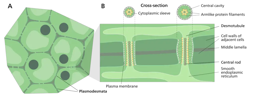
Plasmodesmata have a complicated structure comprised of a number of unique parts. The primary plasmodesmata components are:
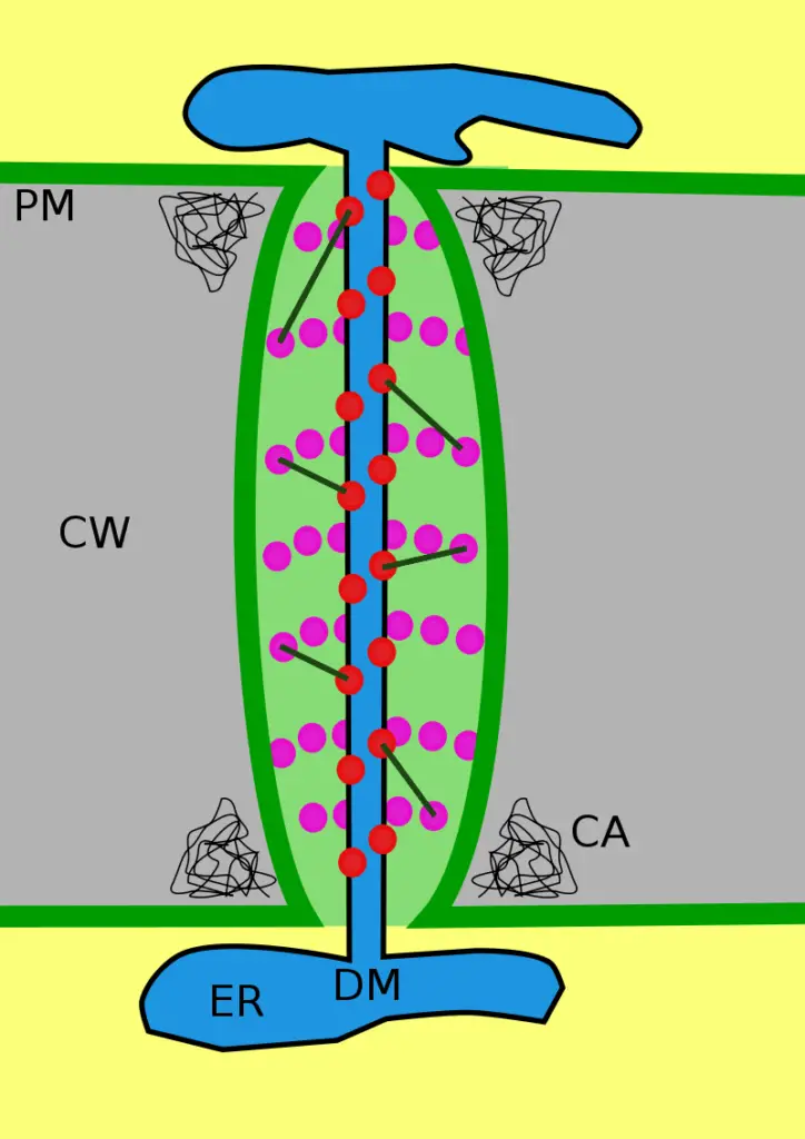
- Desmotubule: The desmotubule is a tubular structure that connects the endoplasmic reticulum of two adjacent cells and passes across the middle of the plasmodesma. In most instances, the desmotubule goes through the middle of plasmodesmata. The desmotubule is a dense rod or thinner cylindrical structure related to the smooth endoplasmic reticulum of neighboring cells. The endoplasmic reticulum of the linked cells gives rise to desmotubules. Now known as desmotubules, its original name was the axial component. Between the outside and interior of the desmotubule and cylindrical plasma membrane, respectively, is the cytosolic annulus. It appears to be constructed on each end of plasmodesmata. Between desmotubules and the plasma membrane are eight to ten microchannels. In electron microscopic images of plasmodesmata, the plasma membrane appears as a three-part structure with a width of 7.2 nm, and the dense central rod has a radius of 1.4 nm. The width of the faint ring is 2,2 nm. It encompasses the central rod.
- Cytoplasmic Sleeve: This is a channel that traverses the plasmodesma and is bordered by the plasma membranes of two neighboring cells. A cytoplasmic sleeve is a fluid-filled region bordered by the cell membrane (plasmalemma) that is a continuous extension of the cytosol.Myosin-like proteins and actin filaments are located within the cytoplasmic sleeve, and it is believed that proteinaceous spike-like projections regularly positioned within the cytoplasmic sleeve generate nanochannels of different sizes. The cytoplasmic sleeve facilitates the movement of ions and molecules via plasmodesmata. Smaller molecules and amino acids can flow through it via diffusion.
- Plasma Membrane: This is the outermost layer of the plasmodesma and is formed of a lipid bilayer.
- Pectin: Pectin is a substance composed of carbohydrates that occupies the gap between the plasma membrane and the desmotubule.
- Proteins: Plasmodesmata include numerous proteins, including callose synthase, which regulates plasmodesmal permeability and the passage of signaling chemicals between cells.
The precise structure of plasmodesmata can change based on the kind of plant tissue, developmental stage, and surrounding environment. Researchers continue to investigate the intricate structure and function of plasmodesmata in plant cells.
Formation of Plasmodesmata
- As parts of the endoplasmic reticulum are stuck across the middle lamella when new cell wall is generated between two recently separated plant cells, primary plasmodesmata are created.
- They eventually create the intercellular cytoplasmic connections. At the formation location, the wall is not thickened further, and pits, or depressions, are produced in the wall.
- Normal pairing of pits between adjacent cells. Plasmodesmata can also be placed between non-dividing cells in existing cell walls (secondary plasmodesmata).
Primary plasmodesmata
- At the phase of cellular division in which the endoplasmic reticulum and the new plate fuse together, primary plasmodesmata are formed; this leads in the production of a cytoplasmic pore (or cytoplasmic sleeve). Desmotubule, also known as the appressed ER, develops concurrently with the cortical ER.
- Both the appressed ER and the cortical ER are densely packed, leaving no margin for luminal fluid. It is proposed that the ER that has been compressed serves as a membrane transport pathway in plasmodesmata.
- When filaments of the cortical ER become entangled during the development of a new cell plate in terrestrial plants, plasmodesmata are formed.
- It is suggested that the appressed ER arises due to the interaction of ER and PM proteins and the pressure exerted by a thickening cell wall. Frequently, primary plasmodesmata are found in regions where the cell wall appears to be thinner.
- This is because the number of main plasmodesmata reduces as a cell wall increases. Secondary plasmodesmata are formed in order to increase plasmodesmal density during cell wall development.
- Several degrading enzymes and ER proteins are believed to encourage the development of secondary plasmodesmata, however this mechanism is not yet entirely understood.
Signaling and Transport Mechanisms of Plasmodesmata
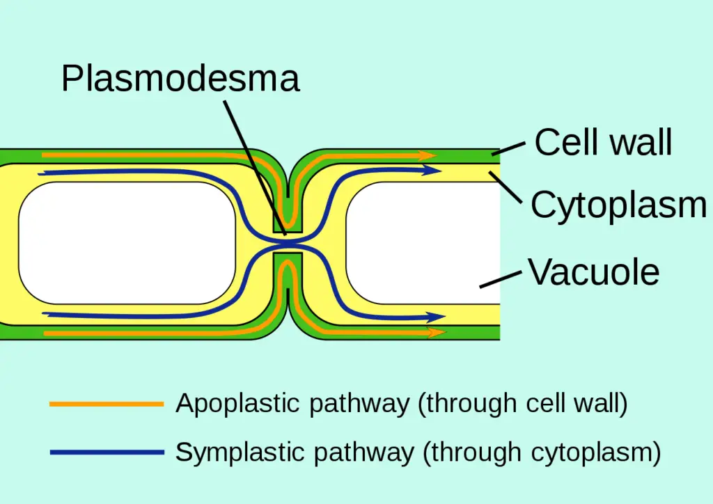
Microinjection of tiny fluorescently labelled probes has been used in traditional investigations of plasmodesmal transport to investigate the passive transport processes of plasmodesmata. In the beginning, these probes were used to define the size-exclusion limits of plasmodesmata, or the largest molecule that can passively pass through plasmodesmata. Analysis of the cytoplasmic sleeve and the spokes that constrict the neck region plasmodesmata revealed that the average width of channels allowing molecules to passively travel through a plasmodesmal annulus is about 3 nm, and that the average size exclusion limit is molecules weighing about 800-1000 Da.
Variations on passive transport
- Yet, recent research has demonstrated that size exclusion limits during passive transport can vary greatly between species and even cell types.
- The size exclusion limit of fluorescently tagged dextrans is just 1 kDa in tobacco mesophyll cells but 7 kDa in trichome cells.
- Wang and Fisher (1994) conducted an additional research using typically apoplastic probes such as Lucifer Yellow and incubating slices of crease tissue from developing wheat grains in these solutions.
- Plasmodesmata were responsible for the dye’s entry into the symplastic gap, and their width was measured to be 6.2 nm in all cells of this tissue except those of the pericarp (twice the normal exclusion limit).
- Why do studies report such a wide range of values for the parameters describing passive diffusion through plasmodesmata? The partial closure of plasmodesmata, maybe due to callose blocking the transport routes, may be the result of the wound response triggered in the plant cell by the microinjection technique used to exogenously add tiny probe molecules.
- Different plant species may have distinctively varied compositions in the neck region of their plasmodesmata, which could account for the variations in size exclusion limits for passive transport.
Active transport of macromolecules
- Some macromolecules designated for intercellular transfer are transported by active transport mechanisms through plasmodesmata.
- Several investigations have demonstrated that plasmodesmata can expand/dilate form an electron-lucent sleeve encircling normal-sized plasmodesmata, hence altering their dimensions.
- This enlargement would allow larger molecules to be transported through the cytoplasmic sleeve. The mechanism or trigger of this plasmodesmal dilatation is essentially unknown; however, it has recently been hypothesised that proteins in the neck region may be involved in this process.
- The link of cytoskeletal components with trafficking/targeting molecules to the plasmodesmata and with the energetics of active transport is a significant discovery concerning active transport through plasmodesmata made recently.
- The endoplasmic reticulum has been shown to be strongly connected with microtubules, and viral movement proteins (described below) have been shown to track through the plant cell along these microtubule.
- Actin and tubulin have been demonstrated to bind viral movement protein, which travels itself via plasmodesmata throughout the plant. Consequently, cytoskeletal elements aid in directing various macromolecules to plasmodesmata, and actin is believed to help relax the sphincter elements of the neck area to permit transport of these bigger molecules.
- Myosin may also function as a cytoskeletal motor to provide the energy for active plasmodesmal transport. Myosin is an ATP-dependent protein found in numerous plant species that creates directed movement (as in flagellar motion).
- Substantial ATPase activity has been proven in plasmodesmata, therefore myosin is a prime candidate for helping active transport and may be the electron microscopically observed spokes connecting the desmotubule to the cytoplasmic annulus throughout the plasmodesmata.
Viruses and Plasmodesmata
- The intercellular movement of plant viruses through plasmodesmata constitutes a subgroup of active plasmodesmal transport.
- There is considerable evidence that the viruses alter the plasmodesmata to allow big viral particles (many times larger than the normal size exclusion limit) to move between cells.
- Studies of the tobacco mosaic virus, which contains a mobility protein (MP) that promotes the transport of a non-virion version of the virus across cells, have deduced the fundamental mechanism behind this type of viral transport. The proposed mechanism is as follows:
- The viral movement protein is expressed.
- The MP interacts with viral RNA
- This interaction enables the RNA to unfurl into a linear rod.
- The RNA and the bound MP then move to the plasmodesmata.
- The MP interacts with plasmodesmata to augment the size exclusion limit.
- The viral RNA can transfer to the neighbouring cell via the plasmodesmata.
- The viral movement protein contains three functional domains: one for RNA binding, one for cooperative RNA binding, and one for plasmodesmal interaction.
- It has been demonstrated that the viral movement protein is acting on plasmodesmata directly to increase their size exclusion limits, as fluorescently-labeled large dextrans (between 9 and 35 kDa) were found to move between cells in transgenic plants expressing the movement protein, and no endogenous metabolism of the dextrans was detected.
- The study of viral movement proteins will continue to help researchers clarify the mechanisms of plasmodesmal targeting and transport more precisely.
Types of Plasmodesmata
When it comes to shape and function, plasmodesmata come in a wide variety of forms. The following are examples of some of the most common kinds:
- Simple plasmodesmata: The vast majority of plant cells have simple plasmodesmata. The cytoplasm of two neighboring cells is joined by a thin passageway.
- Simple plasmodesmata: Branched plasmodesmata are similar in structure to simple plasmodesmata, but instead of being a single channel within the cell wall, they branch off into multiple smaller channels. This improves cellular communication by facilitating the movement of chemicals.
- Ring-type plasmodesmata: These plasmodesmata have a structure that wraps like a ring around the desmotubule, hence the name. Phloem cells are found in plants and play a role in the long-distance transport of signaling chemicals and nutrients.
- Branched-ring plasmodesmata: Some specialized plant tissues have a hybrid form of plasmodesmata known as branched-ring plasmodesmata. Several scientists believe their primary function is to facilitate the intercellular transfer of macromolecules like proteins and nucleic acids.
- Sieve plasmodesmata: Located in the phloem’s sieve elements, sieve plasmodesmata are characterized by their complicated structure, which consists of a sieve plate and a callose sleeve. For example, they play a role in the long-distance transport of carbohydrates and other nutrients.
Variables such as plant age, tissue type, and environmental circumstances all have a role in determining the precise type of plasmodesmata present in each given plant cell or tissue. Plasmodesmata come in a wide variety of forms and play an essential role in the health of plant cells and tissues, allowing for a wide variety of processes to occur.
Roles of Plasmodesmata During Development
- Symplastic transport: Plasmodesmata facilitate the movement of water, nutrients, and signaling molecules between adjacent cells, enabling symplastic transport throughout the plant. This is important for the coordinated growth and development of tissues and organs.
- Cell fate determination: Plasmodesmata allow for the exchange of regulatory molecules such as transcription factors, which can influence the fate of neighboring cells. This is important for specifying cell identities during embryogenesis and organogenesis.
- Cell differentiation: Plasmodesmata can regulate the differentiation of cells by allowing the exchange of small molecules such as microRNAs and hormones. This is important for the differentiation of specific cell types, such as the formation of root hairs or trichomes.
- Defense signaling: Plasmodesmata can allow for the movement of defense-related molecules, such as pathogenesis-related proteins, between cells in response to pathogen attack. This can enable the rapid and coordinated response of neighboring cells to a pathogenic threat.
Plasmodesmata Functions
- Intercellular transport: Plasmodesmata serve as channels that allow for the movement of various molecules and substances, such as nutrients, hormones, signaling molecules, and RNA, between adjacent plant cells. This intercellular transport is critical for plant development, growth, and response to environmental stimuli.
- Communication: Plasmodesmata also enable direct communication between adjacent plant cells. They allow for the exchange of information and signaling molecules, which is important for the coordination of plant tissues and organs.
- Defense: Plasmodesmata play a role in plant defense by controlling the movement of viruses, pathogens, and toxins between plant cells. They can also seal off a cell that has been infected, preventing the spread of the infection to other cells.
- Development: Plasmodesmata are essential for plant development, helping to regulate cell division, differentiation, and organogenesis. They also play a role in maintaining the balance between different parts of the plant, such as the roots and shoots.
- Nutrient exchange: Plasmodesmata play a key role in the exchange of nutrients between adjacent plant cells. They allow for the transport of ions, sugars, amino acids, and other molecules that are essential for plant growth and development.
- Signaling: Plasmodesmata are involved in the transmission of signals between plant cells, which can trigger a variety of physiological responses. For example, plasmodesmata can allow for the transmission of electrical signals, which are important for rapid responses to environmental stimuli.
- Stress response: Plasmodesmata can respond to various environmental stresses, such as drought, salt, or pathogen attack, by altering their structure and function. This can help to limit the spread of damage or infection to adjacent cells, and may also trigger signaling pathways that help the plant to adapt to the stress.
- Plant-microbe interactions: Plasmodesmata play a role in interactions between plants and microbes, such as mycorrhizal fungi or nitrogen-fixing bacteria. They can facilitate the exchange of nutrients or signaling molecules between the two organisms, and may also help to prevent the spread of pathogens.
FAQ
What are plasmodesmata?
Plasmodesmata are microscopic channels that connect adjacent plant cells and allow for the transport of molecules and communication between them.
How are plasmodesmata formed?
Plasmodesmata are formed when specific regions of the cell wall are removed, creating a channel between adjacent cells.
What is the function of plasmodesmata?
The primary function of plasmodesmata is to facilitate the movement of water, nutrients, signaling molecules, and other substances between adjacent plant cells.
What types of molecules can pass through plasmodesmata?
Small molecules such as ions, sugars, and amino acids can pass through plasmodesmata, as well as larger molecules such as proteins and RNA molecules.
How are plasmodesmata regulated?
The opening and closing of plasmodesmata are regulated by various factors, including developmental signals, environmental cues, and plant hormones.
Can viruses and pathogens move through plasmodesmata?
Yes, some viruses and pathogens can move through plasmodesmata to infect neighboring cells, which can contribute to the spread of diseases within the plant.
How do plasmodesmata contribute to plant growth and development?
Plasmodesmata play an important role in plant growth and development by enabling cell-to-cell communication, transport of nutrients and signaling molecules, and regulation of cell differentiation and fate.
How are plasmodesmata related to plant stress responses?
Plasmodesmata are involved in plant stress responses, as they allow for the movement of stress-related signaling molecules between cells, enabling the plant to respond to environmental challenges.
How can plasmodesmata be visualized and studied?
Plasmodesmata can be visualized and studied using various techniques, such as electron microscopy, fluorescent microscopy, and live cell imaging.
Are plasmodesmata unique to plants?
Yes, plasmodesmata are unique to plants and are not found in other organisms. However, similar structures called gap junctions are found in animals and allow for cell-to-cell communication.
References
- https://micro.magnet.fsu.edu/cells/plants/plasmodesmata.html
- https://www.news-medical.net/life-sciences/What-are-Plasmodesmata.aspx
- https://www.cell.com/current-biology/pdf/S0960-9822(08)00093-6.pdf
- https://study.com/learn/lesson/what-is-plasmodesmata.html
- https://www.thoughtco.com/plasmodesmata-the-bridge-to-somewhere-419216
- https://www.biologyonline.com/dictionary/plasmodesmata

