Table of Contents
What is Prophase?
- Prophase, derived from the Ancient Greek terms “προ-” (pro-) signifying ‘before’ and “φάσις” (phásis) denoting ‘appearance’, is a pivotal initial phase in the eukaryotic cell division process. This stage is integral to both mitosis and meiosis.
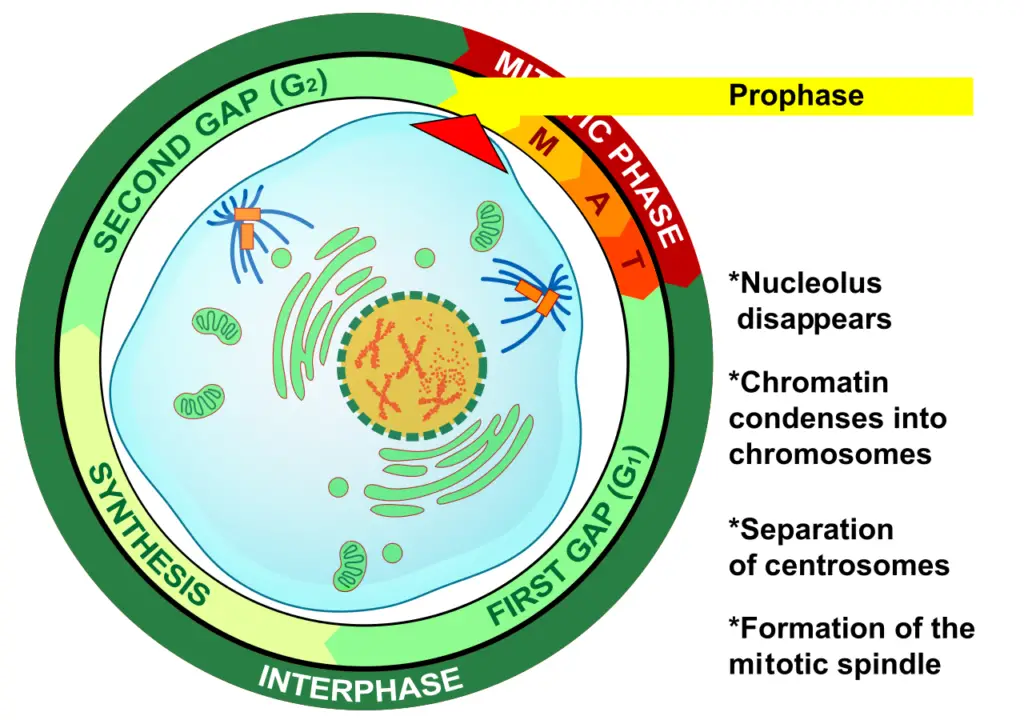
- Following the interphase, wherein DNA replication transpires, the cell enters prophase. The hallmark events of prophase encompass the condensation of the chromatin network and the subsequent disappearance of the nucleolus.
- In the intricate dance of cell division, prophase sets the stage by orchestrating the organization and movement of various cellular components. The chromosomes, previously dispersed as chromatin, undergo condensation, becoming more visually distinct under microscopic observation.
- Concurrently, the centrioles, integral components of the centrosome, segregate. The centrosome, an essential organelle, governs the microtubules within the cell. As these centrioles part ways, they establish microtubule centers for the impending daughter cells.
- These microtubules, in collaboration with associated proteins, function as molecular motors, facilitating the movement of organelles, chromosomes, and other cellular structures. The onset of prophase, therefore, is characterized by the repositioning of these molecular motor centers and the compaction of chromosomes.
- To encapsulate, prophase is a critical juncture in the cell cycle, marking the commencement of cell division. It is characterized by the intricate reorganization of chromosomal material and the strategic positioning of cellular machinery to ensure the fidelity of cell division. This phase, steeped in both mitotic and meiotic contexts, is a testament to the precision and elegance inherent in biological processes.

Definition of Prophase
Prophase is the initial stage of cell division in eukaryotes, characterized by the condensation of chromosomes, the separation of centrioles, and the breakdown of the nuclear envelope.
What Happens in Prophase?
- During the prophase stage of cell division, several critical events transpire to ensure the accurate segregation of genetic material. Initiated post the DNA synthesis in interphase, prophase is marked by the condensation of chromatin structures, which are intricate assemblies of DNA and proteins. This condensation process results in the transformation of these chromatin structures into discernible chromosomes.
- Each chromosome, an organized entity of DNA, assumes an ‘X’ configuration, termed as sister chromatids. These chromatids are essentially identical DNA replicas, conjoined at a specific locus known as the centromere. This union ensures the fidelity of genetic information as the cell prepares for division.
- Concurrently, specialized structures called mitotic spindles begin to emerge at the cell’s opposing ends. Composed of elongated protein filaments called microtubules, these spindles play a pivotal role in the subsequent stages of cell division. Their primary function is to facilitate the separation of the sister chromatids, ensuring that each resulting daughter cell inherits an accurate copy of the genetic material.
- In essence, prophase sets the stage for the meticulous division of genetic content, orchestrating the transformation of chromatin into chromosomes and establishing the machinery for their eventual segregation.
Pointers to prophase
Prophase, a pivotal stage in cell division, is marked by several distinct events that prepare the cell for the subsequent phases of mitosis. Here are the salient pointers to prophase:
- Chromosomal Configuration: Under microscopic observation, chromosomes in prophase manifest as X-shaped entities, a result of their condensation. This X-structure represents the replicated chromosome, consisting of two sister chromatids.
- DNA Condensation: The DNA molecules undergo significant condensation, enhancing their visibility. This transformation leads to the transition from chromatin to distinct chromosomes.
- Chromosomal Alterations: As prophase progresses, chromosomes not only condense but also experience a reduction in size, accompanied by a marked increase in thickness.
- Nuclear Envelope Disintegration: The nuclear envelope, which encapsulates the genetic material, undergoes degradation and eventually disappears.
- Nucleolus Disappearance: Concurrently, the nucleolus, a spherical structure within the nucleus, disintegrates and vanishes.
- Release of Genetic Material: The replicated genetic content housed within the mother cell’s nucleus is readied for distribution between the two forthcoming daughter cells.
- Cytoskeleton Disassembly: The cell’s cytoskeletal framework undergoes disassembly, further preparing the cell for division.
- Sister Chromatids: The replicated chromosomes, now in their characteristic X-shape, are termed sister chromatids. These chromatids remain interconnected at a region called the ‘centromere’.
- Mitotic Spindle Formation: A significant feature of prophase is the emergence of the mitotic spindle apparatus at the cell’s poles. This is attributed to the reorganization of microtubules. These spindles, occasionally visible as thread-like entities under the microscope, play a crucial role in the subsequent separation of sister chromatids, particularly in the anaphase stage.
In essence, prophase sets the stage for cell division by orchestrating a series of cellular and molecular events, ensuring the precise distribution of genetic material to the daughter cells.
Prophase in cell cycle
- Prophase occupies a significant position within the cell cycle, serving as a bridge between the preparatory phases and the active stages of cell division. To understand its role, it’s essential to contextualize prophase within the broader framework of the cell cycle.
- Following the S-phase of the interphase, wherein chromosomes undergo replication, cells may occasionally enter a quiescent phase. This phase, rather than being a mere pause, can be a period of cellular rest or may be influenced by other cellular factors. It’s crucial to note that cells do not invariably transition from interphase to the division phase instantaneously.
- Upon receiving the requisite signals for cell division, the cell embarks on the journey of mitosis or meiosis, with prophase being the inaugural phase. At this juncture, the cell has committed to the division process, aiming to produce daughter cells.
- Prior to prophase, the genetic material within the cell is dispersed in a relaxed state, known as chromatin, within the nucleus. The onset of prophase heralds the condensation of this chromatin into organized structures, facilitating the efficient maneuvering of DNA molecules in the subsequent stages of cell division. This organizational shift is paramount for the seamless progression of both mitotic and meiotic divisions.
- In the grand scheme of the cell division sequence, prophase is succeeded by metaphase, anaphase, and culminates in telophase. Each phase is meticulously orchestrated, ensuring the accurate and efficient division of the cell’s genetic material. In essence, prophase sets the stage, preparing the cell for the intricate dance of division that follows.
Staining and Microscopy
Staining and microscopy are indispensable techniques in the realm of cellular biology, offering a window into the microscopic world that remains invisible to the naked eye. These methods enable scientists to delve deep into the intricacies of cellular structures and processes, such as the dynamic movements of chromosomes during cell division.
Microscopy, as the name suggests, involves the use of microscopes to magnify and examine minute biological specimens. However, the inherent transparency of many cellular components poses a challenge to direct visualization. This is where the art of staining comes into play. Staining involves the application of specific dyes to biological samples, enhancing contrast and rendering certain structures or molecules more discernible under the microscope.
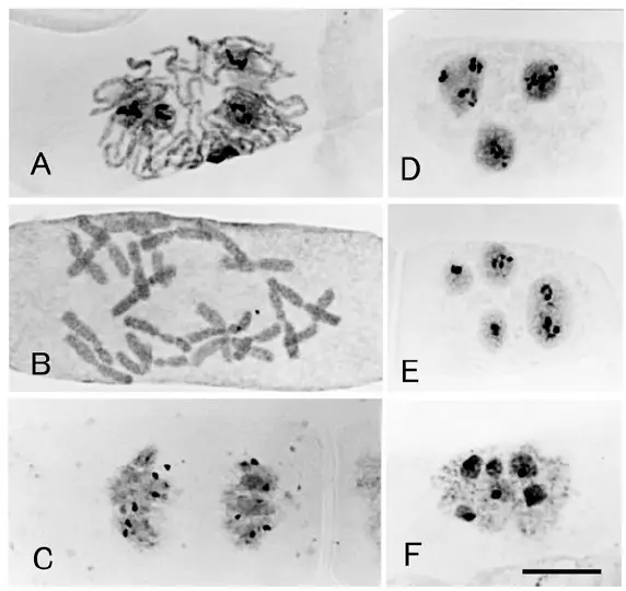
One of the pivotal stages where staining proves invaluable is during the prophase of cell division, be it mitotic or meiotic. As chromatin condenses into distinct chromosomes, the ability to observe this transformation and subsequent chromosomal movements becomes crucial. Both somatic cells, involved in mitotic division, and germinal cells, participating in meiotic division, can be stained to visualize these processes.
Among the myriad of staining techniques available, a few stand out for their specificity and utility:

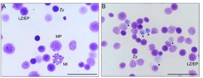
- Giemsa Stain: This versatile stain, composed of methylene blue, eosin, and Azure B, is particularly adept at staining nucleic acids due to its affinity for DNA’s phosphate groups. The resulting G-banding pattern, a hallmark of Giemsa staining, has been instrumental in generating karyograms and detecting chromosomal anomalies. Beyond its application in histopathology, Giemsa stain is also employed in studying fungi, bacteria, and yeast. Typically, nuclei stained with Giemsa adopt a dark blue hue, while the cytoplasm varies from pink to light blue.
- Silver Stain: Gaining prominence in recent times, silver staining offers another avenue for visualizing stages of cell division, including prophase. The stain binds to certain cellular components, rendering them visible under the microscope.
In summary, the synergy of staining and microscopy has revolutionized our understanding of cellular processes. By illuminating the hidden world within cells, these techniques continue to unravel the mysteries of life at the microscopic level.

Prophase in Mitosis
Mitosis, a fundamental process in the cell cycle, ensures the accurate division of replicated genetic material into two daughter cells. Prophase, the initial phase of mitosis in animal cells and the second in plant cells, sets the stage for this intricate dance of chromosomes.
1. Chromosome Condensation:
- Initiated post the DNA synthesis in interphase, prophase witnesses the condensation of DNA.
- The entangled DNA molecules from interphase undergo a structural transformation, compacting into distinct entities called chromatins.
- This condensation facilitates the subsequent separation of identical sister chromatids, a process termed chromatid resolution.
- The resulting chromosomes are robust and resilient, safeguarding them from potential damage during mitosis.
- Central to this condensation are the condensin complexes, comprising condensins and topoisomerase. While condensins are pivotal for chromosome separation and maintenance, topoisomerase, particularly topoisomerase 2, aids in resolving DNA topological challenges.
- The culmination of this process results in two sister chromatids, X-shaped structures interconnected at the centromere.
2. Centrosome Dynamics:
- Centromeres, equipped with microtubules, play a crucial role in chromosome movement.
- These microtubules, with the aid of tubulins, direct the centromeres’ movement.
- Post replication in interphase, centromeres, driven by associated motor proteins, migrate to the cell’s opposite poles.
- These motor proteins, harnessing ATP’s chemical energy, facilitate this movement along the microtubules.
3. Formation of Mitotic Spindles:
- As the centromeres separate, mitotic spindles, fibrous structures composed of microtubules, emerge.
- In cells devoid of centrioles, chromosomes orchestrate the assembly of the mitotic apparatus.
- In plants, the formation of these spindles differs slightly, with microtubules congregating at the cell’s poles, subsequently forming the spindle apparatus.
- These spindles play a pivotal role in segregating sister chromatids as the cell cycle progresses.
4. Nucleoli Disintegration:
- As prophase advances, the nucleoli, responsible for ribosome synthesis, begin to disintegrate, signaling the impending breakdown of the nucleus.
- This transition redirects the cell’s metabolic energy towards the mechanisms of cell division.
- Concurrently, the chromosomes undergo final condensation, becoming highly compacted.
- The nuclear envelope disintegrates, liberating the chromosomes, and some mitotic spindles commence the capture of these chromosomes.
In essence, prophase in mitosis lays the groundwork for the meticulous separation of genetic material, ensuring the fidelity of cell division and the preservation of genetic information across generations.
Prophase in Meiosis
Meiosis, a specialized form of cell division, is instrumental in producing gametes, ensuring genetic diversity across generations. Unlike mitosis, meiosis encompasses two sequential divisions, making it a more extended process. The prophase stage in meiosis is particularly intricate, occurring in two distinct phases: Prophase I and Prophase II.
1. Prophase I:
- Prophase I stands out as the most complex phase in meiosis, setting it apart from mitosis.
- During this phase, homologous chromosomes, each consisting of two sister chromatids, align closely in pairs. This phenomenon is termed synapsis.
- This alignment facilitates the exchange of genetic material between non-sister chromatids, a process known as genetic recombination or crossing over.
- This exchange is pivotal for generating genetic diversity, ensuring that offspring inherit a unique combination of genes from both parents.
- The culmination of Prophase I results in tetrads, groups of four chromatids, which subsequently move to the metaphase plate.
2. Prophase II:
- Following the completion of the first meiotic division, cells enter the second meiotic cycle, commencing with Prophase II.
- This phase mirrors the mitotic prophase in many aspects.
- Chromosomes, already condensed from the previous cycle, become more visible and start to move towards the metaphase plate.
- Unlike Prophase I, there’s no pairing of homologous chromosomes or crossing over in this phase, making it relatively straightforward.
In summation, the prophase stages in meiosis are pivotal for ensuring genetic variation and the accurate segregation of chromosomes. While Prophase I introduces genetic diversity through crossing over, Prophase II readies the chromosomes for the final division, leading to the formation of genetically unique gametes.
Prophase I
Prophase I stands as a pivotal stage in meiosis, characterized by the intricate interactions of homologous chromosomes and the subsequent exchange of genetic material. This phase is further subdivided into five distinct stages, each with its unique processes and characteristics.
1. Leptotene:
- Marking the onset of Prophase I, Leptotene witnesses the condensation of replicated chromosomes.
- These chromosomes, now more compact and distinguishable, resemble strings adorned with bead-like structures termed chromomeres.
- Intriguingly, each sister chromatid anchors itself to the nuclear envelope during this phase.
2. Zygotene (or Zygonema):
- A significant phase, Zygotene is characterized by the close association of homologous chromosomes, forming pairs in a process termed synapsis.
- These paired chromosomes, or tetrads, consist of four chromatids.
- The synaptonemal complex, a zipper-like structure formed by coiled chromatids, plays a crucial role in facilitating synapsis, ensuring the chromosomes remain aligned.
3. Pachytene:
- This stage is marked by the genetic crossover between non-sister chromatids of homologous chromosomes, resulting in chiasmata formation.
- The synaptonemal complex, having completed its role in synapsis, now enables the exchange of genetic material, introducing variations by interchanging maternal and paternal genetic elements.
- Despite the separation of sister chromatids, the homologous chromosomes remain tethered, forming a dense structure known as the synaptonemal complex.
4. Diplotene:
- As the synaptonemal complex disintegrates, the homologous chromosome pairs remain interconnected at the chiasmata.
- The repulsion between chromosomal arms causes them to drift apart, yet the chiasmata hold them together.
- This phase witnesses the phenomenon of terminalization, where chiasmata migrate towards the chromatid ends, becoming microscopically visible.
5. Diakinesis:
- Culminating Prophase I, Diakinesis prepares the cell for the subsequent metaphase.
- The chromosomes, having undergone further condensation, are discernible as tetrads or bivalents under microscopic observation.
- As terminalization concludes, chiasmata position themselves at the chromatid extremities.
- Concurrently, the nucleolus and nuclear envelope disintegrate, freeing the centrioles and facilitating the formation of the mitotic spindle.
In essence, Prophase I in meiosis is a meticulously orchestrated sequence of events, ensuring the accurate exchange and segregation of genetic material. This phase not only underpins the genetic diversity observed in sexually reproducing organisms but also sets the stage for the subsequent phases of meiosis.
Prophase II
Prophase II marks the commencement of the second meiotic division, characterized by a series of cellular events that prepare the cell for subsequent stages of meiosis II.
1. Chromosomal Condensation:
- As Prophase II initiates, there is a notable condensation of chromosomes. This process transforms the elongated chromatin fibers into more compact, distinguishable structures, facilitating their subsequent alignment and separation.
2. Nuclear Envelope Disintegration:
- Concurrent with chromosomal condensation, the nuclear envelope undergoes disintegration. This breakdown removes the barrier between the cytoplasm and the chromosomes, enabling the formation and action of the spindle apparatus.
3. Centrosomal Migration:
- A pivotal event in Prophase II is the movement of centrosomes. These organelles, crucial for spindle formation, begin to migrate towards opposite poles of the cell.
4. Spindle Formation:
- As the centrosomes position themselves, microtubules emanate from them, forming the spindle apparatus. These spindle fibers, or microtubules, play a cardinal role in capturing and aligning chromosomes at the cell’s equatorial plane.
5. Chromosomal Capture:
- The spindle fibers extend towards the chromosomes, attaching to their centromeres. This interaction ensures the accurate alignment and eventual separation of sister chromatids during the subsequent phases of meiosis II.
6. Haploid vs. Diploid Chromosome Number:
- A salient feature distinguishing Prophase II from the prophase of mitosis is the chromosomal number. While mitotic prophase operates with a diploid set of chromosomes, Prophase II is characterized by a haploid set, a consequence of the separation of homologous chromosomes during meiosis I.
7. Chromosomal De-condensation in Telophase I:
- Prior to the onset of Prophase II, telophase I witnesses the de-condensation of chromosomes. This relaxation of chromosomal structure is transient, as the chromosomes undergo re-condensation in Prophase II, preparing them for the ensuing meiotic division.
In summation, Prophase II is a meticulously coordinated phase in meiosis II, ensuring the cell is primed for the accurate segregation of sister chromatids. This phase, with its distinct haploid chromosomal number, underscores the genetic reduction pivotal to sexual reproduction.
Prophase I arrest
In the intricate journey of human female gametogenesis, a unique and prolonged pause, known as the “Prophase I arrest,” plays a crucial role in ensuring the timely and orderly maturation of oocytes. This arrest is not merely a halt but a strategic conservation of the oocytes until the onset of puberty.
1. Initiation of Gamete Preparation:
- The genesis of female gametes, or oocytes, commences during fetal development. As the female fetus develops, the germinal cells embark on the process of meiosis, aiming to produce mature oocytes for future reproductive events.
2. Arrest at Diplotene Sub-stage:
- However, this meiotic journey is not continuous. As the female fetus nears birth, the germinal cells, specifically the oocytes, experience an arrest in their meiotic progression. This pause occurs at the diplotene sub-stage of Prophase I, a specific point in the meiotic cycle.
3. Conservation until Puberty:
- The Prophase I arrest serves as a protective mechanism, ensuring that the oocytes remain in a state of suspended animation and do not advance towards maturation prematurely. This conservation persists throughout childhood, safeguarding the oocytes until the female reaches reproductive maturity.
4. Resumption of Meiosis at Puberty:
- The onset of puberty marks a significant hormonal shift in the female body. With the surge in the levels of Luteinizing Hormone (LH), the arrested oocytes receive the requisite signal to resume their meiotic journey. This hormonal cue propels the oocytes through the remaining stages of Prophase I.
5. Second Arrest at Metaphase II:
- However, the journey of the oocyte is punctuated by another pause. Post-ovulation, the oocyte progresses through meiosis but halts once again, this time at Metaphase II. This arrest remains until the oocyte encounters a sperm cell.
6. Completion of Meiosis during Fertilization:
- The Metaphase II arrest is only lifted upon fertilization. When a sperm cell penetrates the oocyte, it triggers the completion of meiosis. If fertilization does not occur, the oocyte, now termed a secondary oocyte, undergoes degeneration during the menstrual cycle.
In essence, the Prophase I arrest is a testament to the intricate orchestration of female gametogenesis. By strategically pausing the maturation of oocytes, it ensures that the female reproductive system is primed for potential fertilization events during the reproductive years.
Differences in Plant and Animal Cell Mitotic Prophase
Mitosis, a fundamental process of cell division, exhibits certain distinctions when observed in plant and animal cells. While the overarching mechanism remains conserved, the nuances in the prophase stage, particularly concerning mitotic spindle formation, highlight the inherent differences between these two cell types.
1. Role of Centrioles:
- Centrioles are cylindrical structures that play a pivotal role in organizing microtubules during mitosis in animal cells. These structures coalesce to form centrosomes, which serve as the primary microtubule-organizing centers in animal cells.
2. Centrosome Dynamics in Animal Cells:
- During the prophase of mitosis in animal cells, centrosomes undergo division and subsequently migrate to opposite poles of the cell. This migration facilitates the organization and orientation of the mitotic spindle, ensuring accurate chromosome segregation.
3. Absence of Centrioles in Plant Cells:
- Contrary to animal cells, plant cells do not possess centrioles. This absence raises the question: how do plant cells orchestrate the formation and organization of the mitotic spindle without these pivotal structures?
4. Alternative Mechanisms in Plant Cells:
- Despite the lack of centrioles, plant cells have evolved alternative microtubule-organizing mechanisms. These mechanisms enable plant cells to form and organize the mitotic spindle in a manner analogous to animal cells. The exact nature of these mechanisms remains a subject of ongoing research, but it is evident that plant cells have developed efficient strategies to ensure proper spindle formation and function in the absence of centrioles.
5. Cytokinesis Distinctions:
- Beyond prophase, another notable difference between plant and animal mitosis is observed during cytokinesis. While plant cells form a cell plate that eventually gives rise to the cell wall, animal cells undergo cleavage, resulting in the division of the parent cell into two distinct daughter cells.
In conclusion, while the core principles of mitosis are conserved across plant and animal cells, the intricacies of spindle formation during prophase underscore the adaptability and diversity of cellular mechanisms. The absence of centrioles in plant cells and their alternative strategies for spindle organization exemplify the evolutionary adaptations that cells have undergone to ensure accurate and efficient cell division.
Cell checkpoints
- Cell checkpoints play a pivotal role in maintaining the integrity of cellular processes, particularly during the intricate stages of cell division. These checkpoints serve as surveillance mechanisms, ensuring that each phase of the cell cycle progresses accurately.
- One of the most intricate stages where checkpoints are crucial is Prophase I during meiosis, observed in both plant and animal cells. The essence of these checkpoints lies in their ability to monitor and rectify any discrepancies in the DNA structure and function. Specifically, the meiotic checkpoint network is an intricate system designed to oversee the repair of double-strand breaks, regulate chromatin structure, and control the movement and pairing of chromosomes.
- This network is especially vital in ensuring the accurate recombination of genetic material. One of the primary pathways within this network is the meiotic recombination checkpoint. Its primary function is to prevent the cell from advancing to metaphase I if there are unresolved errors stemming from recombination.
- By doing so, it ensures that genetic material is accurately exchanged and that any potential anomalies are addressed before cell division progresses. In essence, cell checkpoints, especially those active during meiosis, are fundamental in preserving the fidelity of genetic information, ensuring that cells divide and reproduce in a manner that maintains the stability and health of an organism.
Importance of Prophase
Prophase, as the initial stage of both mitosis and meiosis, holds paramount importance in the process of cell division. Its significance can be understood through the following points:
- Chromosomal Condensation: During prophase, the chromatin fibers undergo condensation to form distinct, visible chromosomes. This condensation is crucial as it facilitates the subsequent orderly separation of genetic material.
- Formation of Sister Chromatids: Each chromosome replicates to form two sister chromatids, which are essential for the equal distribution of genetic material to the daughter cells.
- Initiation of the Spindle Apparatus: The centrosomes, present in animal cells, start moving to opposite poles of the cell, initiating the formation of the spindle apparatus. This structure is vital for the alignment and separation of chromosomes during cell division.
- Nuclear Envelope Breakdown: The disintegration of the nuclear envelope ensures that the spindle fibers can access and interact with the chromosomes, preparing them for alignment at the metaphase plate.
- Homologous Pairing in Meiosis: Specifically, in prophase I of meiosis, homologous chromosomes pair up, allowing for genetic recombination. This process increases genetic diversity, which is essential for evolution and adaptation.
- Crossing Over: Still in prophase I of meiosis, segments of non-sister chromatids may exchange places. This crossing over results in genetic variation, ensuring that offspring have a unique combination of genes.
- Checkpoint Activation: Prophase also serves as a point where the cell checks the integrity of its genetic material. If any damage is detected, the cell can halt the process, repair the damage, or, in extreme cases, undergo programmed cell death.
- Energy Conservation: By condensing the chromosomes and breaking down certain structures like the nuclear envelope, the cell conserves energy, which can be redirected towards the later stages of cell division.
- Prevention of Genetic Errors: The processes that occur during prophase, especially the checkpoints, ensure that genetic errors are minimized. This is crucial for the proper functioning of the organism and prevention of diseases like cancer.
- Establishment of Polarity: The movement of centrosomes to opposite poles establishes a polarity essential for the directional pull on chromosomes during the subsequent stages of cell division.
In summary, prophase sets the stage for the precise and orderly distribution of genetic material, ensuring the proper functioning and survival of cells and, by extension, the entire organism.
Quiz
Which phase of mitosis immediately precedes prophase?
a) Telophase
b) Anaphase
c) Metaphase
d) Interphase
During prophase, what happens to the nuclear envelope?
a) It thickens
b) It dissolves
c) It multiplies
d) It remains unchanged
Chromosomes become visible during which phase of mitosis?
a) Telophase
b) Prophase
c) Metaphase
d) Anaphase
Which structures are responsible for organizing the mitotic spindle in animal cells during prophase?
a) Lysosomes
b) Centrioles
c) Ribosomes
d) Endoplasmic reticulum
What is the primary function of the mitotic spindle?
a) DNA replication
b) Protein synthesis
c) Separation of chromosomes
d) Cellular respiration
In which phase of mitosis do chromosomes first become condensed?
a) Anaphase
b) Telophase
c) Prophase
d) Metaphase
Which protein complex plays a key role in chromosome condensation during prophase?
a) Actin
b) Tubulin
c) Condensin
d) Collagen
During prophase, the chromosomes are made up of two identical structures called:
a) Centromeres
b) Telomeres
c) Sister chromatids
d) Nucleotides
Which enzyme helps in the relaxation and unlinking of supercoils formed during interphase?
a) Helicase
b) DNA polymerase
c) Topoisomerase
d) Ligase
In plant cells, prophase is the __ phase of mitosis.
a) First
b) Second
c) Third
d) Fourth
FAQ
What is prophase in mitosis?
Prophase is the first stage of mitosis where the chromosomes condense and become visible, the nuclear envelope breaks down, and the spindle apparatus begins to form.
How is prophase different from other stages of mitosis?
Prophase is distinct because it’s the phase where chromosomes first become visible and the nuclear envelope starts to dissolve. It sets the stage for the alignment and separation of chromosomes in the subsequent phases.
Why do chromosomes condense during prophase?
Chromosomes condense during prophase to facilitate their orderly separation and distribution to the two daughter cells. Condensed chromosomes are easier to move without tangling or breaking.
What structures are responsible for organizing the mitotic spindle during prophase in animal cells?
Centrioles are responsible for organizing the mitotic spindle in animal cells during prophase.
Do plant cells have prophase?
Yes, plant cells undergo prophase during mitosis, but they lack centrioles. Instead, they have other microtubule organizing centers to help form the spindle apparatus.
How long does prophase last?
The duration of prophase can vary depending on the cell type and organism. However, in a typical human cell, prophase might last anywhere from 30 minutes to several hours.
What happens to the nucleolus during prophase?
The nucleolus, which is responsible for ribosome production, begins to disappear or disintegrate during prophase.
Why is the nuclear envelope important during prophase?
The nuclear envelope encloses the nucleus and separates the DNA from the cytoplasm. Its breakdown during prophase allows the spindle fibers to access and interact with the chromosomes.
How can one differentiate between early and late prophase?
Early prophase is characterized by the beginning of chromosome condensation. By late prophase, the chromosomes are fully condensed, the nucleolus has disappeared, and the nuclear envelope is breaking down.
Is prophase involved in both mitosis and meiosis?
Yes, prophase is a stage in both mitosis and meiosis. However, in meiosis, prophase occurs twice (Prophase I and Prophase II) and is more complex during the first round (Prophase I) due to events like crossing over.
References
- Ishishita, S., Inui, T., Matsuda, Y., Serikawa, T., & Kitada, K. (2013). Infertility associated with meiotic failure in the tremor rat (tm/tm) is caused by the deletion of spermatogenesis associated 22. Experimental Animals, 62(3), 219–227. https://doi.org/10.1538/expanim.62.219
- Hizume, M. (2014). Chromosome analysis in Cupressus sempervirens L., Cupressaceae sensu stricto. Chromosome Botany, 9, 125-128.
- Grey, C., & de Massy, B. (2021). Chromosome Organization in Early Meiotic Prophase. Frontiers in Cell and Developmental Biology, 9, 688878. https://doi.org/10.3389/fcell.2021.688878
- Greenstein, D. (2005). Control of oocyte meiotic maturation and fertilization. In WormBook: The Online Review of C. elegans Biology. WormBook. https://www.ncbi.nlm.nih.gov/books/NBK19690/figure/A3722/
- Sanchez, A. D., & Feldman, J. L. (2017). Microtubule-organizing centers: from the centrosome to non-centrosomal sites. Current Opinion in Cell Biology, 44, 93–101. https://doi.org/10.1016/j.ceb.2016.09.003


