Table of Contents
What is Protists?
Protists are a group of eukaryotic organisms belonging to the Kingdom Protista. They are distinct from animals, plants, and fungi. While all protists are eukaryotes with a nucleus and cellular organelles, they can vary widely in their characteristics. The majority of protists are unicellular, although there are a few multicellular protists, with kelps or brown algae being the only known examples.
The Kingdom Protista includes three main groups of protists: protozoa (animal-like protists), algae (plant-like protists), and slime molds and water molds (fungus-like protists). Protozoa are usually motile and display animal-like behaviors, while algae are typically photosynthetic and exhibit plant-like features. Slime molds and water molds share characteristics with fungi.
Protists inhabit a variety of environments, with many living in water or damp terrestrial habitats. Some protists also act as parasites. One interesting aspect of protists is that they are believed to be the common ancestral link between plants, animals, and fungi. It is hypothesized that these three major groups of organisms branched out from protists during the process of evolution. Therefore, protists are considered the predecessors to plants, animals, and fungi, and they represent some of the earliest eukaryotes.
The concept of protists has evolved over time. Initially, organisms were classified into three kingdoms: animal, plant, and mineral. However, in the 1860s, John Hogg introduced the idea of the kingdom Protoctista, which included unicellular plants and animals. Later, Ernst Haeckel proposed the term “Protist,” leading to a classification system with three biological kingdoms: plants, animals, and protists. Eventually, Herbert Copeland expanded the protist kingdom to include nucleated eukaryotes such as diatoms, green algae, and fungi.
Whittaker’s classification system later established fungi as a separate kingdom, resulting in the current five-kingdom classification: (1) Fungi, (2) Animalia, (3) Plantae, (4) Protista, and (5) Monera (prokaryotes).
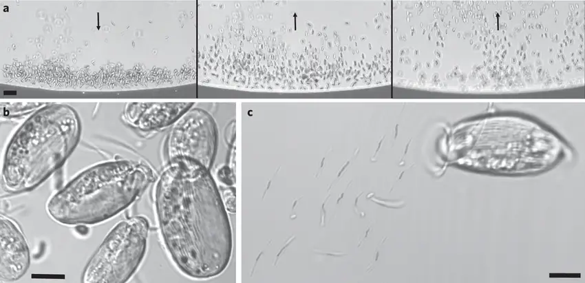
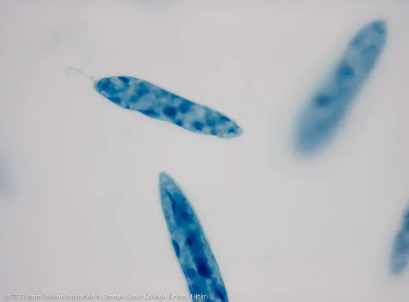
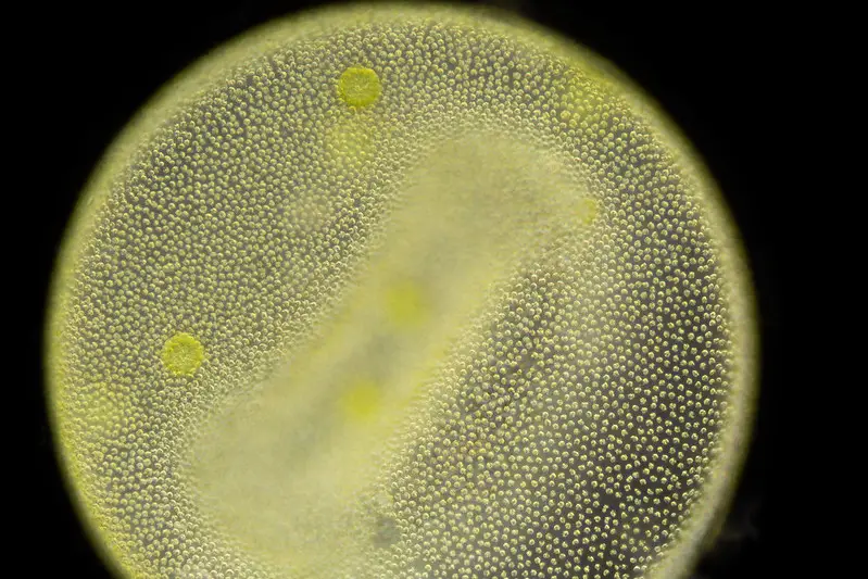
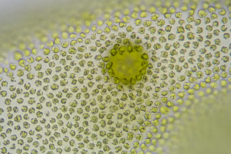
Some examples of protists include Amoeba proteus, Paramecium pentaurelia, Stentor coeruleus, Euglena gracilis, Colonial volvox, and Slime mold Physarum polycephalum.
In summary, protists are a diverse group of eukaryotic organisms that do not fit into the categories of animals, plants, or fungi. They play a crucial role in the evolutionary history of life on Earth, being considered the ancestors of more complex organisms.
Characteristics of Protists
Protists are a diverse group of eukaryotic organisms with various unique features. Here are the key characteristics that all protists share:
- Eukaryotic Organisms: Protists are eukaryotes, meaning they have cells with a membrane-enclosed nucleus. This distinguishes them from prokaryotes, such as bacteria, which lack a defined nucleus.
- Presence of Mitochondria: Protists possess mitochondria, which are organelles responsible for energy production through cellular respiration. This characteristic is common among eukaryotes and plays a crucial role in providing energy for various cellular processes.
- Parasitic Capabilities: Some protists can be parasitic in nature, like Trypanosoma protozoa. These organisms live on or within other living organisms, often causing harm to their hosts.
- Habitat: Protists are typically found in aquatic environments, such as lakes, rivers, and oceans. However, they can also exist in soil or other moist environments.
- Unicellular and Multicellular Forms: While the majority of protists are unicellular, there are exceptions. For instance, kelps, which belong to the Kingdom Protista, are multicellular protists and can grow to impressive heights, with examples like Giant Kelp reaching up to 100 feet.
- Presence of Nucleus and Membrane-Bound Organelles: Protists have a nucleus, a central control center for the cell’s genetic material, and various membrane-bound organelles that compartmentalize and carry out specific functions within the cell.
- Autotrophic, Heterotrophic, or Symbiotic: Protists can exhibit various nutritional strategies. Some are autotrophic, meaning they can produce their own food through photosynthesis, like certain types of algae. Others are heterotrophic, relying on consuming other organisms or organic matter for nutrition. Additionally, some protists engage in symbiotic relationships, where they live in close association with other organisms, often providing mutual benefits.
- Locomotory Organs: Many protists possess locomotory organs that enable them to move. These may include cilia and flagella, which are hair-like structures used for propulsion. Others use a pseudopodium, a temporary extension of the cell, for movement.
- Reproduction: Protists typically reproduce asexually through methods like binary fission or budding. However, in rare cases or under specific conditions, some protists can undergo sexual reproduction.
In conclusion, protists are a fascinating and diverse group of eukaryotic organisms with a wide range of characteristics. Despite their differences, they all share the common trait of being eukaryotic and play essential roles in various ecosystems as autotrophs, heterotrophs, and even parasites. Their presence in diverse environments, from water bodies to soil, underscores their adaptability and importance in the natural world.
Types of Protists
Protists are a diverse group of eukaryotic organisms that can be classified into three main types based on their characteristics and functions:
- Animal-like Protists (Protozoa): Animal-like protists, also known as protozoa, are heterotrophic organisms, meaning they obtain their nutrition by consuming other organisms or organic matter. They are typically motile, possessing structures like cilia, flagella, or pseudopodia that allow them to move. Some examples of animal-like protists include Amoeba, Paramecium, and Euglena. These organisms exhibit various animal-like behaviors and are often found in aquatic environments.
- Plant-like Protists (Algae): Plant-like protists, also known as algae, are autotrophic organisms capable of carrying out photosynthesis. They use sunlight to produce their own food and are an essential part of aquatic ecosystems. Algae come in various forms, from microscopic unicellular species to large multicellular forms like seaweed. Some common examples of plant-like protists include Spirogyra, Chlamydomonas, and Diatoms.
- Fungi-like Protists (Slime Molds and Water Molds): Fungi-like protists are heterotrophic organisms that share some characteristics with fungi, particularly the presence of cell walls in their cells. They reproduce by forming spores. Slime molds and water molds are examples of fungi-like protists. Slime molds, like Physarum polycephalum, are known for their unique life cycle and behavior. Water molds, such as Phytophthora infestans, can be plant pathogens and cause diseases in crops.
It is important to note that the classification of protists can be complex and ever-changing due to the vast diversity within this kingdom. Various factors, such as shape, size, nuclear structures, and cytoplasmic organelles, contribute to their classification. As a result, the taxonomy of protists continues to be refined as new research and discoveries are made.
One of the most common and practical approaches to categorizing protists is based on their nutrition and motility. This approach simplifies the understanding of protist diversity and helps scientists study and identify these fascinating organisms more effectively.
In conclusion, protists encompass a wide array of organisms, and they can be broadly categorized into animal-like, plant-like, and fungi-like protists based on their characteristics and nutritional strategies. Their importance in ecosystems, their roles as primary producers or consumers, and their unique behaviors make them a critical group of organisms to study and understand in the field of biology.
Requirement for Observing Protist Under Microscope
To observe protists under a microscope, you will need the following essential tools and materials:
- A Microscope: Use a compound light microscope with a magnification of at least 40x. Compound microscopes are ideal for observing small, transparent organisms like protists. They allow you to view the cellular structures and movements of protists in detail.
- Wet Mount Slide: Prepare a wet mount slide to hold the protist sample. A wet mount slide is created by placing a drop of water or saline solution on a clean glass slide. The liquid helps keep the protists alive and allows them to move freely during observation.
- Coverslip: After placing the water or saline solution on the slide, carefully position a thin piece of glass called a coverslip over the drop. The coverslip prevents the water from evaporating and keeps the protists in place, allowing for a clear view under the microscope.
- Protist Sample: Collect a sample of protists from a suitable source. Pond water, soil, or even your own saliva can contain various types of protists. Using a dropper or pipette, transfer a small amount of the sample to the center of the wet mount slide.
- Protist Identification Guide: Having a protist identification guide is beneficial, especially if you are new to observing these microorganisms. A guide can help you identify and differentiate the different types of protists based on their characteristics and features.
Procedure for Observing Protist Under Microscope
Observing protists under a microscope can be an exciting and educational experience. Follow these steps to successfully observe and identify protists:
- Gather Your Materials: Collect all the necessary materials, including a compound light microscope, a wet mount slide, a coverslip, a protist sample, and a protist identification guide if available.
- Prepare the Wet Mount Slide: Place the wet mount slide on a clean surface. Add a drop of water or saline solution to the center of the slide using a dropper or pipette.
- Add the Protist Sample: Using the same dropper or pipette, transfer a small amount of the protist sample to the drop of water or saline solution on the wet mount slide. Mix the sample gently with the liquid.
- Place the Coverslip: Carefully position a coverslip over the drop of water and protist sample. To avoid trapping air bubbles, place one edge of the coverslip on the slide and then slowly lower it over the sample.
- Secure the Coverslip: Gently press down on the coverslip to ensure that it is in contact with the water or saline solution and that the sample is spread evenly.
- Set Up the Microscope: Place the prepared wet mount slide on the microscope stage. Secure it in place using the stage clips or the slide holder.
- Adjust the Microscope to Low Power: Start with the lowest magnification (e.g., 40x) to get a general overview of the protist sample. Adjust the focus and lighting as needed to obtain a clear image.
- Locate Protists: Use the microscope’s stage controls to move the slide around and locate areas where protists are concentrated. Protists may be dispersed unevenly, so exploration is essential.
- Increase the Magnification: Once you have located a protist, gradually increase the magnification to get a closer look at its structures and movements. Adjust the focus as needed to keep the protist in clear view.
- Observe Protist Movements: Observe the protist’s movements carefully. Note if it uses flagella, cilia, pseudopods, or other means of locomotion.
- Identify the Protist: If you have a protist identification guide, use it to identify the protist based on its characteristics and features. Compare your observations to the guide to make an accurate identification.
- Record Your Findings: Take notes or use a microscope camera to capture images or videos of the protists you observe. Document any unique features, behaviors, or movements for further study.
- Cleanup: After observing the protists, carefully clean the wet mount slide and coverslip. Properly dispose of the protist sample according to safety guidelines.
Observing protists under a microscope offers an opportunity to explore the fascinating microscopic world and gain insights into the diversity and behaviors of these essential organisms. Proper handling and care of the microscope and slides ensure accurate and enjoyable observations.


Additional tips for observing protists under a microscope
- Fresh Sample: Protists are delicate organisms and can die quickly, especially when exposed to air. Therefore, it is essential to use a fresh protist sample for observation. Collect the sample just before preparing the wet mount slide to ensure the protists are alive and active during the observation.
- Low-Power Objective: Begin your observation using a low-power objective, such as 40x or 100x. This will provide a broader view of the protist sample, making it easier to locate and identify protists. Once you have found the protists of interest, you can then increase the magnification for a closer look.
- Appropriate Magnification: While increasing the magnification allows you to examine the protists in greater detail, be cautious not to go too high. Excessive magnification may lead to a smaller field of view, making it challenging to follow the movements of the protists. Use the optimal magnification that provides a clear view without sacrificing visibility.
- Observe Movement: Take note of the protist’s movement patterns. Some protists use flagella (whip-like appendages), cilia (hair-like structures), or pseudopods (temporary extensions of the cell) for locomotion. Movement characteristics can be useful in identifying different types of protists and understanding their behaviors.
- Use a Protist Identification Guide: If you are not familiar with the different types of protists, having a protist identification guide can be immensely helpful. The guide will contain information about various protists’ characteristics, enabling you to identify the protists accurately based on what you observe under the microscope.
- Take Your Time: Observing protists can be challenging, especially if they are small and actively moving. Be patient and take your time to explore the slide thoroughly. Adjust the focus and lighting as needed to get the best view of the protists.
- Record Your Observations: Keep a notebook or record your observations electronically. Document the features, movements, and any distinguishing traits you observe in each protist. Taking notes will aid in reviewing your findings and discussing them with others.
- Practice Makes Perfect: The more you practice observing protists under the microscope, the more proficient you will become. Gradually, you’ll develop the skills to identify different types of protists and recognize their unique characteristics.
By following these additional tips, you can enhance your experience of observing protists under a microscope. With careful preparation, proper techniques, and a curious mind, you can uncover the fascinating world of these tiny yet remarkable organisms.
Additional safety precautions
- Avoid Touching the Coverslip: When handling the coverslip, be cautious not to touch it with your fingers. Touching the coverslip can introduce contaminants to the protist sample, affecting the accuracy of your observation. Always handle the coverslip by its edges or use a pair of forceps for placement.
- Practice Good Hygiene: If you are using a protist sample collected from an outdoor source, such as pond water or soil, wash your hands thoroughly with soap and water after handling the sample. This precaution helps prevent any potential exposure to harmful microorganisms.
- Watch Eye Safety: If your microscope has a high-powered objective, like 100x, be mindful of the distance between your eyes and the lens. Getting too close to the lens can result in injury to your eyes. Always keep a safe distance from the microscope eyepiece during use.
- Use a Microscope Cover: When observing protists, always use a microscope cover. The cover helps maintain a consistent environment on the slide, preventing the protist sample from drying out, which could lead to inaccurate observations.
- Proper Disposal of Protist Samples: Once you have finished observing the protists, dispose of the sample properly. Depending on the type of protist sample and local regulations, you may need to flush it down the sink with tap water or add household bleach or isopropanol to the culture before disposal. Follow appropriate disposal procedures to protect the environment and prevent potential contamination.
Additional Tips for Microscope Safety:
- Wear Safety Glasses: Always wear safety glasses or goggles when using a microscope. This essential safety gear protects your eyes from any accidental spills or breakage of glass components.
- Handle the Microscope with Care: Avoid bumping or dropping the microscope, as it can damage the lenses or other delicate parts. Always carry the microscope with both hands, ensuring a stable grip.
- Inspect the Microscope: Before use, inspect the microscope to ensure it is in proper working condition. Check for any signs of damage or malfunctions. If you notice any issues, do not use the microscope and notify a supervisor or lab manager immediately.
- Follow Laboratory Safety Guidelines: Adhere to any specific safety guidelines set by your institution or laboratory when using the microscope. This may include proper waste disposal, use of personal protective equipment, and handling hazardous materials.
By following these additional safety precautions, you can ensure a safe and enjoyable experience when observing protists under a microscope. Prioritizing safety not only protects you but also helps maintain the accuracy and integrity of your observations.
FAQ
What is a protist?
Protists are a diverse group of eukaryotic microorganisms that are not classified as plants, animals, or fungi. They display various characteristics and behaviors, making them an intriguing subject for microscopic observation.
How can I collect a protist sample for observation?
You can collect a protist sample from various sources, such as pond water, soil, or even a water droplet from a plant leaf. Use a dropper or pipette to transfer a small amount of the sample to a wet mount slide.
What magnification should I use to observe protists?
Start with a low-power objective, such as 40x, to get a general view of the protist sample and locate protists. Once located, you can increase the magnification gradually to observe their structures and movements in greater detail.
How can I identify different types of protists?
Using a protist identification guide can be helpful in identifying different types of protists. The guide will contain information about their characteristics, shapes, and movements, assisting you in accurate identification.
What are some common locomotion methods of protists?
Protists can move using structures like flagella (whip-like appendages), cilia (hair-like structures), or pseudopods (temporary extensions of the cell). Their unique locomotion methods are essential for their survival and ecological roles.
How do I prevent the protist sample from drying out?
Using a microscope cover or ensuring a tight seal between the coverslip and the wet mount slide can help prevent the protist sample from drying out during observation.
Are there any safety precautions I should follow when observing protists under a microscope?
Yes, it’s essential to wear safety glasses, avoid touching the coverslip with your fingers, and practice proper hygiene when handling protist samples. Additionally, always follow your laboratory’s safety guidelines.
Can I observe protists in my saliva?
Yes, you can observe protists in your saliva. Collect a small sample of saliva and prepare a wet mount slide as you would with other protist samples. Saliva may contain various protists, providing a unique opportunity for observation.
How can I maintain the health of the protist sample during observation?
To keep the protist sample healthy and active during observation, ensure you use a fresh sample, and do not expose it to prolonged periods of air exposure. Additionally, maintain the proper temperature and moisture levels for the protists to thrive.
Can I observe protists from different sources in the same slide?
Yes, you can combine protist samples from different sources in the same slide. However, be cautious about contamination, and avoid overcrowding the slide, as it may hinder your ability to observe individual protists clearly.


