Table of Contents
What is Algae?
- Algae (singular: alga) are diverse, photosynthetic organisms that belong to the group of protists in the Kingdom Protista. They are simple, autotrophic organisms that convert sunlight, carbon dioxide, and water into organic compounds through the process of photosynthesis. Algae can be found in various aquatic environments, including freshwater and marine habitats, as well as some moist terrestrial environments.
- There is a wide range of algae species, and they come in various sizes and colors, including green, red, brown, and golden hues. Some algae are unicellular, existing as individual cells, while others are multicellular, forming colonies or larger structures.
- Algae play a crucial role in the ecosystem as primary producers, producing a significant portion of the Earth’s oxygen and serving as the base of many food chains in aquatic environments. They also contribute to the carbon cycle by sequestering carbon dioxide from the atmosphere during photosynthesis.
- Beyond their ecological importance, algae have practical applications in various industries. For instance, certain types of algae are used as food sources for humans (e.g., seaweed) and as additives in food products. They are also used in the production of biofuels, pharmaceuticals, and various biotechnological processes.
- Due to their ability to grow rapidly under the right conditions, some species of algae can cause harmful algal blooms (HABs) that produce toxins harmful to marine life and can have negative impacts on human health, fisheries, and tourism. Monitoring and managing algae blooms are essential to mitigating their potential adverse effects.
- Overall, algae are incredibly diverse and biologically important organisms that have a significant impact on both natural ecosystems and human activities.
Requirement for observation of Algae Under Microscope
- Microscope with Adequate Magnification: The first and foremost requirement is a microscope with a magnification of at least 40x or higher. This level of magnification enables researchers and enthusiasts to observe the intricate details of algae cells, their structures, and various morphological features. Higher magnification power, such as 100x, may also be necessary to study smaller and more intricate algae species.
- Slide and Coverslip: A clean and appropriately sized glass microscope slide is necessary to hold the algae sample. The slide provides a flat and stable surface for placing the specimen, facilitating ease of observation. A coverslip is then placed over the sample to prevent distortion, protect the specimen, and reduce evaporation during the examination.
- Algae Sample: An algae sample containing the target organism is crucial for observation. The sample can be collected from various sources, including water bodies, aquatic environments, or laboratory cultures. It is essential to handle the sample carefully to avoid contamination and ensure the algae’s natural state is preserved.
- Water or Mounting Medium: A drop of water is typically used to suspend the algae sample on the slide. The water serves as a mounting medium, allowing the specimen to spread evenly and become visible under the microscope. Alternatively, specific mounting media can be used to enhance visibility and preserve the algae’s natural appearance.
- Staining Solution (Optional): While not always necessary, a staining solution may be used to enhance specific features of the algae, making them more distinguishable under the microscope. Stains can provide contrast and help differentiate various cell structures or aid in species identification. Care should be taken to use appropriate stains that do not alter the algae’s cellular characteristics significantly.
- Light Source: A reliable light source is essential for illuminating the algae sample. Most microscopes come with built-in light sources, either in the form of a built-in lamp or an external light attachment. Proper illumination ensures a clear and well-defined image of the algae cells, enabling better analysis and identification.
Algae Sample Collection Procedure
Algae sample collection is a crucial step in studying these diverse organisms. To ensure a successful and accurate collection, follow this unique procedure:
- Choose the Sampling Location: Select a suitable location to collect the algae sample. Algae can be found in various habitats, including ponds, lakes, rivers, streams, oceans, and even on wet surfaces. Consider the research objectives and target algae species when deciding on the sampling site.
- Collecting the Algae Sample: Use appropriate tools to collect the algae. For free-floating algae, use a soft brush to gently brush the algae from the surface. Alternatively, use forceps to pick up individual algae cells or filaments. Be careful not to contaminate the sample or damage fragile algae structures during collection.
- Securely Store the Sample: Place the collected algae in a clean and sealable container. Properly label the container with essential information, including the location of collection, date, and any other relevant details. This labeling aids in proper sample documentation and analysis.
- Timely Transportation to the Laboratory: Transport the algae sample to the laboratory as soon as possible. Prompt transportation helps preserve the sample’s integrity and prevents potential changes or degradation that may occur with prolonged storage.
Tips:
- Collect samples from various locations to obtain a representative sample of algae diversity in the area.
- When collecting algae from a water source, sample at a depth of at least 10 cm to ensure representation of free-floating algae.
- Before collecting algae from a surface, clean it to remove contaminants that could interfere with identification.
Safety Precautions:
- Wear gloves during algae collection to protect against harmful bacteria or parasites in the water.
- When collecting algae from marine environments, be aware of potential hazards such as jellyfish or stingrays.
Storage:
- Store the sample in a cool, dark place to preserve its condition until processing.
- The sample can be stored for up to 24 hours before processing.
Processing:
- Upon returning to the laboratory, process the sample. This may involve staining, mounting on a slide, or preparing for microscopic analysis.
- The processing methods will depend on the algae type and the identification techniques being used.
Identification:
- After processing, identify the algae using a microscope, field guide, or online resource. Carefully examine the algae’s morphology, characteristics, and any specific features to determine the species accurately.
Following this comprehensive algae sample collection procedure ensures the acquisition of reliable data, enabling researchers to gain valuable insights into the diverse and fascinating world of algae.
Procedure for observation of Algae Under Microscope
- Microscope Preparation: Begin by setting up the microscope on a stable surface. Ensure that the microscope is clean and free from dust or debris. Adjust the focus and magnification to the lowest setting to provide a clear starting point for the observation.
- Algae Sample Collection: Collect a sample of algae from a suitable source. Algae can be found in various aquatic environments such as ponds, lakes, streams, or even in a water bottle or aquarium. Use a clean container or sample collection kit to scoop or extract a small amount of algae-rich water.
- Preparing the Slide: Take a clean glass microscope slide and place it on a flat surface. Using a dropper or a pipette, transfer a small drop of the algae sample onto the center of the slide. Be cautious not to add too much liquid to avoid spillage and distortion.
- Applying the Coverslip: Gently lower a coverslip over the drop of liquid containing the algae. Position the coverslip at a slight angle to avoid trapping air bubbles. Gradually lower the coverslip to cover the entire specimen smoothly. This step helps create a flat and uniform sample for observation.
- Optional Staining (If Desired): If greater contrast and detail are required, you may consider using a staining solution. Apply a small amount of the appropriate staining solution to the edge of the coverslip. The stain will spread under the coverslip due to capillary action, enhancing specific features of the algae cells.
- Microscopic Observation: Carefully place the prepared slide onto the microscope stage. Start with the lowest magnification objective (e.g., 40x) to locate and center the algae. Gradually increase the magnification as needed to observe finer details. Adjust the focus to obtain a clear and crisp image.
- Note-taking and Sketching: As you observe the algae, take notes on their characteristics, such as size, shape, and any distinctive features. You may also create sketches to capture the morphology of the observed algae accurately. These notes and sketches will be valuable for documentation and further analysis.
- Clean-up and Preservation: After the observation is complete, carefully remove the slide from the microscope stage. Clean the slide and coverslip with a suitable cleaning solution to ensure they are ready for future use. If you wish to preserve the algae sample, consider sealing the coverslip edges with clear nail polish or specialized mounting media.
Observation
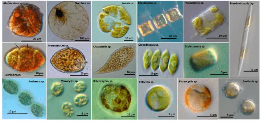
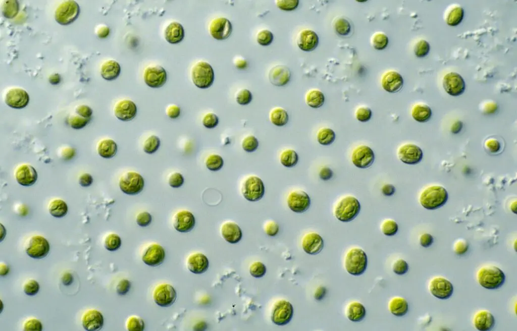
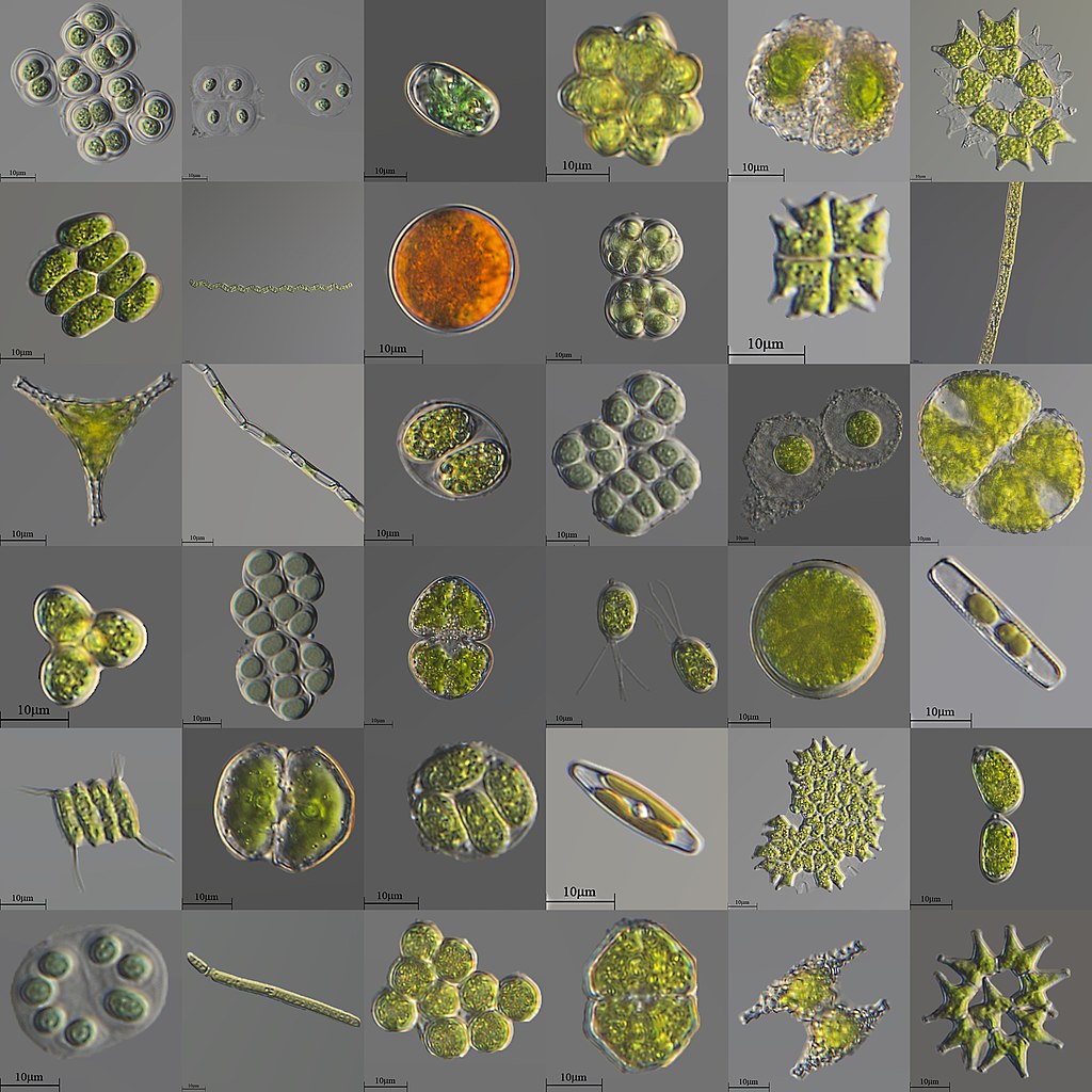
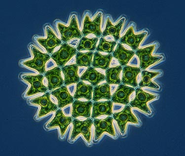
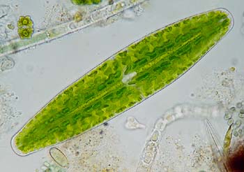
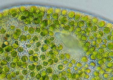
- Chlorophyta Algae: Chlorophyta, commonly known as green algae, are prevalent and easily recognizable due to their green coloration resulting from the presence of chlorophyll. These algae can be observed under the microscope in both single-celled and multicellular forms, displaying a wide range of shapes, such as spheres, filaments, and sheets. Their cellular structures and chloroplasts containing chlorophyll pigments are clearly visible, allowing for a detailed study of their photosynthetic capabilities.
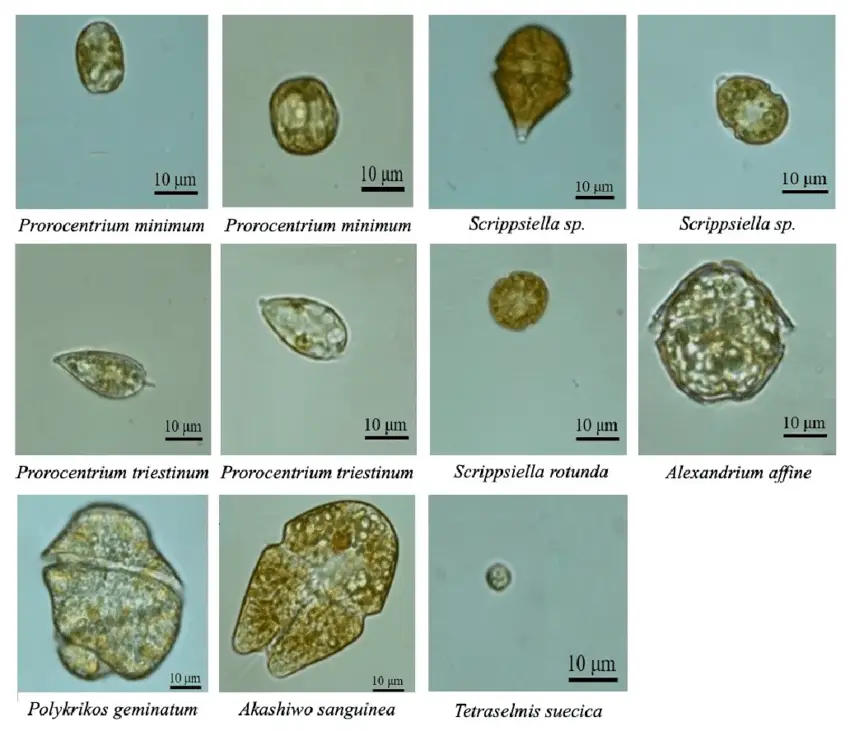
- Dinoflagellates Algae: Dinoflagellates are a distinct group of algae characterized by their unique whip-like structures called flagella, which facilitate movement. Under the microscope, dinoflagellates can be observed as single-celled organisms or forming colonies. Their striking colors, including red, green, and brown, add to their allure. The presence of flagella is evident during microscopic observation, providing insights into their locomotion mechanisms and distinctive cell structures.
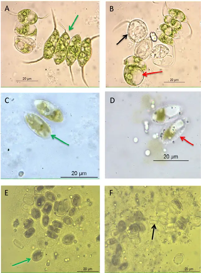
- Diatoms Algae: Diatoms are remarkable algae that showcase intricate silica shells, known as frustules, which come in various shapes and sizes. Their exquisite and delicate frustules can be admired under the microscope, offering a mesmerizing view of their geometric patterns. Diatoms can be found in both single-celled and colonial forms and are commonly encountered in both marine and freshwater environments.
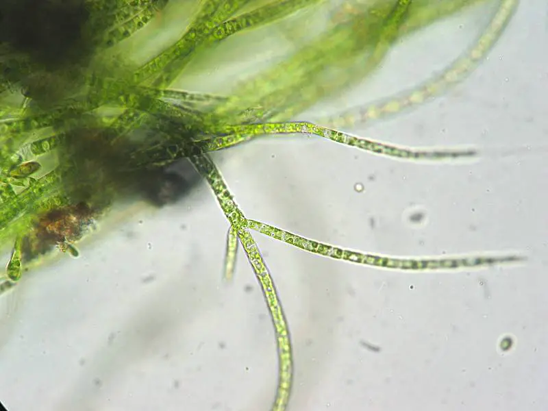
- Cyanobacteria (Blue-Green Algae): Cyanobacteria, also known as blue-green algae, exhibit a unique blue-green coloration due to the presence of the photosynthetic pigment phycocyanin. Under the microscope, cyanobacteria appear as single-celled or multicellular organisms, presenting diverse shapes like spheres, filaments, and sheets. Their blue-green pigmentation and distinct cellular structures are readily observable, making them stand out during microscopic examination.
To observe algae under the microscope, one can follow a simple procedure: collect a sample of algae from a suitable source, prepare a microscope slide with the sample and a drop of water, and gently lower a coverslip to create a flat and uniform sample. The use of staining solutions may enhance specific features, if desired. Observations should begin with low magnification to locate and center the algae, gradually increasing magnification for finer details.
During the observation process, keen note-taking and sketching of the observed algae can provide a valuable record for future reference and analysis. Through this microscopy journey, researchers, students, and enthusiasts gain a profound appreciation for the beauty and complexity of the microscopic algae world, fostering a deeper understanding of their ecological significance and contributions to the natural environment.
Safety precautions
- Wear Protective Gear: When collecting algae samples, wear gloves to protect your hands from potential harmful bacteria or parasites present in the water. Gloves act as a barrier, minimizing the risk of skin contact and contamination.
- Avoid Lens Contamination: Refrain from touching the microscope lens with your fingers. Fingerprints and oils on the lens can obstruct clarity and adversely affect the observation. Instead, use lens cleaning tools designed for microscopes to maintain lens integrity.
- Handle Staining Solutions with Caution: If you opt to use staining solutions for enhanced observation, follow the safety instructions provided with the solution diligently. Some stains may contain toxic components, so it is crucial to take necessary precautions and avoid any direct exposure.
- Proper Disposal of Algae Samples: After observing algae samples, dispose of them responsibly. If collected from natural water sources, return the sample to its original location to prevent disruption to the ecosystem. If the samples are hazardous or non-biodegradable, dispose of them according to local regulations.
- Work in a Well-Ventilated Area: Certain algae species can produce toxins, which may be harmful if inhaled. Ensure that you work in a well-ventilated space to minimize potential respiratory risks. Conduct observations in a controlled environment with proper airflow.
- Thoroughly Wash Hands: After handling algae samples, remove your gloves and wash your hands thoroughly with soap and water. This practice minimizes any potential contamination or transfer of algae or pathogens to other surfaces.
- Maintain Microscope Cleanliness: Keep the microscope clean and sanitized. Regularly wipe down the microscope surfaces, including the stage and eyepieces, with a damp cloth after each use. This routine prevents the spread of bacteria and ensures a clear view during subsequent observations.
FAQ
What is the best magnification for observing algae under a microscope?
The best magnification for observing algae typically ranges from 40x to 400x. Lower magnifications are suitable for getting an overview of larger algae structures, while higher magnifications are necessary to study finer details of smaller algae species.
Do I need to use a staining solution to observe algae under the microscope?
Staining solutions are not always necessary but can be helpful for enhancing certain features or distinguishing different cell structures. However, many algae can be observed without staining, especially if they have distinct pigmentation or cell characteristics.
How should I prepare algae samples for microscope observation?
To prepare algae samples, place a drop of the sample on a clean microscope slide, cover it with a coverslip to create a flat and uniform sample, and ensure there are no air bubbles. Staining can be added if desired.
What type of microscope should I use to observe algae?
A compound light microscope is commonly used for observing algae. It provides sufficient magnification and allows for the examination of cellular structures and other features of algae.
Can I observe live algae under the microscope?
Yes, live algae can be observed under the microscope, especially if they are collected from a freshwater source. However, some marine algae may require special techniques to maintain their viability during observation.
What are some common types of algae I can observe under the microscope?
Common types of algae that can be observed under the microscope include diatoms, green algae (Chlorophyta), dinoflagellates, and blue-green algae (Cyanobacteria).
Can I differentiate between algae species using a microscope alone?
While a microscope can help identify certain algae species based on their morphological features, additional techniques such as DNA analysis or specialized staining may be required for precise species identification.
How can I avoid contaminating my algae samples during collection and observation?
Wear gloves during sample collection to avoid contamination. Also, make sure to clean the microscope thoroughly between observations to prevent cross-contamination.
Can I preserve the algae samples for future observation or analysis?
Yes, you can preserve algae samples for future observation by using specialized preservatives or storing them in suitable containers in a cool, dark place.
Why is it important to take notes and make sketches during algae observation?
Note-taking and sketching are crucial for documentation and analysis. They help record important observations, characteristics, and any unique features of the algae, aiding in future reference and research.


