Table of Contents
- Whittacker grouped living organisms into five kingdoms: Kingdom Monera, Kingdom Protista, Kingdom Fungi, Kingdom Plantae, and Kingdom Animalia.
- All eukaryotic unicellular creatures constituted the kingdom Protista. The holozoic heterotrophic protists are known as Protozoa. In contrast to the unicellular protozoans, Kingdom Animalia includes all multicellular hetertrophic holozoic animals, commonly known as metazoa.
- Metazoa are embryonic multicellular heterotrophic mobile creatures. The gametes are generated by a unique structure composed of germ cells and somatic cells (they are not intracellular).
- In addition to being multicellular, the cells are in tight-junction communication, which aids in coordinating and separates the entire multicellular system from the external environment.
- Collagen fibres are widespread among metazoans and serve to hold cells together. Over the whole animal kingdom, sperms are monoflagellated.
Origin of Parazoa
- An ancient genus of creatures, sponges are distinguished by their unique filter-feeding system. The significant events of their origin and evolution occurred throughout the Precambrian period.
- Sponges have traditionally not been viewed as real animals, but rather as being on the threshold between colonialility and full multicellularity. Hence, the word Parazoa has been used to this group of animals.
- The absence of true tissues and organs, the mobility of their cells, the absence of apparent body axes in adult sponges, and their ability to regenerate from dissociated cells, along with the prevalence and significance of monociliated cells, choanocytes, strongly suggest a direct protistan ancestry (i.e. unicellular protists gave rise to the sponges).
- Sponges (Phylum Porifera) are the phylogenetically earliest and most fundamental form of metazoa, with fossil records reaching back to over 600 million years ago (mya) (Archaeocyathus as fossil) and they are still alive today.
- Fossils of sponges were abundant during the Cambrian period, but their quantity and diversity peaked during the Cretaceous. The origin of sponges is a matter of contention.
- It was first postulated at the end of the nineteenth century that choanoflagellates, a group of non-pigmented flagellates, might be related to sponges, a multicellular animal group.
- The basis for proposing this connection is that choanoflagellate cells are very similar to the so-called choanocyte cells of sponges. Proterospongia sp. is closely linked to colonial flagellate protozoans – choanoflagellates and shares their characteristics (similarities).
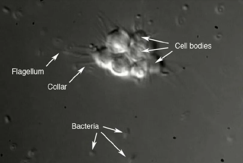
Choanoflagellate: Link to sponges
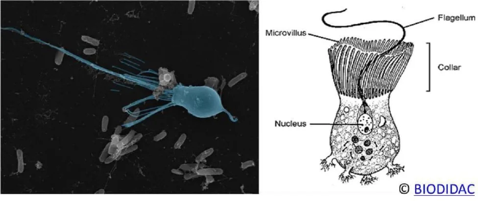
- Choanoflagellates are collared flagellate prevalent in practically all aquatic ecosystems. These are ovoid to spherical cells with a single, forward-directed flagellum ringed by 30–40 tentacles in an inverted cone-shaped collar.
- The flagellum beats with a base-to-tip undulation that generates a forward-directed water stream that attracts bacteria. Many species of choanoflagellates cling to surfaces with stalks.
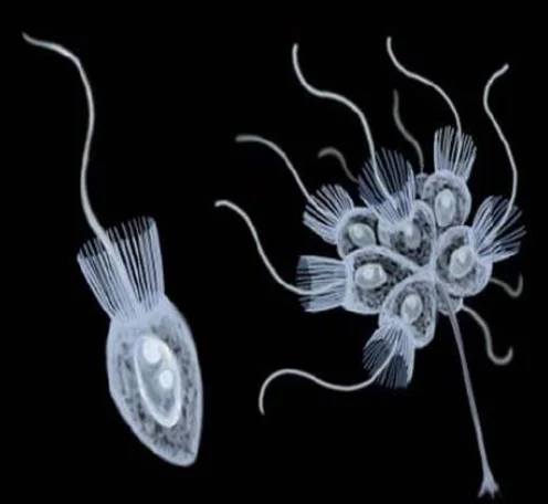
Sponges’ internal chambers are bordered by a series of specialised cells known as choanocytes (choano = collar; cyte =cell). These are the cells that feed sponges. Each choanocyte, like choanoflagellates, possesses a collar of tentacles enclosing a single anterior flagellum. By undulating dozens of flagella, choanocytes generate a stream of water that sucks in water from outside the animal. The water is subsequently evacuated by an exhalant hole after passing through the internal chambers. Choanocytes are responsible for transferring oxygen-rich water to the sponge’s internal cavity and bringing food items, specifically bacteria, to the surface of the collars, where they can be consumed. Hence, choanoflagellates and sponge choanocytes are surprisingly similar in structure and function; both utilise a flagellum to produce water flow and a collar of microvilli to capture prey microorganisms.
Debate about origin of Sponges
- The origin of sponges has been the subject of considerable controversy and differing opinions. It was first considered a distinct entity. A putative evolutionary relationship between choanoflagellates and sponges was originally mentioned in the middle of the nineteenth century.
- French researcher Dujardin (1841), who was interested in protozoa and their evolution, proposed that sponges were colonial Infusoria — the name then used to designate tiny organisms.
- However, following the introduction of the term Protozoa by the German biologist Golfuss (1817) and the subsequent exclusive use of this term by another German scientist, von Siebold (1845), for single-celled organisms, sponges came to be regarded as the crucial link between Protozoa and more complex animals.
- Henry James-Clark (1868) was initially prompted by the apparent resemblance between choanoflagellates and poriferan collar cells (choanocytes) to view sponges as highly specialised choanoflagellate colonies distinct from animal ancestry (James-Clark, 1868).
- With the following discovery of collar cells in numerous non-poriferans (such as Cnidaria and Echinodermata), Porifera re-entered Metazoa, although the debate continued. Haeckel claimed that sponges ought to be classified as Metazoa.
- While he did not consider sponges to be in the straight evolutionary line of animals. Saville-Kent (1880), a contemporary British protozoologist, believed that the sponges as colonial Protozoa descended straight from choanoflagellates due to the similarities of their collared cells.
- On the belief that it was the “missing link” between choanoflagellates and sponges, he named the colonial choanoflagellate he discovered Proterospongia (protero = primitive or original). Proterospongia is regarded as the initial evolutionary stage of porifera.
- Choanocytes are found on the internal lining of porifera, however they are found on the exterior of Proterospongia.
- This is a point of contention, as Choanocytes are found on the internal lining of porifera, but on the exterior of Protero However, this criticism was refuted by the consensus that choanocytes become internal only in the later stages of sponges’ life cycles and are responsible for the evolution of canal networks in Porifera. Evolutionary changes in sponges mostly involve the development of their canal system and skeleton.
Similarities and Dissimilarities
Similarities and Differences with Unicellular Protista
| Similarities with Unicellular Protista | Differences with Unicellular Protista |
| Absence of tissue organisation grade. | Organization of comparable cells in distinct bodily layers with a shared function (outer pinacoderm and inner choanoderm). |
| Cells of choanocytes are present. | A group of cells that have similar tasks. |
| Intracellular skeleton secretion | Their basic developmental pattern (as in metazoans) includes steps such as fertilisation of sperm and egg to produce zygote, cleavage and formation of multicellular blastula, gastrulation as invagination generating amphiblastula larva, and development of germ layers. |
| Nutrition based on phagocytosis and intracellular digestion. | |
| Absence of tight junctions between cells; hence, cellular coordination and communication. |
Similarities and Differences with Metazoan (Cnidaria)
| Similarities with Metazoan (Cnidaria) | Differences with Metazoan (Cnidaria) Metazoans with many cells are poriferans. Nonetheless, because they are too simple and primitive to be considered real metazoans; |
| Similar to cnidarians in that they are both sessile, diploblastic, and acoelomate. | Absence of animal tissue or superior organisational level with division of labour. |
| Both reproduce asexually and form colonies by budding. | Somatic and germinal bodily parts are not distinguished. |
| Parenchymula larva of sponges resembles planula of Cnidaria. | Minimal cellular differentiation. There is no formation of the three embryonic germinal layers, and the developed germ layers can be reversed. |
| Each cell is autonomous and independent, i.e., they are cell aggregates without cooperation. | |
| Amoeboid cells are remarkable for its totipotency (cell fate determination is not rigid). | |
| Animals lack an anterior-posterior axis. | |
| Spongocoel is distinctive and not digestive; it is merely the expanded chamber of a distinctive and specialised water canal system. |
Theories of Origin of Metazoa
Metazoan animals are multicellular, mitochondrial eukaryotes. Metazoa currently includes all creatures with differentiated tissues, such as nerves and muscles. They emerged roughly 700 million years ago from protists. The birth and evolution of metazoa involve two fundamental transitions:
- Transformation of unicellular sponges into multicellular sponges with cell aggregate-level body organisation; and
- Transformation of multicellular sponges into the modular progenitor of bilateralia.
There are two prominent ideas on the origin of metazoans, however one, the syncytial theory, has been criticised. The colonial theory, developed by Ernst Haeckel in 1874, posits that multicellular animals are descended from a colonial predecessor. This is consistent with the theory that choanoflagellates, a colonial protist group, generated the colonies from which multicellular creatures initially emerged. This evolution occurred somewhere during the Precambrian epoch; in 1946, the earliest known fossils of animals were discovered in Precambrian rocks. Due to their delicate bodies, fossil records of early metazoans are scarce. Late Pre-Cambrian fossil records date back 600 million years (from the Edicara hills of Australia). Hence, there is no direct evidence of the genesis of metazoans and their ancestry, and the study of the origin of metazoans is dependent on existing taxa. Zoologists believe, however, that metazoans originated from a common ancestor consisting of unicellular protistans/protozoans. Significant hypotheses on the origin of Metazoa from protozoa include: –
- Syncytial theory – Metazoan animals originated from multinucleate protocilliates, according to the Syncytial theory.
- Classical colonial theory – metazoan creatures developed from colonial choanoflagellate.
- Polyphyletic theory – According to the polyphyletic view, metazoans originated from numerous origins, including choanoflagellates and protocilliates.
1. Syncytial Ciliate Theory of Evolution of Metazoa
- The Syncytial Theory of Metazoan Origin was first postulated by Jovan Hadzi (1953) and Earl D. Hanson (1958).
- Their hypothesis was supported by similarities between multinucleate ciliates and acoelomate flatworms.
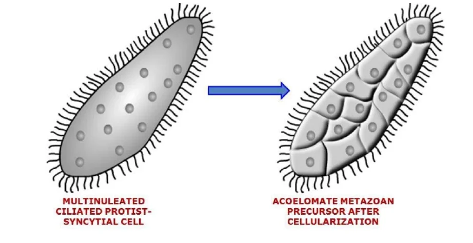
- Syncytial describes a multinucleate state characterised by the absence of cell membranes between neighbouring nuclei. This idea is referred to as the syncytial theory because it emphasises that metazoans (acoelomate flatworms) developed from the segmentation of multinucleated ciliates.
- Afterwards, a cell membrane formed surrounding the nucleus, and distinct cell types differentiated architecturally and functionally.
Proposition
According to Hadzi and Hanson, the origin of metazoans was a bilaterally symmetrical, multinucleate ciliate that adopted a benthic lifestyle with its mouth groove facing the food source. Over time, the surface nuclei were separated by the creation of the cell membrane. The creation of cellular epidermis covering the inner syncytial mass and additional alterations led to the formation of a creature resembling an acoelous flatworm.
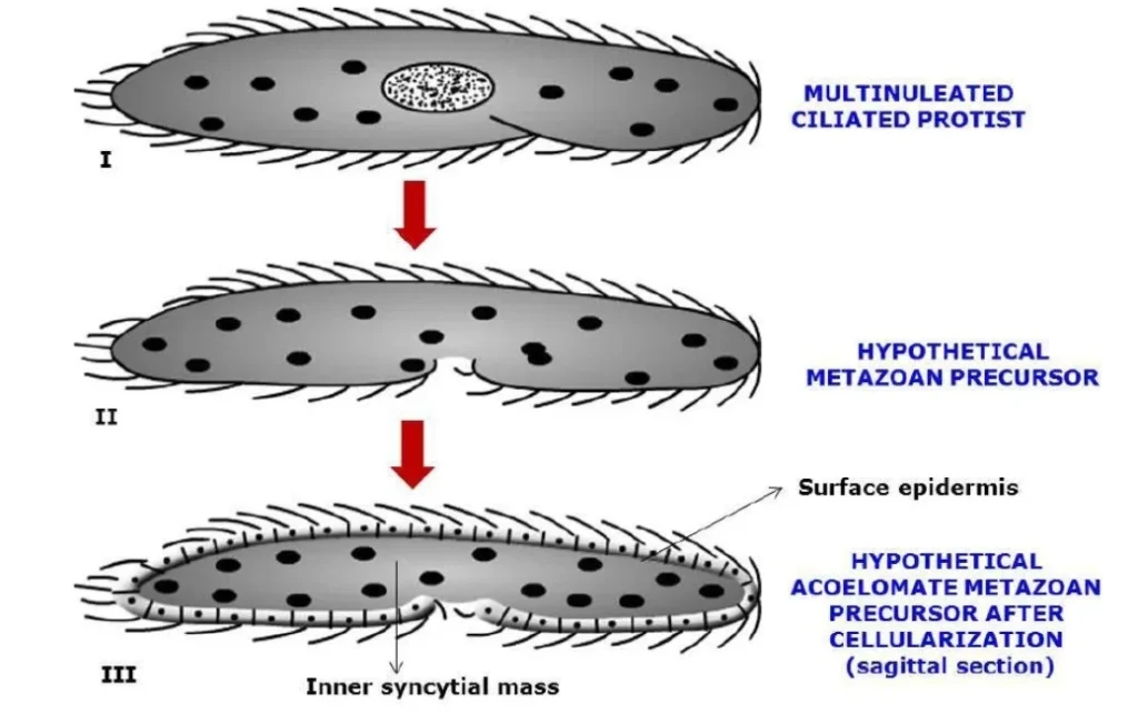
Support
- In terms of bilateral symmetry (multinucleate ciliates resemble acoelomate turbellarians like Convoluta), antero-posterior axis, size, mouth placement, and surface ciliation, ciliated protists and acoel-flatworms share similarities.
- The early multinucleate stage of both slime moulds and insects (Drosophila) undergoes cellularization to produce a multicellular organism.
Disadvantage of Syncytial Ciliate Theory of Evolution of Metazoa
- The idea claims that the first metazoan was an acoelomate triploblastic flatworm, but fails to explain the origin of the more basic Cnidaria and Ctenophora.
- It is unable to account for the radial symmetry of coelenterates/radiata.
- No indication of cellularization was discovered during the ontogeny of lower metazoans; nevertheless, superficial cellularization (cleavage in some arthropods) in insects is caused by centrolecithal yolk.
- It does not explain the genesis of flagella in metazoans or the general presence of flagellated sperm in metazoans. Ciliates do not develop flagellated sperm cells equivalent to those of vertebrates.
- According to this idea, the bilateral symmetry of acoelomates should represent the primitive symmetry of metazoans, whereas radial symmetry should be secondary and degenerate.
- The morphological and gene sequence-based phylogenetic research does not indicate a link with multinucleate ciliates.
In light of more refined phylogenetic analysis and gene sequence analysis, this proposal by Hadzi and Hanson was not universally accepted despite much research.
2. Colonial Theory of Evolution of Metazoa
The Colonial or Flagellate Hypothesis is the most widely recognised explanation for the origin of metazoa. The theory was first offered by Ernst Haeckel in 1874, and Otto Butschli later proposed an offshoot version (1883). Metschnikoff made modifications (1887). L.H. Hyman later renewed this theory (1940). There are a number of variants of this theory:
I. HAECKEL’S THEORY OF GASTREA
The colonial theory is mostly based on the Gasterea Theory of metazoan ancestry proposed by Haeckel (1874).
Proposition
- Metazoan originated through the aggregation of solitary protistan cells into small, hollow, spherical, flagellate colonies, comparable to Volvox with a specific anterior-posterior axis.
- The hypothetical organism’s name was Blastea, and it was compared to the blastula or coeloblastula stages of existing metazoans.
- The basic multicellular organism Blastea consisted of a hollow sphere of flagellated cells. The differentiation of somatic and reproductive cells, as well as the performance of functions like as motility via flagella, reproduction, transport, and absorption via cell membranes.
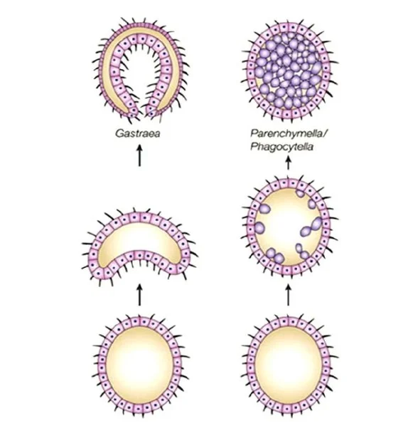
- Blastea transformed into the double-walled sac Gastrea through invagination at the posterior pole.
- According to Haeckel, Gastrea was the hypothesised progenitor of all metazoans, which is symbolised by the gastrula stage in the embryonic development of Metazoa.
- The two layers of the gastrea are conserved as ectoderm and endoderm in all Metazoa. Ectoderm is responsible for perception and motility, whereas endoderm is responsible for food intake and digesting for the entire colony.

Support
- The majority of Haeckel’s idea is founded on embryological evidence.
- The blastea and gastrea phases of ancient metazoans are mirrored by the blastula and gastrula embryonic stages of higher metazoans, according to Haeckel’s Recapitulation theory.
- Similarity in structure between the ancestral gastrea and lower metazoans such as hydrozoan cnidarians and porifera.
- There are numerous living instances of colonial aggregates of flagellated cells, including a. Algae b. Choanomastigotes
Disadvantages of Haeckel’s Theory Of Gastrea
- Issues with ontogeny-phylogeny analogy. Examples used resemble embryonic stages, not adults.
- Endoderm is not necessarily created through invagination, but it can be formed by ectodermal cells that have wandered inward. In the instance of Cnidaria, which is structurally most similar to the gastrea, the Parenchymula larva is generated through multipolar ingression and not by cell invagination. In addition, gastrulas in cnidarians are solid and not hollow.
- Radial symmetry is considered a primitive trait by Haeckel, but it is also observed in echinoderms and some lower chordates, where the larval form is bilaterally symmetrical and the adult form is radially symmetrical (adaptation to sedentary lifestyle).
II. METSCHNIKOFF’S REVISED THEORY OF GASTREA
The Theory of Gastrea by Haeckel (1874) was modified by Metchnikoff (1887).
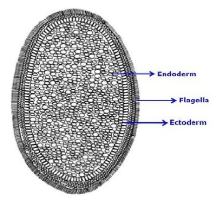
Proposition
- Metchnikoff observed that the original form of gastrulation in cnidarians is ingression (inwandering of ectodermal cells) and not invagination, as Haeckel’s theory predicted. Hence, he proposed that blastea underwent ingression rather than invasion.
- He also discovered that ingression resulted in the cnidarian-typical solid gastrula.
- Planuloid was the name of the ancestor, and The Planuloid Theory was the name of the theory.
- It was an ovoid, solid mass of cells with flagella on the exterior that was radially symmetrical. This was similar to the free-swimming planula larva of coelenterates, therefore it was dubbed the planuloid ancestor, from which all other metazoans arose. It explains the radial symmetry of cnidarians. There was no mouth, and phagocytosis from the outside carried in food.
Support
- Accounting for Cnidarian solid gastrula.
- The metazoan ancestor has a similar structure to the cnidarian planula.
Disadvantages
- There are no extant representatives other than planuloid cnidarians.
III. OTTO BUTSCHLI’S THEORY OF PLAKULA
The Theory of Plakula was proposed by Butschli (1883).
Proposition
- Otto Butschli (1883) believed that no model could explain the purpose of the evolution of the digestive layer, which is more likely to be related with absorption than the exterior layer.
- According to the Plakula theory, the majority of metazoans after sponges were a disc-shaped mass of cells with two layers of flagellated cells, with the ventral layer containing digesting cells and the dorsal layer containing protective and reproductive cells.
- These were benthic organisms that crawled on substrate and have a modified ventral surface for absorption.
- It crouches to collect and digest food.
- May produce further forms with a more persistent cavity.
- Suggested sequence: creeping existence -> bilateral symmetry -> bilaterogastrea
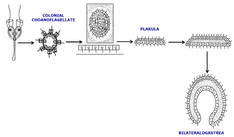
Support
- Trichoplax adhaerens was identified shortly after Butschli’s theory was proposed.
- Comparable in look, habits, and feeding behaviour.
- Grell later amended the hypothesis (1969, 1985).
- Marine ecologists also backed the theory because primordial species were benthic and ate from the ventral surface.
Disadvantages
- Does not explain Cnidarian radial symmetry.
- The jury is still out, although the existence of >bilaterogastrea adds credence to the concept of plakula’s ancestor.
- According to phylogenetic study, Trichoplax is a cnidarian and not a primate.
IV. HYMEN’S COLONIAL THEORY
Hymen (1940) endorsed Haeckel’s notion of hollow blastaea, but added that, as a result of multipolar ingression, hollow blastaea became solid and radially symmetrical Parenchymula/stereogastrula of Metschnikoff (1881), so generating the planuloid metazoan ancestor (similar to planula larva of coelenterates).
Proposition
- Similar to Haeckel’s blastea, Metazoa evolved from a colony of hollow spherical flagellates.
- It demonstrates the distinction between somatic and germ cells.
- The differentiation of somatic cells into locomotor-perceptive and nutritive cell types.
- The hollow sphere is transformed into solid, ovoid, radially symmetrical parenchymula or stereogastrula through multipolar ingression (inwandering of nutritive cells).
- This structure lacks a mouth and digestive system; food is collected by outside flagellated cells and transferred to inner nutritive cells for intracellular digestion.
- This hypothesised progenitor was referred to as a planuloid ancestor due to its resemblance to the planula larva of coelenterates.

MODERN COLONIAL OR FLAGELLATE THEORY
Proposition
- It emphasises that metazoans evolved from colonial flagellate protozoa – choanoflagellates.
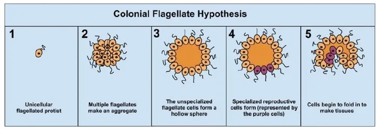
- According to the colonial theory, the pre-metazoans (flagellate protozoan) comprised of a hollow spherical colony with flagellated cells on the surface (which aided in motility and eating) e.g. choanoflagellate colony – Proterospongia haeckeli (called Blastaea by Haeckel, 1874).
- The colony originated from a cell that had repeated mitotic divisions, however the daughter cells did not separate during cell division.
- These offspring cells were deeply entrenched in a proteinaceous ECM that encircled and held them together. The gelatinous ECM also occupied a significant portion of the sphere’s interior.
- Similar to extant choanoflagellates, each cell possessed a single flagellum with a collar. Within the ECM’s centre mass, several cells lost their flagella and dispersed. They are able to produce flagellated cells and other types of cells, such as gametes.
- In addition, there was structural and functional specialisation of colony cells. Haeckel (1874) related this colony to metazoan blastula/coeloblastula and designated it Blastaea.
- It has cellular differentiation in the form of flagellated somatic cells (locomotion and eating) and non-flagellated inner totipotent reproductive cells in the extracellular matrix (ECM) (archaeocytes in sponges and interstitial cells of coelenterates). This Blastaea underwent invagination to become a didermic sac-like organism known as the gastraea with gut (endodermal) and is considered the ancestor of all metazoans. This gastraea was supposed to be the potential ancestor of metazoans akin to the gastrula stage of embryonic development in metazoans.
Support
- Similar to several stages of metazoan embryology, flagellates have a propensity to form compact colonies.
- Comparability between choanoflagellates, choanocytes of sponges, and collar cells of Cnidarians and Echinoderms.
- Choanoflagellates are the only known protozoan whose cells are remarkably similar to those of metazoans.
- The presence of flagella in multicellular creatures, i.e. cells with a single flagellum and a collar of microvilli, is ubiquitous in metazoan animals such as sponges, jellyfishes, sea anemones, etc., as well as in their sperms.
- The attachment of cells to extracellular matrix (ECM) is essentially universal in metazoans.
- In some colonial flagellates, cellular specialisation, i.e. the formation of eggs and sperm, has evolved. The flagellated sperms are present in all metazoans.
- Differentiation of somatic and germinal cells in phytoflagellates such as Volvox.
- According to molecular and morphological evidence, choanoflagellates and metazoans belong to the protozoan taxon Opisthokonta as sister taxa. Comparatively, the present choanoflagellate colony – Proterospongia haeckeli mimics putative pre-metazoans very closely.
Pro-metazoan (hypothetical primitive metazoan) superiority over Pre-metazoan – Choanoflagellates
- Cell surfaces are in close proximity and in constant touch, enabling intercellular communication. This also served to separate the internal ECM from the external environment, thereby providing an independent function.
- Polarization of the anterior-posterior axis of the body.
- Cellular specialisation is fostered by layer separation and body polarity.
- Origin and evolution of massive body size and its complexity through cellular replication, specialisation, and integration of similar specialised units with shared structure and function. Hence, this theory emphasises the monophyletic origin of metazoans, from sponges and cnidarians to higher taxa that are all connected to another animal species.
Polyphyletic theory of metazoan origin
This idea emphasises the origin and evolution of various kingdom groups. Animalia is polyphyletic, meaning that flatworms (planarians) originated from the cellularization of syncytial multinucleated unicellular ciliate protozoans, and sponges originated independently from choanoflagellates.
Summary
- There are unicellular protozoans and multicellular metazoans in the animal kingdom. Metazoans developed from colonies of choanoflagellate protozoa.
- The first metazoans were sponges belonging to the phylum Porifera. Parazoans/poriferans are the simplest multicellular animals in terms of their organisation and occupy the lowest place in the Metazoa lineage.
- Due to their simplicity and lack of tissue and organ organisation, sponges/Porifera were previously classified as Parazoa and believed to be a branching offshoot of the evolutionary tree of metazoan animals.
- Yet, their morphological and physiological integration and division of labour at the cellular level of organisation are more evolved than colonial protozoa.
- Moreover, they have a unique system of water canals for feeding, excretion, and breathing via water current.
- Based on recent molecular, biochemical, and morphological research, it has been determined that parazoans are the most fundamental and primitive metazoans in the evolution of multicellular animals.
- The origin and evolution of metazoans (Parazoa and Eumetazoa) from unicellular protists have been explained by two primary theories: the syncytial and colonial theories.
References
- King N, Rokas A. Embracing Uncertainty in Reconstructing Early Animal Evolution. Curr Biol. 2017 Oct 9;27(19):R1081-R1088. doi: 10.1016/j.cub.2017.08.054. PMID: 29017048; PMCID: PMC5679448.
- Barnes, R.D. (1982). Invertebrate Zoology, V Edition. Holt Saunders International Edition.
- Borchiellini, C., Manuel, M., et.al., 2001. Sponge paraphyly and the origin of Metazoa. Journal of Evolutionary Biology. 14: 171-179.
- Nielsen, C., 2001. Chapter 2, Kingdom Animalia (= Metazoa). in: Animal evolution: interrelationships of the living phyla, 2nd ed. Oxford Univ. Press, New York.
- Brusca, Richard C. and Gary J. Brusca. 2003. Invertebrates, Second Edition. Sinauer Associations, Sunderland, Massachusetts.
- Barnes, R.S.K., Calow, P., Olive, P.J.W., Golding, D.W. and Spicer, J.I. (2002). The Invertebrates: A New Synthesis, III Edition, Blackwell Science.
- King, N., 2004. The unicellular ancestry off Animal Development. Developmental Cell. 7: 313- 325.
- Muller, W.E.G. (2003). The origin of Metazoan complexty: Porifera as integrated Animal. Integr. Comp. Biology., 43:3-10.
- Ruppert, E.E. and R.D. Barnes. 1994. Evolution of metazoans/Epithelial and connective tissues/Metazoan life cycles and development. Pp. 69-73, in: Invertebrate zoology, 6th ed. Saunders College Publishing, Philadelphia.
- https://courses.lumenlearning.com/suny-biology1/chapter/animal-phylogeny-2/
- https://study.com/academy/lesson/parazoa-definition-and-characteristics.html
- http://palaeos.com/metazoa/classification.html
- https://www.biologydiscussion.com/animals-2/body-development-of-animals/32406
- https://comenius.susqu.edu/biol/202/animals/parazoa/porifera/porifera.htm
- https://www.iaszoology.com/parazoa/
- https://egyankosh.ac.in/bitstream/123456789/16480/1/Unit-3.pdf
- https://en.wikipedia.org/wiki/Parazoa
- https://biocyclopedia.com/index/general_zoology/mesozoa_and_parazoa.php
- https://www.sciencedirect.com/topics/earth-and-planetary-sciences/metazoan
- https://www.studyandscore.com/studymaterial-detail/status-of-parazoa
- https://dhingcollegeonline.co.in/attendence/classnotes/files/1603901134.pdf
- http://searchill.weebly.com/uploads/1/2/7/3/12738281/lecture_7.pdf
- https://apsche.ap.gov.in/Pdf/Zoology%20Sem%20I.pdf


