Table of Contents
What is Phylum Porifera? – Definition of Phylum Porifera
Phylum Porifera refers to a group of simple, sessile aquatic animals commonly known as sponges. Sponges lack true tissues and organs and have a porous body structure with numerous channels and chambers that allow water to flow through them. They feed by filtering small particles from the water and are considered to be the simplest multicellular animals. Sponges are found in a variety of marine and freshwater environments and play important ecological roles in their ecosystems.
- Sponges are aquatic sessile (sedentary) invertebrates. They are a successful and diverse group that inhabits both marine and freshwater environments. R. E. Grant classified sponges in the phylum Porifera in 1830. (L. pore bearing animals).
- Due to the peculiar cellular organisation of these organisms, Huxely (1875) and Sollas (1894) considered classifying them separately from Metazoa. Tuzet (1963) noted that despite the fact that sponges had the most primitive characteristics, they were regarded diversified animals produced throughout the evolution of Metazoa and were classified as Parazoa.
- Numerous kinds of sponges have an endo skeleton formed of spicules composed of calcium carbonate or silica. Spicules appear in a variety of shapes and sizes; their kind, shape, combination, and arrangement aid in identifying a specimen.
- Sponges are ecologically, commercially, and evolutionarily significant. Sponges can function as bioremediators and indicators of environmental quality. In the past, sponges were utilised as household objects, for personal cleanliness, pain alleviation, disease treatment, and in art.
- Recent interest in sponges is largely attributable to their ability to produce novel bioactive chemicals, which may have medicinal and antifouling benefits.
- In addition, their skeletal architectures have sparked additional interest due to their exceptional optical and mechanical capabilities, which may enable the future production of new materials.

General Characteristics of Phylum Porifera
Sponges are the first multicellular organisms with body/level organisation at the cellular level. Some organs or tissues are missing.
- All are aquatic, the most being marine and a few being freshwater (family Spongillidae),
- Whether solitary or colonial, everyone leads a sedentary lifestyle.
- The shape of the body may be vase-like, cylindrical, tubular, cushion-like, branching, etc.
- Display either radial symmetry or asymmetries. Between the outer pinacoderm (dermal epithelium) and inner choanoderm (gastral epithelium) is a gelatinous non-cellular matrix (mesohyl).
- Mesohyl is composed of skeletal structures and free amoeboid cells.
- Body Cells are loosely arranged and do not form distinct layers; hence, they are not considered to be fully diploblastic.
- Many incurrent pores (ostia) through which saltwater enters the bodily cavity.
- Body cavity comprised of canals and chambers (Canal system) that facilitate the movement of seawater.
- Seawater leaves the body cavity through one or more excurrent pores (osculum) (Canal system).
- Choanocytes or flagellated collar cells typically line the canal system’s chambers.
- The skeleton is composed of calcareous or siliceous spicules, protein – spongin fibres, or both, or it is absent.
- Intracellular digestion.
- Specific Without respiratory and excretory organs, the canal system is responsible for respiration and excretion.
- There is a primitive nervous system with poorly formed neurons.
- All sponges are hermaphrodite, but cross-fertilization does occur. Reproduction asexually by means of buds or gemmules. Sexual reproduction involves the development of ova and sperm.
- Shows a highly developed capacity for regeneration.
- The cut is holoblastic.
- Amphiblastula or parenchymula are free-swimming, ciliated larvae that develop during embryogenesis.
Morphology of Sponges / Structure of Phylum Porifera
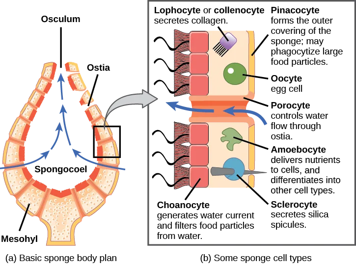
- The simplest sponges have the morphology of a cylinder with a big central hollow, the spongocoel, occupying the cylinder’s interior. Many pores in the body wall allow water to enter the spongocoel. Water entering the spongocoel is expelled through the osculum, a huge shared orifice.
- Nonetheless, sponges exhibit a variety of body morphologies, including differences in the size of the spongocoel, the number of osculi, and the location of the cells that filter food from the water.
- While sponges (with the exception of hexactinellids) lack tissue-layer organisation, they do possess diverse cell types with specific roles. Pinacocytes, which are epithelial-like cells, constitute the outermost layer of sponges and enclose a substance known as mesohyl that resembles jelly.
- Mesohyl is an extracellular matrix composed of a collagen-like gel containing suspended cells with a variety of activities. The gel-like consistency of mesohyl functions as an endoskeleton and preserves the tubular structure of sponges.
- In addition to the osculum, sponges have many pores on their bodies called ostia that allow water to enter. Porocytes, single tube-shaped cells that govern the flow of water into the spongocoel, generate the ostia in certain sponges. In other sponges, ostia are created by folds in the sponge’s body wall.
- Choanocytes (also known as “collar cells”) are found in a variety of locations depending on the type of sponge, but they invariably border the inner parts of a water-flowing area (the spongocoel in simple sponges, canals within the body wall in more complex sponges, and chambers scattered throughout the body in the most complex sponges).
- Choanocytes tend to coat particular inner sections of the sponge body that encompass the mesohyl, whereas pinacocytes line the exterior of the sponge. The shape of a choanocyte is essential to its function, which is to generate a water current within the sponge and to capture and consume food particles by phagocytosis.
- See the visual similarities between sponge choanocytes and choanoflagellates (Protista). This resemblance shows that sponges and choanoflagellates share a recent common ancestor and are closely related. The cell body is encased in mesohyl and has all organelles necessary for normal cell operation. However, projecting into the “open space” within the sponge is a mesh-like collar formed of microvilli with a single flagellum at its centre.
- The cumulative action of the flagella from all choanocytes assists the passage of water through the sponge, bringing water in through the various ostia, into the choanocyte-lined spaces, and out through the osculum (or osculi).
- In the interim, food particles, such as watery bacteria and algae, are captured by the sieve-like collar of the choanocytes, slide down into the cell’s body, are ingested via phagocytosis, and are enclosed in a food vacuole. Choanocytes will then differentiate into sperm for sexual reproduction, dislodging from the mesohyl and exiting the sponge through the osculum as water is discharged.
- Amoebocytes (or archaeocytes) are the second essential cell type in sponges; they are so termed because they travel across the mesohyl in an amoeba-like manner. Amoebocytes have multiple purposes, including transporting nutrients from choanocytes to other cells in the sponge, producing eggs for sexual reproduction (which remain in the mesohyl), transporting phagocytosed sperm from choanocytes to eggs, and developing into more specialised cell types.
- Collencytes and lophocytes generate the collagen-like protein to preserve the mesohyl, sclerocytes produce spicules in some sponges, and spongocytes produce spongin in the majority of sponges. These cells create collagen to maintain the mesohyl’s consistency.
- Depending on the species of sponge, sclerocytes release tiny spicules made of calcium carbonate or silica into the mesohyl of certain sponges. These spicules assist to impart additional rigidity to the sponge’s body. Moreover, the exterior presence of spicules may deter predators. Spongin, an additional form of protein, may also be present in the mesohyl of some sponges.
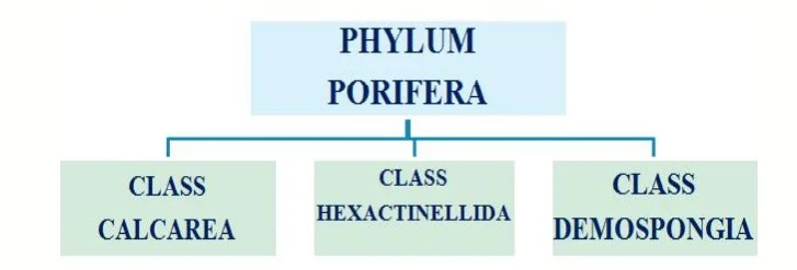
- The presence and content of spicules/spongin are distinguishing characteristics of the three sponge classes: Class Calcarea has calcium carbonate spicules and no spongin, class Hexactinellida has six-rayed siliceous spicules and no spongin, and class Demospongia has spongin and may or may not have spicules; if they are present, they are siliceous. Spicules are particularly prevalent in the class Hexactinellida, which consists of glass sponges. Some of the spicules may acquire gigantic sizes (relative to the average size range of glass sponges, 3 to 10 mm), as shown in the 3 m-long Monorhaphis chuni.

Digestion in Phylum Porifera
- Sponges lack sophisticated digestive, respiratory, circulatory, and reproductive systems. When water travels through the ostia and out the osculum, their food is trapped.
- Bacteria less than 0.5 microns in size are consumed via phagocytosis by choanocytes, which are the major cells involved in nourishment. Pinacocytes are capable of phagocytosing particles larger than the ostium.
- Amoebocytes carry food from cells that have consumed food particles to cells that have not. With this form of digestion, in which food particles are digested within individual cells, the sponge pulls water through diffusion for this type of digestion. This method of digestion is limited by the fact that food particles must be smaller than individual cells.
- All other primary bodily functions of the sponge (gas exchange, circulation, and excretion) are carried out by diffusion between the cells lining the openings of the sponge and the water passing through those pores. All sponge cell types receive oxygen from water by diffusion.
- Carbon dioxide is also emitted into seawater by diffusion. In addition, nitrogenous waste created as a consequence of protein metabolism is expelled into the water as it travels through the sponge via diffusion by individual cells.
Reproduction in Phylum Porifera
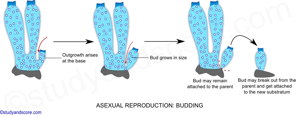
- Sponge reproduction is both sexual and asexual. Fragmentation (when a piece of the sponge breaks off, settles on a new substrate, and develops into a new individual) or budding are typical forms of asexual reproduction (a genetically identical outgrowth grows from the parent and eventually detaches or remains attached to form a colony).
- The development of gemmules is an uncommon form of asexual reproduction that is exclusive to freshwater sponges. Gemmules are inverted sponge-like structures created by adult sponges that are resistant to the environment.
- An inner layer of amoebocytes is surrounded by a layer of collagen (spongin) that may be strengthened by spicules in gemmules. The collagen typically present in the mesohyl becomes the outermost layer of defence.
- Gemmules in freshwater sponges may endure unfavourable environmental conditions, such as temperature fluctuations, and serve to repopulate the area once environmental conditions settle.
- Gemmules are able to connect to a substratum and produce new sponges. Gemmules are a good method of colonisation for sessile organisms due to their ability to tolerate harsh conditions, desiccation resistance, and lengthy periods of dormancy.
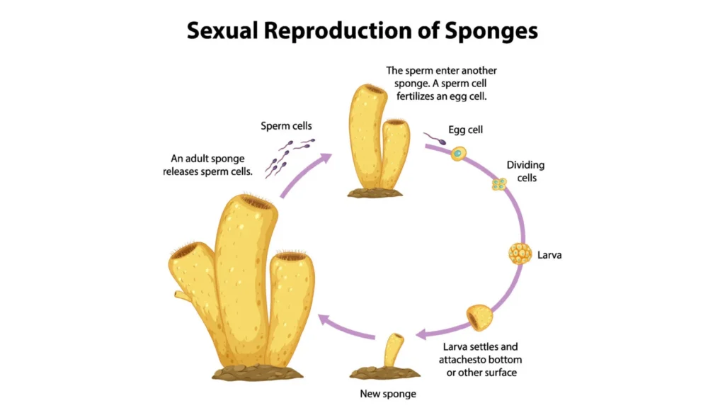
- Sponges engage in sexual reproduction when gametes are produced. Sponges are monoecious (hermaphroditic), meaning that a single individual can concurrently generate both gametes (eggs and sperm).
- In some sponges, gamete generation can occur throughout the year, whereas in others, sexual cycles are dependent on water temperature. Moreover, sponges can become progressively hermaphroditic, generating oocytes first and spermatozoa subsequently.
- Oocytes develop from amoebocytes and are maintained in the spongocoel, while spermatozoa develop from choanocytes and are expelled through the osculum. As observed in certain species, spermatozoa ejection may be a coordinated and timed action.
- Spermatozoa conveyed by water currents are able to fertilise oocytes in the mesohyl of other sponges. Inside the sponge, early larval development takes place, and free-swimming larvae are eventually expelled via the osculum.
Locomotion of Phylum Porifera
- Adult sponges are generally sessile and spend their entire lives adhering to a stable substratum. They do not exhibit large-scale movement like other free-swimming sea invertebrates.
- Nonetheless, sponge cells are able to crawl along substrata due to organisational plasticity. In experimental conditions, scientists have demonstrated that on a physical support, distributed sponge cells have a leading edge for directed movement.
- It has been hypothesised that this restricted creeping movement aids sponges in adapting to microenvironments close to the attachment site. It must be noted, however, that this movement pattern has only been observed in laboratory settings and not in natural sponge environments.
Classification of Phylum Porifera
The classification of Phylum porifera is primarily based on skeleton types. This phylum has been categorised in a number of different ways, but the classifications proposed by Hyman in 1940 and Burton in 1967 are of great significance. The phylum Porifera consists of three classes:
Class 1: Calcarea
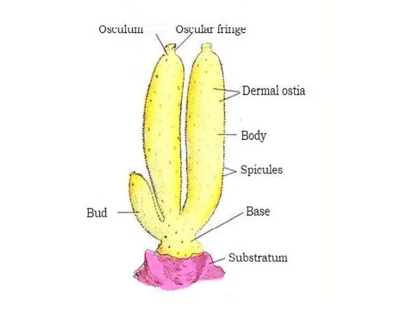
- These are little calciferous sponges (10cm in height). Isolated or colonial.
- The body is cylindrical or vase-shaped.
- The skeleton is composed of calcareous spicules with one, three, or four rays.
- All are aquatic animals.
- The structure of the body may be Asconoid, Syconoid, or leuconid. e.g. Leucosolenia, Scypha, Granita, Scycon.
Class 2: Hexactinellids
- Rarely exceeding one metre in length, sponges are typically of a middling size.
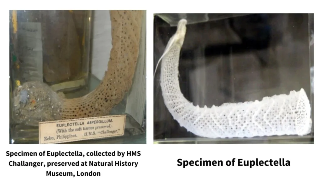
- They are often referred to as glass sponges. The body may be cylindrical, funnel- or cup-shaped.
- The canal system is quite typical and Syconoid in body organisation.
- They are found in the deep ocean. exemplified by Euplectella (Venus flower basket) and Hyalonema (Glass- rope sponge).
Class 3: Desmospongiae
- Individual or colonial
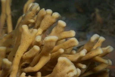
- Cup or vase-shaped body
- The size of Demospongiae was both tiny and enormous.
- Spicules are monaxon or tetraxon in appearance.
- The canal system is of the leuconoid variety.
- All are marine poriferans with the exception of freshwater sponges of the family Spongillidae, including Chalina, Microciona, and Haliscara.
Skeletal system in sponges
Sponges have an endoskeleton that gives their bodies structure and form. The skeletons of all sponges are implanted in the mesenchyme. The skeleton is composed of distinct spicules, interlacing spongy fibres, or both. The skeleton provides support and protection for the soft body portions of sponges. The skeleton is also the basis for classifying sponges into classes such as Calcarea, Hexactanellida, and Desmospongia.
Spongin
Spongin is an organic, sticky, elastic material whose chemical makeup mimics silk. It is a sulfur-containing scleroprotein that is chemically related to collagen. Several types of spongin fibres are found in Desmospongia. It may exist as a bonding agent between siliceous spicules. It can be found in the form of branching fibres that contain siliceous spicules. In Keratosa, spicules are missing and only spongin is produced.
Development of spongin
Spongin fibres are released by spongioblast cells, which are flask-shaped mesenchyme cells. During development, the spongioblast cells are organised in rows, and their spongin rods combine with neighbouring cells to create a fibre. After secreting a given amount of spongin, the spongioblasts vacuolate and are ultimately degraded.
Spicules
Structure and types
Spicules are small crystalline structures that provide stiffness and shape to sponges. Spicule consists of radiating spines or rays from a point. They are secreted by scleroblast cells, which are specialised mesenchymal amoebocytes. The following are examples of several types of spicules:
Based on the sort of deposit on the organic matter core: All types of spicules have an organic core that is surrounded by either calcium carbonate or colloidal silica. Consequently, there are two types of spicules:
- Calcareous spicules: Calcareous spicules are composed of calcium carbonate or calcite as its organic component. This is a feature of the class Calcarea sponges.
- Siliceous spicules: Colloidal silica or Silicon is the organic component of this form of spicules. These kinds of spicules are characteristic of the class Hexactanellida sponges.
Spicules can be large or little according to their size and function. Consequently, there are two types of spicules:
- Megascleres: Megascleres are the largest spicules that make up the sponge’s primary structure.
- Microscleres: They are the microscopic spicules that occur interstitially.
Based on the number of axes and rays: Spicules can appear in various shapes, including basic rods, forks, anchors, shovels, stars, plumes, etc. The morphologies of spicules depend on the number of axes and rays present. Thus, they can be categorised as follows:
- Monaxon: These spicules are created by growth along a single axis in monaxon. They may be straight, resembling needles or rods, or curved. Their terminations might be pointy, hooked, or knobby. Both calcareous and siliceous monaxons exist.
- Triaxon: These triaxon spicules contain three axes that intersect at right angles to create six rays. Thus, it is also known as a hexactinal spicule. These triaxon spicules are characteristic of the class Hexactanellida glass sponges.
- Polyaxon: These are the spicules of a polyaxon, which have numerous equal rays emanating from a central point. They can be grouped to give the appearance of stars. Microscleres are seen alongside polyaxon spicules.
Development of spicules
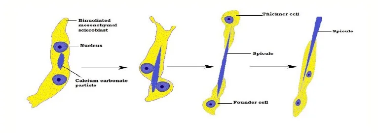
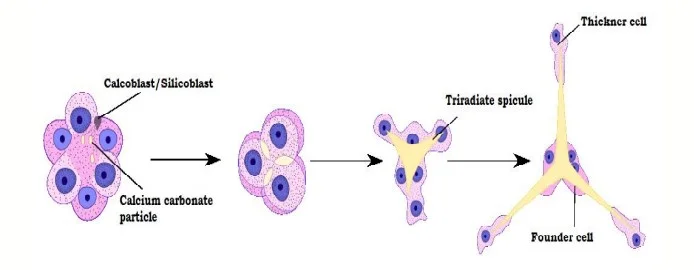
Calcareous spicules are secreted by sclerocytes, a specialised type of cell. These sclerocytes are produced from mesenchymal scleroblasts with two nuclei. A set of two sclerocytes secretes a monaxon spicule or each ray of the triradiate spicule.

One of these sclerocytes functions as a thickening cell, while the other serves as the founder cell. The creation of the spicule is initiated by the deposition of a calcium carbonate particle between the two nuclei of binucleated mesenchymal cells. This particle grows, separating the two nuclei, resulting in the formation of two sclerocytes. Now, the thickening cell deposits an additional layer of calcium carbonate, increasing the spicule’s thickness. After the spicule is fully developed, both the thickening cell and founder cell migrate into the mesenchyme. The calcoblast is the scleroblast that secretes a calcareous spicule, while the silicoblast is the scleroblast that secretes a siliceous spicule.
Economic Importance of Phylum Porifera
The sponge phylum Porifera has great economic significance. Here are several examples:
- Bath sponges: Bath sponges are most frequently used for bathing and cleaning. Many commercial sponges are collected in the wild or cultivated in aquaculture facilities.
- Biomedical research: Several species of sponges produce bioactive substances with potential medical applications, including as anti-cancer, anti-inflammatory, and antimicrobial characteristics, according to biomedical studies. These chemicals are being studied for therapeutic development purposes.
- Food source: In some regions of the globe, certain types of sponges are consumed as a delicacy.
- Aquaculture: Certain species of sponges can be cultivated in aquaculture facilities and sold for a variety of applications, including aquarium filtration and water treatment.
- Habitat creation: Sponges provide essential habitat for a wide range of marine species, including fish and invertebrates. Moreover, they can prevent erosion by stabilising the seafloor.
- Tourism: Sponges are a great draw for divers and snorkelers, thereby contributing to the local economy through tourism.
The economic value of sponges underscores the need for sustainable management and conservation approaches to ensure their future availability and use.
Examples of Porifera
Sycon
- These are solitary or colonial marine sponges found adhering to rocks in shallow waters.
- Many spores cover the body’s cylindrical form. The radial canal is composed of cells with flagella.
- Water enters the body through the Ostia and travels through the prosopyles to the radial canals. These species are capable of both sexual and asexual reproduction.
Cliona
- They are commonly found in coral skeletons, mollusk shells, and other calcareous structures and are also known as Boring Sponges.
- They are coloured green, purple, or pale yellow.
- Leuconoid sponges are distinguished by their canal system, and they reproduce both asexually and sexually.
Hylonema
- These aquatic organisms are also known as glass rope sponges.
- The body is circular or oval with root tufts that are twisted.
- There are little amphidiscs in the skeleton.
Spongilla
- They are prevalent in ponds, streams, and lakes, where they grow on submerged vegetation and sticks.
- The body wall consists of a thin dermis with Ostia pores.
- They possess a canal system like a rhagon. They reproduce both sexually and asexually.
Euplectella
- These are located in deep waters and are also known as Venus flower baskets.
- The cylindrical, long, and curving body is embedded in the mud at the ocean floor.
- The canal system is a straightforward synconoid kind.
- The skeleton is composed of fused siliceous spicules that create a three-dimensional network with parietal gaps.
FAQ
What are the characteristics of phylum Porifera?
The characteristics of phylum Porifera, or sponges, include:
1. Lack of true tissues and organs
2. Asymmetrical or radially symmetrical body plan
3. Porous body structure with numerous channels and chambers called canals and choanocytes, respectively
4. Water flow through the sponge’s body, facilitated by flagellated cells called choanocytes
5. Ability to filter small particles from the water for food
6. Asexual and sexual reproduction
7. Sessile lifestyle, anchored to a substrate
8. Presence of spicules or spongin fibers for structural support, which can be made of calcium carbonate, silica, or protein fibers depending on the species
9. Occurrence in marine and freshwater environments at various depths and temperatures
10. Ecologically important roles such as providing habitat, filtering water, and serving as a food source for other organisms.
Where are the sponges found?
Sponges are found in a variety of marine and freshwater environments, including oceans, coral reefs, and rivers.
What is the mode of nutrition of Poriferans?
Poriferans are filter feeders, meaning they obtain their nutrition by filtering small particles, such as plankton and bacteria, from the water that flows through their bodies.
Why are Poriferans confused to be plants instead of animals?
Poriferans may be confused with plants because they are sessile, lack organs, and have a simple body structure. However, they are considered animals because they are multicellular and lack chloroplasts, which are found in plants and enable photosynthesis.
How are sponges important?
Sponges are important ecologically because they provide habitat for other organisms, filter water, and serve as a food source for other animals. Additionally, some species of sponges contain bioactive compounds that have potential medical and industrial applications.
Why are sponges considered to be animals?
Sponges are considered animals because they are multicellular, have specialized cells, and lack chloroplasts. They are also capable of asexual and sexual reproduction.
Name the different types of sponges.
There are three main types of sponges: demosponges, calcareous sponges, and hexactinellid sponges. Demosponges are the most common and diverse type, accounting for about 90% of all sponge species.
The classification of Porifera is based on?
The classification of Porifera is based on their skeletal structure, which is determined by the type and arrangement of spicules (small, needle-like structures) and spongin fibers that make up their body.
Give a few examples of Poriferans.
Some examples of Poriferans include:
1. Bath sponges (class Demospongiae)
2. Glass sponges (class Hexactinellida)
3. Calcareous sponges (class Calcarea)
4. Neptune’s cup sponge (genus Cliona)
5. Tube sponge (genus Callyspongia)
6. Elephant ear sponge (genus Agelas)
7. Yellow tube sponge (genus Aplysina)
8. Venus flower basket (genus Euplectella)
9. Barrel sponge (genus Xestospongia)
10. Fire sponge (genus Tedania).
References
- Manconi, R., & Pronzato, R. (2015). Phylum Porifera. Thorp and Covich’s Freshwater Invertebrates, 133–157. doi:10.1016/b978-0-12-385026-3.00008-5
- Hill, M. S., & Hill, A. L. (2009). Porifera (Sponges). Encyclopedia of Inland Waters, 423–432. doi:10.1016/b978-012370626-3.00186-1
- Manconi, R., & Pronzato, R. (2016). Phylum Porifera. Thorp and Covich’s Freshwater Invertebrates, 39–83. doi:10.1016/b978-0-12-385028-7.00003-2
- Students, K. S. C., & Biology, B. 381 T. M. (2022, August 24). Phylum Porifera. https://bio.libretexts.org/@go/page/46811
- https://unacademy.com/content/cbse-class-11/study-material/biology/phylum-porifera/
- https://manoa.hawaii.edu/exploringourfluidearth/biological/invertebrates/phylum-porifera
- https://ucmp.berkeley.edu/porifera/porifera.html
- https://www.bio.fsu.edu/~bsc2011l/Summer2009/02%2009%20Sum%20MT%20Porifera.pdf
- https://www.embibe.com/exams/phylum-porifera/
- https://collegedunia.com/exams/phylum-porifera-biology-articleid-7318
- https://www.geneseo.edu/~bosch/InvSponges.pdf
- https://infinitylearn.com/surge/biology/phylum-porifera/
- https://www.toppr.com/guides/biology/animal-kingdom/phylum-porifera/
- https://www.vedantu.com/biology/phylum-porifera
- https://blogs.ubc.ca/mrpletsch/2019/01/10/phylum-porifera/
- https://www.aakash.ac.in/important-concepts/biology/phylum-porifera
- https://www.geeksforgeeks.org/phylum-porifera-features-characteristics-classification-examples/
- https://www.austincc.edu/sziser/Biol%201413/LectureNotes/lnexamII/Phylum%20Porifera.pdf
- https://www.digitalatlasofancientlife.org/learn/porifera/
- https://courses.lumenlearning.com/suny-wmopen-biology2/chapter/phylum-porifera/
- https://www.shapeoflife.org/phylum-porifera
