Table of Contents
What is Germ layer?
- Germ layers play a fundamental role in the process of embryonic development, giving rise to all the tissues and organs that make up an animal’s body. They are the primary layers of cells that form during gastrulation, the transformation of a blastula or blastocyst into a gastrula.
- During gastrulation, cells from the inner cell mass of the blastocyst or blastula undergo morphogenetic movements, shifting and rearranging themselves to new positions. This cellular movement is crucial for the formation of the three germ layers: ectoderm, mesoderm, and endoderm.
- The ectoderm is the outermost germ layer and gives rise to various structures, including the epidermis, nervous system, sensory organs, and other related tissues. It forms the skin, hair, nails, and the lining of the mouth and anus. Additionally, it develops into the brain, spinal cord, and peripheral nerves, playing a vital role in the body’s communication and coordination.
- The mesoderm, which lies between the ectoderm and endoderm, develops into tissues such as muscle, bone, connective tissue, blood vessels, kidneys, and reproductive organs. It contributes to the formation of the circulatory system, skeletal system, and the majority of the body’s internal organs.
- The endoderm is the innermost germ layer and gives rise to the lining of the digestive tract, respiratory system, liver, pancreas, and other associated organs. It forms the epithelial linings of organs and participates in the production of secretory glands, such as the glands of the gastrointestinal tract.
- The fate of the germ layers is highly conserved among triploblastic animals, meaning that the three germ layers develop into specific tissues and organs regardless of the animal’s species. This common developmental process is crucial for understanding the similarities and differences across different organisms.
- The concept of germ layers was first observed by Caspar Friedrich Wolff in the 18th century, who noticed the organization of the early embryo into leaf-like layers. Heinz Christian Pander further advanced the understanding of germ layers in 1817 when he discovered the three primary germ layers while studying chick embryos. Robert Remak’s work between 1850 and 1855 refined the concept, associating each layer with specific structures in the developing organism.
- The terminology we use today, including the terms “ectoderm,” “mesoderm,” and “endoderm,” was introduced by scientists such as Thomas Huxley and Edwin Ray Lankester in the 19th century. These terms have become fundamental in the field of embryology and developmental biology.
- In summary, germ layers are the primary layers of cells that form during gastrulation and give rise to all the tissues and organs in an animal’s body. The ectoderm, mesoderm, and endoderm each contribute to specific structures, providing the necessary foundation for organogenesis and the overall development of an organism.
Fate of The germ layers
1. Ectoderm
- The ectoderm is one of the three germ layers that forms during gastrulation and plays a crucial role in the development of various tissues and organs. It gives rise to several important structures and systems in the body.
- One of the primary contributions of the ectoderm is the formation of the epidermis, which is the outermost layer of the skin. Additionally, it gives rise to epidermal derivatives such as epidermal glands, including sweat glands and oil glands. The ectoderm is responsible for the development of hair, nails, and tooth enamel as well.
- The ectoderm is also involved in the formation of the nervous system. It gives rise to the neural tube, which develops into the brain and spinal cord. Motor neurons, responsible for transmitting signals from the central nervous system to muscles and glands, are also derived from the ectoderm. The neural crest, a specialized group of cells derived from the ectoderm, contributes to the development of the peripheral nervous system. It gives rise to various structures, including sensory neurons, autonomic ganglia, and the adrenal medulla. Additionally, melanocytes, the pigment-producing cells responsible for skin, hair, and eye color, are derived from the neural crest.
- Several important organs and structures of the sensory system also originate from the ectoderm. The ectoderm forms the lens of the eye, cornea, retina, iris, and ciliary muscles. It contributes to the development of the internal ear, which is involved in hearing and balance. Furthermore, the ectoderm is responsible for the formation of the nasal and olfactory epithelia, which are involved in the sense of smell.
- Certain endocrine structures are also derived from the ectoderm. The medulla of the adrenal gland, as well as the posterior and intermediate lobes of the pituitary gland, are formed from the ectoderm. Additionally, the pineal gland, which regulates certain biological rhythms, is also derived from the ectoderm.
- The ectoderm also contributes to the development of the epithelium of the foregut and hindgut. Furthermore, it is involved in the formation of various glands, including mammary glands, salivary glands, lacrimal glands (tear glands), and the enamel of teeth.
- It is important to note that while most of the anterior pituitary is derived from the ectoderm, a small portion called Rathke’s pouch gives rise to this structure.
- In summary, the ectoderm is a germ layer that plays a critical role in the development of diverse structures and systems. It gives rise to the epidermis, nervous system, sensory organs, certain endocrine structures, epithelia, and glands. The ectoderm’s contributions are essential for the proper functioning and coordination of various physiological processes in the body.
2. Mesoderm
- The mesoderm is one of the three germ layers that forms during gastrulation and plays a crucial role in the development of various tissues and organs. It gives rise to a diverse range of structures throughout the body.
- One of the major contributions of the mesoderm is the formation of the dermis, which is the layer of skin located beneath the epidermis. The dermis contains various connective tissues, blood vessels, and sensory receptors, providing support and nourishment to the skin. Additionally, the mesoderm gives rise to the muscles in the body, with the exception of the iris and ciliary muscles of the eye.
- Connective tissues throughout the body, such as tendons, ligaments, and cartilage, are also derived from the mesoderm. These tissues provide structural support and flexibility to various organs and joints. The mesoderm further contributes to the formation of adipose tissue, which functions as a protective cushion and energy storage.
- Several vital organs and systems develop from the mesoderm. The kidneys, responsible for filtering waste products from the blood and maintaining fluid balance in the body, are formed from this germ layer. The gonads, which give rise to the testes in males and ovaries in females, are also derived from the mesoderm. Furthermore, the mesoderm plays a role in the formation of the notochord, a flexible rod-like structure that provides support during early development.
- The mesoderm is crucial for the development of the circulatory system. It gives rise to the heart, blood vessels, and lymph vessels, which collectively ensure the transport of oxygen, nutrients, and immune cells throughout the body. The mesoderm also contributes to the formation of urinary and reproductive ducts, which play essential roles in excretion and reproduction, respectively.
- Most of the skeletal system, including bones and cartilage, is derived from the mesoderm. These structures provide support, protection, and allow for movement in the body. The mesoderm is also involved in the formation of coelomic epithelium, which lines the coelomic cavities in the body.
- Other structures that originate from the mesoderm include the pericardium and pleura, which are protective membranes surrounding the heart and lungs, respectively. The dentine of teeth, a hard tissue that forms the bulk of the tooth, is also derived from the mesoderm. Additionally, the mesoderm contributes to the development of mesenteries, which are membranous sheets that hold organs in place within the body cavity.
- The mesoderm plays a role in the formation of the sclera and choroid of the eyes, providing structural support and nourishment to the eyeball. It also contributes to the wall of the gut, except for its lining, ensuring proper function and support of the digestive system.
- In summary, the mesoderm is a germ layer that gives rise to a wide range of tissues and organs in the body. It forms the dermis, muscles (excluding certain eye muscles), connective tissues, kidneys, gonads, notochord, circulatory system, skeletal system, coelomic epithelium, and various other structures. The mesoderm’s contributions are vital for the proper functioning and development of the body’s systems and provide support, protection, and movement throughout the organism.
3. Endoderm
- The endoderm is one of the three germ layers that forms during gastrulation and plays a crucial role in the development of various organs and tissues. It gives rise to important structures within the body.
- The endoderm primarily forms the epithelial lining of the digestive tract, with the exception of parts of the mouth, pharynx, and terminal portion of the rectum, which are lined by the ectoderm. It contributes to the formation of the lining of the esophagus, stomach, small intestine, and colon. The endoderm is also responsible for the development of glands associated with the digestive tract, including the pancreas, liver, gastric glands, and intestinal glands. These glands play crucial roles in digestion and the secretion of digestive enzymes.
- Several important organs and structures in the body are derived from the endoderm. The inner layer of the tympanic membrane, as well as the lining of the middle ear, is formed by the endoderm. The respiratory system also originates from the endoderm, with the formation of the trachea, bronchi, and lungs. The bladder and a portion of the urethra are derived from the endoderm, contributing to the urinary system.
- The endoderm is involved in the formation of certain endocrine glands as well. It gives rise to the follicle lining of the thyroid gland, which is responsible for the production and release of thyroid hormones. The thymus, an organ involved in immune system function, is also derived from the endoderm.
- In summary, the endoderm is a germ layer that gives rise to the epithelial lining of the digestive tract, excluding specific regions. It contributes to the formation of glands associated with the digestive system, such as the pancreas and liver. Additionally, the endoderm forms the inner layer of the tympanic membrane, lining of the middle ear, trachea, bronchi, lungs, bladder, urethra, thyroid gland, and thymus. The endoderm’s contributions are essential for the proper functioning and development of the digestive, respiratory, urinary, and endocrine systems within the body.
4. Neural crest
- The neural crest is a unique structure derived from the ectoderm during embryonic development. Although it is not classified as a germ layer, its significance is often likened to that of a fourth germ layer due to its crucial role in the formation of various tissues and organs in the body.
- The neural crest emerges along the borders of the neural plate, which eventually gives rise to the central nervous system. As the neural tube forms, some cells located at the edges undergo a process known as delamination, where they detach from the neural tube and migrate to different regions of the embryo. These migratory cells are referred to as neural crest cells.
- The neural crest is a highly dynamic and multipotent population of cells with remarkable developmental potential. It undergoes extensive migration throughout the developing embryo, distributing cells to various regions and contributing to the formation of diverse tissues and structures.
- One of the significant contributions of the neural crest is its involvement in the development of the peripheral nervous system. Neural crest cells give rise to sensory neurons, autonomic neurons, and glial cells found in peripheral nerves. They play a crucial role in connecting the central nervous system to the rest of the body, transmitting sensory information and coordinating autonomic functions.
- In addition to the peripheral nervous system, the neural crest is involved in the formation of numerous other tissues and organs. It contributes to the development of craniofacial structures, including bones and cartilage of the skull, facial muscles, and connective tissues of the head and neck region. The neural crest is also responsible for the formation of melanocytes, the pigment-producing cells that give color to the skin, hair, and eyes.
- Furthermore, the neural crest plays a role in the development of several structures in the cardiovascular system. It contributes to the formation of smooth muscle cells in the walls of blood vessels, as well as the connective tissue and valves of the heart. Neural crest cells also contribute to the development of certain structures in the digestive system, such as ganglia in the gastrointestinal tract.
- Importantly, the neural crest has been associated with several developmental disorders and diseases when its migration, proliferation, or differentiation processes are disrupted. Defects in neural crest development can lead to conditions such as craniofacial abnormalities, heart defects, and pigmentary disorders.
- In summary, although the neural crest is not classified as a germ layer, it is derived from the ectoderm and plays a crucial role in embryonic development. It exhibits remarkable migratory and multipotent properties, contributing to the formation of various tissues and structures in the body. The neural crest is involved in the development of the peripheral nervous system, craniofacial structures, cardiovascular system, and pigment-producing cells. Its intricate involvement in embryogenesis highlights its significance and impact on overall human development.
Formation of Three Germ Layers
(i) Formation of Endoderm
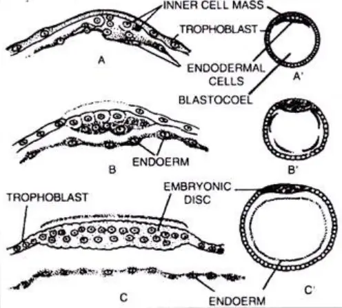
- The formation of the endoderm, one of the primary germ layers, occurs during early embryonic development. It begins with the growth and expansion of the blastocyst, a hollow ball of cells that implants into the uterine wall. Within the blastocyst, there is a group of cells known as the inner cell mass or embryonic knob, which will give rise to various tissues and organs of the developing embryo.
- During this process, some cells from the inner cell mass separate and migrate into the blastocoel, the fluid-filled cavity within the blastocyst. These cells form the endoderm, which is the innermost germ layer. The endoderm differentiates and undergoes further development to give rise to important structures within the body.
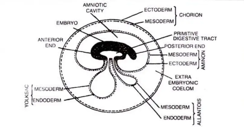
- The cells of the endoderm differentiate into the primitive gut, which is an early form of the digestive tract. A portion of the endoderm gives rise to the alimentary canal, which later develops into the gastrointestinal tract, including the esophagus, stomach, small intestine, and large intestine. This portion of the endoderm is responsible for the formation of the inner lining of these organs.
- Another portion of the endoderm forms the yolk sac. The yolk sac plays a role in early embryonic nutrition and also contributes to the formation of certain structures, such as blood cells and blood vessels. It is important for the early development and nourishment of the embryo before the placenta takes over as the primary source of nutrition.
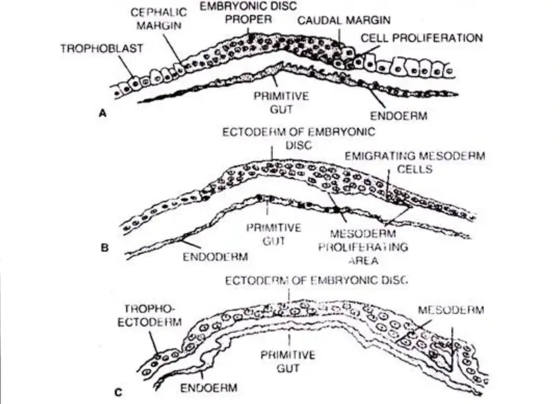
- After the formation of the endoderm, the remaining mass of cells in the inner cell mass organizes into the embryonic disc. The embryonic disc has three distinct regions: the cephalic margin (head region), the embryonic disc proper (the main body region), and the caudal margin (tail region). These regions will further differentiate and give rise to different tissues and organs of the developing embryo.
- In summary, the formation of the endoderm occurs as some cells from the inner cell mass separate and migrate into the blastocoel, where they differentiate into the endoderm. The endoderm then develops into the primitive gut, including the alimentary canal, and also contributes to the formation of the yolk sac. The remaining cells of the inner cell mass form the embryonic disc, which plays a crucial role in the subsequent development of the embryo.

(ii) Formation of Mesoderm
- The formation of the mesoderm, one of the three primary germ layers, occurs during early embryonic development. It originates from the caudal margin, or tail region, of the embryonic disc, which is part of the inner cell mass of the developing embryo.
- During this process, the existing cells within the embryonic disc undergo rapid division, leading to an increase in cell number. As a result, a mass of cells detaches from the embryonic disc to form the mesoderm. This detachment allows the mesoderm to separate and differentiate from the other germ layers, namely the endoderm and ectoderm.
- The mesoderm plays a crucial role in the development of various tissues and organs in the body. It is involved in the formation of structures such as the dermis of the skin, muscles (with the exception of iris and ciliary muscles), connective tissues, kidneys, gonads, notochord, heart, blood and lymph vessels, urinary and reproductive ducts, most of the skeleton, coelomic epithelium, pericardium and pleura, dentine of teeth, cortex of adrenal gland, mesenteries, sclera and choroid of the eyes, and the wall of the gut (excluding its lining).
- The mesoderm also gives rise to other important tissues and systems, including the circulatory system, lymphatic system, genitourinary system, and serous membranes. Additionally, it contributes to the formation of muscles (both smooth and striated), bone, cartilage, adipose tissue, and the notochord.
- The formation of the mesoderm marks an important step in embryonic development. It ensures the generation of the necessary cell types and tissues that will form the foundation for the complex anatomy and physiology of the developing organism. The mesoderm’s ability to differentiate into various cell types and contribute to the formation of a wide range of structures underscores its critical role in embryogenesis.

(iii) Formation of Ectoderm
- The formation of the ectoderm, one of the primary germ layers, occurs during early embryonic development. It takes place after the separation of the mesoderm from the embryonic disc, which is part of the inner cell mass of the developing embryo.
- Once the mesoderm has detached and formed its own layer, the remaining cells of the embryonic disc organize and differentiate to give rise to the ectoderm. This process ensures the formation of the three germ layers: ectoderm, mesoderm, and endoderm, which are fundamental for the development of different tissues and organs in the body.
- The ectoderm plays a crucial role in the development of various structures and systems. It gives rise to the epidermis of the skin, along with its derivatives such as hair, nails, and epidermal glands. The ectoderm is also responsible for the formation of the nervous system, including the brain and spinal cord, as well as peripheral nerves. It contributes to the development of the medulla of the adrenal gland, the posterior and intermediate lobes of the pituitary gland, the pineal gland, and the enamel of teeth.
- Furthermore, the ectoderm forms important sensory structures such as the eyes, including the conjunctiva, cornea, lens, retina, iris, and ciliary muscles, as well as the internal ear and the nasal and olfactory epithelia. It also contributes to the formation of certain glands, including sweat glands, oil glands, mammary glands, salivary glands, and lacrimal glands.
- In summary, the ectoderm is formed from the remaining cells of the embryonic disc after the separation of the mesoderm. It gives rise to a wide range of structures, including the epidermis of the skin, nervous system, sensory organs, certain glands, and various epithelia. The formation of the ectoderm is a crucial step in embryonic development, as it ensures the differentiation and specialization of cells that will ultimately contribute to the formation of diverse tissues and organs in the growing embryo.

FAQ
What are germ layers?
A germ layer refers to the primary layers of cells that form during embryonic development. They are the ectoderm, mesoderm, and endoderm.
How are germ layers formed?
Germ layers are formed through a process called gastrulation. During gastrulation, cells in the inner cell mass of the blastocyst or blastula move and rearrange, giving rise to the three germ layers.
What is the fate of the ectoderm?
The ectoderm gives rise to structures such as the epidermis of the skin, nervous system (including the brain and spinal cord), sensory organs, certain glands, and various epithelia.
What tissues and organs are derived from the mesoderm?
The mesoderm forms tissues and organs such as the dermis of the skin, muscles (except for the iris and ciliary muscles), connective tissues, kidneys, gonads, heart, blood and lymph vessels, urinary and reproductive ducts, most of the skeleton, and serous membranes.
What does the endoderm develop into?
The endoderm develops into the lining of the digestive tract (except for parts of the mouth and pharynx and the terminal part of the rectum), as well as glands associated with the digestive system (e.g., pancreas, liver, gastric glands, intestinal glands). It also forms the lining of the respiratory tract, urinary bladder, urethra, and certain glands like the thyroid, parathyroid, and thymus.
Can the germ layers give rise to different types of tissues?
Yes, each germ layer has the capacity to differentiate into various types of tissues. For example, the mesoderm gives rise to muscle, bone, cartilage, connective tissue, and the circulatory system.
What is the significance of the neural crest?
The neural crest, although derived from the ectoderm, plays a critical role in embryonic development. It gives rise to diverse cell types, including components of the peripheral nervous system, facial cartilage, adrenal medulla, and melanocytes.
Are all animals triploblastic?
No, not all animals are triploblastic. Some animals, like sponges, have only two germ layers (ectoderm and endoderm) and are referred to as diploblastic. Triploblastic animals, including humans, possess a third germ layer, the mesoderm.
What is the developmental significance of germ layers?
Germ layers are essential for the development and organization of tissues and organs in multicellular organisms. They serve as the foundation for subsequent processes such as organogenesis and tissue specialization.
Are the fates of germ layers consistent among all triploblastic animals?
Yes, the fates of the germ layers (ectoderm, mesoderm, and endoderm) are generally consistent among all triploblastic animals. However, there may be some variations and modifications in specific organisms based on their evolutionary adaptations and specialized structures.


