Table of Contents
What is Gram Positive bacteria?
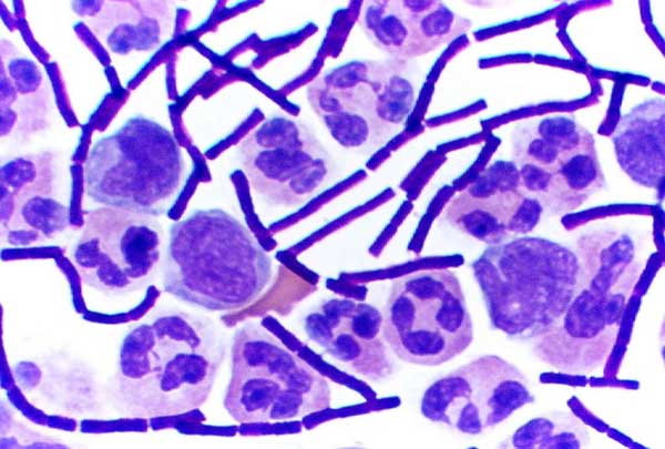
Gram-positive bacteria are a group of bacteria that exhibit a positive result in the Gram stain test, a widely used method for bacterial classification based on cell wall characteristics. When subjected to this staining technique, Gram-positive bacteria retain the crystal violet dye and appear purple under an optical microscope.
The retention of the crystal violet stain by Gram-positive bacteria is attributed to the presence of a thick layer of peptidoglycan in their cell wall. This layer retains the stain even after the decolorization step, where alcohol is used to remove excess dye from the sample. In contrast, Gram-negative bacteria do not retain the crystal violet stain due to the presence of an outer membrane that is degraded by the alcohol, resulting in a porous cell wall unable to retain the dye. Gram-negative bacteria take up a counterstain, such as safranin or fuchsine, and appear red or pink.
The distinction between Gram-positive and Gram-negative bacteria based on their staining properties has practical implications. For example, certain cell wall-targeting antibiotics are more effective against Gram-positive bacteria due to the absence of an outer membrane. The thicker peptidoglycan layer in Gram-positive bacteria makes them more susceptible to the action of these antibiotics.
Gram staining has been a valuable tool in bacterial identification and classification because it helps differentiate bacteria based on their cell wall composition. The cell wall of Gram-positive bacteria is primarily composed of mucin, peptidoglycan, and mucopeptides, which provide structural rigidity to the bacterial cell. The peptidoglycan layer in Gram-positive bacteria is thicker, ranging from 20 to 80 nanometers, and is located outside the cell membrane. In contrast, Gram-negative bacteria have a thinner peptidoglycan layer, approximately 2 to 7 nanometers, sandwiched between an inner cell membrane and an outer membrane composed of lipids, making their cell wall more complex.
By understanding the differences in cell wall structure between Gram-positive and Gram-negative bacteria, scientists can gain insights into their characteristics, behavior, and susceptibility to antibiotics. Gram staining remains a fundamental technique in microbiology and continues to play a crucial role in bacterial identification and classification.

Definition of Gram Positive bacteria
Gram-positive bacteria are a group of bacteria that retain the crystal violet stain in the Gram stain test, appearing purple under a microscope. They have a thick peptidoglycan layer in their cell wall, which contributes to their ability to retain the stain. Gram-positive bacteria lack an outer membrane and are generally more susceptible to certain antibiotics compared to Gram-negative bacteria.
Characteristics of Gram-positive bacteria
Gram-positive bacteria possess distinct characteristics that set them apart from other types of bacteria. These features include:
- Lack of outer membrane: Gram-positive bacteria do not have an outer membrane, which is a characteristic feature of Gram-negative bacteria.
- Thin cytoplasmic lipid layer: They have a thin layer of cytoplasmic lipid membrane compared to Gram-negative bacteria.
- Thick peptidoglycan layer: Gram-positive bacteria have a thick peptidoglycan layer in their cell wall. This layer provides structural support and rigidity to the bacterial cell.
- Abundance of teichoic acid: The peptidoglycan layer of Gram-positive bacteria contains a significant amount of teichoic acid. These acids, including lipoteichoic acid, play a crucial role in bacterial adherence and serve as chelating agents.
- Cross-linking of peptidoglycan: The peptidoglycan chains in Gram-positive bacteria are cross-linked by the action of the bacterial enzyme DD-transpeptidase. This cross-linking contributes to the stability and strength of the cell wall.
- Smaller periplasmic volume: Gram-positive bacteria have a relatively smaller volume of periplasm compared to Gram-negative bacteria. The periplasm is the space between the cytoplasmic membrane and the outer membrane in Gram-negative bacteria.
- Capsule formation: Some Gram-positive bacteria have a strong capsule composed of polysaccharides. This capsule helps protect the bacteria from the host immune system and contributes to their virulence.
- Flagella: While not all Gram-positive bacteria have flagella, those that do typically possess only two basal body rings to support the flagellum, unlike Gram-negative bacteria that have four basal body rings.
- Surface layer (S-layer): Gram-positive bacteria commonly have an S-layer, a surface layer attached to the peptidoglycan layer. The S-layer provides additional protection and structural stability to the bacteria.
In summary, Gram-positive bacteria exhibit these characteristic features, including the absence of an outer membrane, a thick peptidoglycan layer, teichoic acids, and lipoteichoic acids, as well as unique flagellar and capsule arrangements. These characteristics contribute to their distinct physiology and behavior within the bacterial world.
Shape of Gram positive bacteria
Gram-positive bacteria exhibit various shapes that can be observed under a microscope. These shapes provide additional characteristics for defining and categorizing these bacteria. The shapes of Gram-positive bacteria can be broadly classified into two main categories:
- Cocci: Cocci are spherical or oval-shaped bacteria. They have a diameter ranging from 0.5 to 1.0 micrometers. Cocci can occur in different arrangements, such as:
- Pairs: Two cocci arranged side by side, for example, Staphylococcus spp.
- Chains: Cocci arranged in a linear chain-like structure, for example, Streptococcus spp.
- Clusters: Cocci arranged in clusters, for example, Staphylococcus spp.
- Singles: Isolated cocci without any specific arrangement.
- Special arrangements: There are unique arrangements like tetrads, which are groups of four cocci, and sarcina, which are thick-walled cocci occurring in clusters of four or cubes of eight.
Examples of Gram-positive cocci bacteria include Staphylococcus spp., Streptococcus spp., Micrococcus spp., and Sarcina spp.
- Bacilli: Bacilli are rod-shaped bacteria. They have a stick-like appearance with rounded, tapered, square, or swollen ends. Bacilli are typically larger than cocci, with a length ranging from 1 to 10 micrometers and a width of 0.3 to 1.0 micrometers. Examples of Gram-positive bacilli bacteria include Bacillus spp.
It’s important to note that Gram-positive bacteria can also exhibit other special shapes in addition to cocci and bacilli. For instance:
- Tetrad: A specific arrangement of cocci occurring in clusters of fours, forming a square-like structure. An example is Micrococcus spp.
- Sarcina (Octae): Thick-walled cocci arranged in clusters of four or cubes of eight.
These variations in shape provide additional information for the identification and classification of Gram-positive bacteria. By examining their shapes, microbiologists can further characterize and differentiate between different species and strains within the Gram-positive bacterial group.
Cell wall Structure of Gram-positive bacteria
The cell wall structure of Gram-positive bacteria is characterized by several components that provide strength, rigidity, and other functional properties. These components include peptidoglycan, teichoic acid, and a thin lipid layer.

- Peptidoglycan:
- Peptidoglycan, also known as murein, makes up the majority (about 90%) of the Gram-positive bacterial cell wall.
- It plays a crucial role in providing shape, strength, and rigidity to the cell wall.
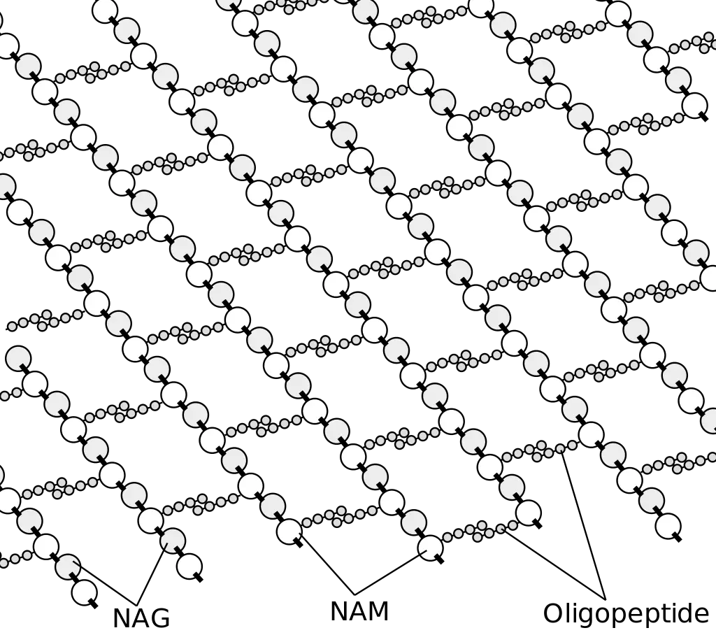
- Peptidoglycan is a high-quality polymer composed of two sugar derivatives, N-acetylglucosamine and N-acetylmuramic acid, as well as a chain of L-amino acids and three unique D-amino acids (D-glutamic acid, D-alanine, and meso-diaminopimelic acid).
- The D-amino acids and L-amino acids form connections with N-acetylmuramic acid, and L-lysine can replace meso-diaminopimelic acid in some cases.
- This interconnection of peptidoglycan subunits contributes to the strength, integrity, and elasticity of the cell wall.
- Peptidoglycan is also permeable, allowing molecules to move in and out of the bacterial cell.
- Teichoic Acid:
- Teichoic acid is a component present in the cell walls of Gram-positive bacteria, but it is absent in Gram-negative bacteria.
- It is a polymer composed of glycerol copolymers and makes up a significant portion (up to 50%) of the total dry weight of the bacterial cell wall.
- Teichoic acid can be directly connected to the peptidoglycan through covalent bonds or to the cell membrane as lipoteichoic acid.
- The direct connection to peptidoglycan occurs through the 6-hydroxyl N-acetylmuramic acid.
- Teichoic acid carries a negative charge and extends to the surface of the peptidoglycan, contributing to the overall negative charge of the bacterial cell wall.
- It plays a role in maintaining the structure of the cell wall and has other functions depending on the bacterial species.
- Lipid:
- Gram-positive bacteria have a thin layer of lipids located below the peptidoglycan layer, comprising about 2-5% of the cell wall.
- This lipid layer functions to anchor the bacterial cell wall.

Together, these components form a complex cell wall structure in Gram-positive bacteria. The peptidoglycan provides strength and rigidity, while teichoic acid contributes to the negative charge and structural integrity of the cell wall. The lipid layer anchors the cell wall and provides additional functionality. Understanding the cell wall structure of Gram-positive bacteria is essential for identifying and targeting specific components for therapeutic interventions.
Gram positive bacteria examples and diseases
Gram-positive bacteria encompass a diverse group of bacteria that can cause various diseases in humans. Here are some examples of Gram-positive bacteria along with the diseases they are associated with:
- Staphylococcus aureus:
- Furuncles, carbuncles (localized skin infections)
- Wound infections
- Cellulitis
- Impetigo
- Respiratory infections (pneumonia)
- Cardiovascular infections (endocarditis, septicemia)
- Musculoskeletal infections (osteomyelitis, arthritis)
- Streptococcus pyogenes:
- Acute pharyngitis or pharyngotonsillitis
- Impetigo
- Erysipelas
- Puerperal sepsis
- Invasive group A streptococcal disease (necrotizing fasciitis/myositis)
- Streptococcal toxic shock syndrome
- Acute rheumatic fever
- Acute glomerulonephritis
- Streptococcus pneumoniae:
- Community-acquired acute bacterial pneumonia
- Adult bacterial meningitis
- Otitis media in children
- Bacteremia/sepsis
- Meningitis
- Bacillus anthracis:
- Pulmonary anthrax (woolsorter’s disease) with hemorrhagic necrosis and edema of the mediastinum
- Cutaneous anthrax with pruritic papule lesions and edema, lymphadenopathy, and systemic symptoms
- Corynebacterium diphtheriae:
- Diphtheria, a localized infection of the upper respiratory tract
- Cardiac conduction defects, myocarditis, and neuritis of cranial nerves in severe cases
- Clostridium botulinum:
- Botulism poisoning characterized by visual disturbances, inability to swallow, speech difficulty, muscle weakness, and respiratory paralysis
- Clostridium tetani:
- Tetanus characterized by muscle spasms, jaw muscle infection, and generalized muscle spasm
- Clostridium difficile:
- Pseudomembranous colitis associated with watery, bloody diarrhea
- Enterococcus faecium and Enterococcus faecalis:
- Infections in immune-compromised individuals such as urinary tract infections, bacteremia/sepsis, endocarditis, biliary tract infection, or intra-abdominal abscesses
- Listeria monocytogenes:
- Listeriosis associated with septicemia and meningitis
- Focal and granulomatous skin lesions
- Asymptomatic vaginal disease in pregnant women, leading to flu-like illness and meningitis in newborns
These examples highlight the wide range of diseases caused by Gram-positive bacteria, emphasizing the importance of understanding their characteristics and implementing appropriate preventive measures and treatment strategies.
Antibiotics for Gram positive bacteria
Gram-positive bacteria are susceptible to various antibiotics, which are essential for treating bacterial infections caused by these pathogens. Here are some commonly used antibiotics and their mechanisms of action against Gram-positive bacteria:
- ß-lactam antibiotics (Amoxicillin, Methicillin, Oxacillin, Ampicillin, Penicillin G):
- Mechanism of action: Disruption of bacterial cell wall synthesis by inhibiting the formation of peptidoglycan, leading to cell lysis.
- Effective against: Staphylococcus aureus, Streptococcus pneumoniae, Streptococcus pyogenes, Corynebacterium diphtheriae, Bacillus anthracis, Clostridium botulinum.
- Vancomycin, Erythromycin, Azithromycin:
- Mechanism of action: Inhibition of cell wall synthesis by preventing the crosslinking of peptidoglycan peptidases.
- Effective against: Staphylococcus spp, Streptococcus spp, Bacillus spp, Clostridium difficile.
- Bacitracin:
- Mechanism of action: Inhibition of cell wall synthesis by preventing the movement of cytoplasmic membrane and peptidoglycan precursors.
- Effective against: Corynebacterium spp, Bacillus anthracis.
- Macrolides (Azithromycin, Clarithromycin):
- Mechanism of action: Inhibition of bacterial protein synthesis by preventing polypeptide elongation at the 50S ribosomes.
- Effective against: Streptococcus pyogenes.
- Cephalosporin:
- Mechanism of action: Inhibition of cell wall synthesis by disrupting peptidoglycan synthesis.
- Effective against: Streptococcus pneumoniae, Bacillus anthracis.
- Aminoglycosides (Gentamicin):
- Mechanism of action: Inhibition of bacterial protein synthesis by producing aberrant peptide chains at the 30S ribosomes.
- Effective against: Staphylococcus aureus, Streptococcus pneumoniae, Streptococcus pyogenes, Enterococcus spp.
- Oxazolidinone:
- Mechanism of action: Inhibition of protein synthesis at the 50S ribosomes.
- Effective against: Enterococcus spp.
- Rifampicin:
- Mechanism of action: Inhibition of nucleic acid synthesis by preventing the transcription of binding DNA-dependent RNA polymerase.
- Effective against: Bacillus anthracis.
- Sulfonamides (Sulfamethoxazole):
- Mechanism of action: Inhibition of folic acid synthesis by blocking the enzyme dihydropteroate synthase.
- Effective against: Listeria monocytogenes.
- Trimethoprim:
- Mechanism of action: Inhibition of folic acid synthesis by blocking the enzyme dihydrofolate reductase.
- Effective against: Listeria monocytogenes.
These antibiotics play a crucial role in combating Gram-positive bacterial infections by targeting specific mechanisms essential for bacterial survival and replication. However, it’s important to note that antibiotic resistance can develop over time, necessitating the judicious use of these medications and ongoing research for the development of new treatment options.
Low G+ C (Firmicutes): General characteristics with suitable examples
Low G+C Gram-positive bacteria, also known as Firmicutes, are a phylum of bacteria characterized by a low guanine-cytosine (G+C) content in their genomic DNA. They exhibit diverse characteristics and include several notable examples. Here are the general characteristics of Firmicutes along with suitable examples:
- Cell Wall Structure:
- Firmicutes have a thick peptidoglycan cell wall that provides structural integrity.
- The peptidoglycan layer is composed of alternating N-acetylglucosamine and N-acetylmuramic acid residues.
- Shape and Morphology:
- Firmicutes exhibit a variety of shapes, including cocci (spherical), bacilli (rod-shaped), and filamentous forms.
- Some examples of Firmicutes with specific shapes include Streptococcus (cocci in chains or pairs) and Bacillus (rod-shaped).
- Metabolism:
- Firmicutes display diverse metabolic capabilities, including fermentative, anaerobic, and some aerobic metabolism.
- They can utilize a wide range of carbon sources and produce various end products such as acids, alcohols, and gases.
- Examples of Firmicutes:
- Staphylococcus aureus: This bacterium is a significant human pathogen responsible for various infections, including skin and soft tissue infections, pneumonia, and septicemia.
- Streptococcus pyogenes: It causes diseases such as strep throat, impetigo, cellulitis, and invasive infections like necrotizing fasciitis and toxic shock syndrome.
- Bacillus subtilis: A model organism extensively studied for its physiology and genetics. It is commonly found in soil and has diverse metabolic capabilities.
- Clostridium difficile: This bacterium is associated with antibiotic-associated diarrhea and pseudomembranous colitis.
- Listeria monocytogenes: It is responsible for the foodborne illness listeriosis, which can lead to septicemia, meningitis, and other severe infections.
- Enterococcus faecalis: Found in the gastrointestinal tract, it can cause opportunistic infections, including urinary tract infections and endocarditis.
These examples illustrate the diversity of Firmicutes and their significance in both pathogenic and non-pathogenic contexts. Firmicutes play essential roles in various ecological niches, including the human microbiota, industrial processes, and environmental cycles. Their unique characteristics and metabolic capabilities contribute to their ecological success and relevance in various fields of study.
| Example Genus | Microscopic Morphology | Unique Characteristics |
|---|---|---|
| Bacillus | Large, gram-positive bacillus | Aerobes or facultative anaerobes; form endospores; B. anthracis causes anthrax in cattle and humans, B. cereus may cause food poisoning |
| Clostridium | Gram-positive bacillus | Strict anaerobes; form endospores; all known species are pathogenic, causing tetanus, gas gangrene, botulism, and colitis |
| Enterococcus | Gram-positive coccus; forms microscopic pairs in culture (resembling Streptococcus pneumoniae) | Anaerobic aerotolerant bacteria, abundant in the human gut, may cause urinary tract and other infections in the nosocomial environment |
| Lactobacillus | Gram-positive bacillus | Facultative anaerobes; ferment sugars into lactic acid; part of the vaginal microbiota; used as probiotics |
| Leuconostoc | Gram-positive coccus; may form microscopic chains in culture | Fermenter, used in food industry to produce sauerkraut and kefir |
| Mycoplasma | The smallest bacteria; appear pleomorphic under electron microscope | Have no cell wall; classified as low G+C Gram-positive bacteria because of their genome; M. pneumoniae causes “walking” pneumonia |
| Staphylococcus | Gram-positive coccus; forms microscopic clusters in culture that resemble bunches of grapes | Tolerate high salt concentration; facultative anaerobes; produce catalase; S. aureus can also produce coagulase and toxins responsible for local (skin) and generalized infections |
| Streptococcus | Gram-positive coccus; forms chains or pairs in culture | Diverse genus; classified into groups based on sharing certain antigens; some species cause hemolysis and may produce toxins responsible for human local (throat) and generalized disease |
| Ureaplasma | Similar to Mycoplasma | Part of the human vaginal and lower urinary tract microbiota; may cause inflammation, sometimes leading to internal scarring and infertility |
High G+C (Actinobacteria): General characteristics with suitable examples
High G+C Gram-positive bacteria, also known as Actinobacteria, are a phylum of bacteria characterized by a high guanine-cytosine (G+C) content in their genomic DNA. They are a diverse group of bacteria with unique characteristics. Here are the general characteristics of Actinobacteria along with suitable examples:
- Cell Wall Structure:
- Actinobacteria have a complex cell wall structure.
- The cell wall contains peptidoglycan, but it also includes other components such as mycolic acids, arabinogalactan, and various sugars.
- Morphology:
- Actinobacteria exhibit a variety of morphologies, including cocci, bacilli, and filamentous forms.
- Some actinobacteria form branching filaments called mycelia.
- Metabolism:
- Actinobacteria have diverse metabolic capabilities.
- They are known for their ability to produce a wide range of secondary metabolites, including antibiotics, enzymes, and pigments.
- Actinobacteria are involved in the decomposition of organic matter and nutrient cycling in various ecosystems.
- Examples of Actinobacteria:
- Streptomyces spp.: This genus is well-known for its ability to produce a vast array of antibiotics, such as streptomycin, tetracycline, and erythromycin. Streptomyces species are abundant in soil and play a crucial role in the decomposition of organic matter.
- Mycobacterium tuberculosis: The causative agent of tuberculosis, one of the deadliest infectious diseases worldwide. It has a unique cell wall structure containing mycolic acids, which contribute to its virulence and resistance to antibiotics.
- Nocardia spp.: These bacteria can cause opportunistic infections in humans, particularly in individuals with weakened immune systems. Nocardia species are commonly found in soil and can cause pulmonary and cutaneous infections.
- Corynebacterium diphtheriae: The bacterium responsible for diphtheria, a respiratory disease characterized by the formation of a pseudomembrane in the throat. It produces a potent exotoxin that can cause severe complications if left untreated.
- Actinomyces spp.: These bacteria form characteristic filamentous structures and can cause chronic infections in humans, such as actinomycosis. They are part of the normal flora in the oral cavity and gastrointestinal tract.
These examples highlight the diversity and significance of Actinobacteria. They play vital roles in various ecological processes, including nutrient cycling, soil health, and the production of valuable secondary metabolites. Actinobacteria have significant medical and biotechnological applications, and their unique characteristics continue to be the subject of scientific investigation.
| Table 1. Actinobacteria: High G+C Gram-Positive | ||
|---|---|---|
| Example Genus | Microscopic Morphology | Unique Characteristics |
| Actinomyces | Gram-positive bacillus; in colonies, shows fungus-like threads (hyphae) | Facultative anaerobes; in soil, decompose organic matter; in the human mouth, may cause gum disease |
| Arthrobacter | Gram-positive bacillus (at the exponential stage of growth) or coccus (in stationary phase) | Obligate aerobes; divide by “snapping,” forming V-like pairs of daughter cells; degrade phenol, can be used in bioremediation |
| Bifidobacterium | Gram-positive, filamentous actinobacterium | Anaerobes commonly found in human gut microbiota |
| Corynebacterium | Gram-positive bacillus | Aerobes or facultative anaerobes; form palisades; grow slowly; require enriched media in culture; C. diphtheriae causes diphtheria |
| Frankia | Gram-positive, fungus-like (filamentous) bacillus | Nitrogen-fixing bacteria; live in symbiosis with legumes |
| Gardnerella | Gram-variable coccobacillus | Colonize the human vagina, may alter the microbial ecology, thus leading to vaginosis |
| Micrococcus | Gram-positive coccus, form microscopic clusters | Ubiquitous in the environment and on the human skin; oxidase-positive (as opposed to morphologically similar S. aureus); some are opportunistic pathogens |
| Mycobacterium | Gram-positive, acid-fast bacillus | Slow growing, aerobic, resistant to drying and phagocytosis; covered with a waxy coat made of mycolic acid; M. tuberculosis causes tuberculosis; M. leprae causes leprosy |
| Nocardia | Weakly gram-positive bacillus; forms acid-fast branches | May colonize the human gingiva; may cause severe pneumonia and inflammation of the skin |
| Propionibacterium | Gram-positive bacillus | Aerotolerant anaerobe; slow-growing; P. acnes reproduces in the human sebaceous glands and may cause or contribute to acne |
| Rhodococcus | Gram-positive bacillus | Strict aerobe; used in industry for biodegradation of pollutants; R. fascians is a plant pathogen, and R. equi causes pneumonia in foals |
| Streptomyces | Gram-positive, fungus-like (filamentous) bacillus | Very diverse genus (>500 species); aerobic, spore-forming bacteria; scavengers, decomposers found in soil (give the soil its “earthy” odor); used in pharmaceutical industry as antibiotic producers (more than two-thirds of clinically useful antibiotics) |
Importance of Gram Positive bacteria
Gram-positive bacteria play a significant role in various aspects of life and have several important contributions. Here are some key points highlighting the importance of Gram-positive bacteria:
- Human Health: Many Gram-positive bacteria are associated with human health, both as beneficial commensals and as pathogens. Some beneficial Gram-positive bacteria, such as those belonging to the genera Lactobacillus and Bifidobacterium, are part of the normal human microbiota and contribute to digestion, nutrient absorption, and immune system regulation. Additionally, probiotic preparations containing Gram-positive bacteria are used to promote gut health. On the other hand, some Gram-positive bacteria can cause infections, such as Staphylococcus aureus, Streptococcus pneumoniae, and Clostridium difficile, necessitating appropriate medical interventions.
- Antibiotic Production: Many important antibiotics used in medicine are produced by Gram-positive bacteria. For example, Streptomyces species, which are Gram-positive, filamentous bacteria, are prolific antibiotic producers. They have contributed to the development of antibiotics such as streptomycin, tetracycline, erythromycin, and vancomycin. These antibiotics have played a crucial role in combating bacterial infections and saving countless lives.
- Bioremediation: Gram-positive bacteria have the ability to degrade or transform various environmental pollutants, making them valuable in bioremediation processes. Some species of Bacillus, Arthrobacter, and Rhodococcus are known for their capacity to degrade hydrocarbons, pesticides, and toxic chemicals, thereby aiding in the cleanup of contaminated environments.
- Industrial Applications: Gram-positive bacteria have diverse industrial applications. For instance, certain species of Lactobacillus and Streptococcus are involved in the production of fermented foods and beverages, including yogurt, cheese, sauerkraut, and sourdough bread. The metabolic activities of these bacteria contribute to the flavor, texture, and preservation of these food products.
- Enzyme Production: Gram-positive bacteria are utilized in the production of various enzymes used in industries. For example, Bacillus species are known for producing amylases, proteases, lipases, and cellulases, which find applications in detergent manufacturing, textile processing, food processing, and biofuel production.
- Agriculture and Soil Health: Gram-positive bacteria play a vital role in soil fertility, nutrient cycling, and plant health. Some species form symbiotic relationships with plants, promoting their growth and providing essential nutrients. For example, Rhizobium species form nodules on the roots of leguminous plants, fixing atmospheric nitrogen and making it available to the plants. Other Gram-positive bacteria, such as Bacillus and Streptomyces, produce antimicrobial compounds that suppress plant pathogens, benefiting agricultural practices.
Overall, Gram-positive bacteria have significant implications in human health, biotechnology, environmental sustainability, and agricultural systems. Their diverse metabolic capabilities, antibiotic production, and ecological contributions make them indispensable in various fields, highlighting their importance in shaping and impacting our lives.


