Table of Contents
What is Penicillium?
- Penicillium, a fascinating group of ascomycetous fungi, holds immense significance in various realms of life, encompassing the natural environment, food production, and the pharmaceutical industry. The genus is renowned for giving rise to the well-known antibiotic drug, penicillin, a medical marvel in combating certain bacterial infections. Furthermore, other species of Penicillium contribute to the artistry of cheese production, enriching our culinary experiences.
- The study of Penicillium under a microscope reveals a captivating life form, readily available for observation within our own refrigerators. Microorganisms such as bacteria, yeasts, and molds are commonly found even in regularly cleaned fridges, but the most fruitful search for Penicillium can be conducted in an uncleaned refrigerator left untouched for two months or more.
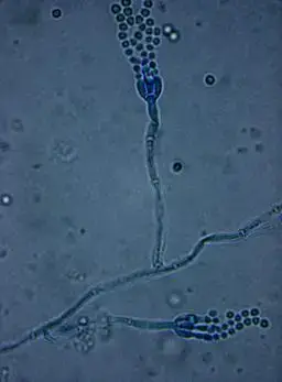
- This saprophytic fungus thrives in various habitats, including soil, air, and decaying organic matter. Commonly referred to as the green or blue mold, Penicillium species exhibit a distinctive appearance, characterized by numerous densely packed brush-like structures known as penicilli, which produce spores called conidia. These spores are formed in dry chains and emanate from the tips of flask-shaped structures called phialides, present on simple or branching structures slightly elongated in shape.
- The identification of Penicillium species often hinges on their unique branching patterns. For instance, some species like P. glabrum remain unbranched, bearing only one cluster of phialides at the top of the stipe. Understanding these morphological features aids researchers and enthusiasts alike in discerning the diverse members of the Penicillium genus.
- At the core of its vegetative body lies the mycelium, a complex network of branched, septate hyphae. These hyphae consist of thin-walled cells housing one or more nuclei, with each septum boasting a central pore necessary for the continuity of cytoplasmic flow.
- Penicillium exhibits intriguing strategies for nutrient absorption. While some mycelia delve deeper into the substratum to acquire food nutrients, others persist on the substrate, forming a mycelial felt. This adaptive behavior ensures the efficient utilization of available resources for the organism’s sustenance.
- Within the mycelium, Penicillium stores food in the form of oil globules, a unique characteristic contributing to its ability to survive and thrive in various ecological niches. This remarkable attribute further enhances its significance in diverse fields of human interest.
- As we delve deeper into the microscopic wonders of Penicillium, our understanding of its ecological roles and industrial applications expands. From being a source of life-saving antibiotics to lending its hand in the art of cheese-making, Penicillium continues to captivate researchers and enthusiasts, unveiling its secrets in the intricate world of fungi. As we appreciate the profound impact of Penicillium in our lives, let us also embrace the wonders of the microscopic universe that surrounds us.

Requirements for Penicillium Microscopy
- Microscope Slide: The foundation of any microscopic examination begins with a high-quality microscope slide. A clean and optically clear slide provides a stable platform for mounting the specimen, enabling precise observations under the microscope.
- Compound Microscope with Power Supply and Illuminants: A compound microscope is indispensable for studying Penicillium in detail. It allows for both low and high magnifications, crucial for examining the intricate structures of the fungi. A reliable power supply ensures consistent illumination, while various illuminants, such as LED or halogen bulbs, offer versatility in lighting conditions.
- Hematoxylin Stain: Staining is a valuable technique to enhance the visibility of Penicillium structures. Hematoxylin, a commonly used stain, imparts a bluish-purple hue to the fungal elements, making them stand out against the background. Staining facilitates a clearer view of the fungal morphology, aiding in species identification and detailed analysis.
- Clean Coverslip: Placing a clean coverslip over the stained sample ensures the preservation of the specimen and prevents distortion during observation. It also flattens the specimen, minimizing the effects of uneven surfaces and enabling sharper focus.
- Forceps: Handling microscopic specimens, especially during the preparation and mounting process, requires precision and care. Fine-tipped forceps allow for delicate manipulation of Penicillium samples without causing damage.
- Oil for Immersion: High-resolution microscopy often involves using oil-immersion objectives. Immersing the objective in a special oil with a refractive index similar to glass optimizes light transmission and resolution, allowing for clearer and more detailed images of Penicillium structures.
Procedure of Penicillium Microscopy
- Sample Collection: Begin by conducting a thorough search in the home refrigerator, as moldy food produce is a common source of Penicillium. With the help of forceps, carefully obtain a small sample of the mold from the food.
- Slide Preparation: Place the obtained mold sample on a clean microscope slide, ensuring that the slide’s surface is free from any debris or contaminants. Proceed to add three drops of freshly prepared hematoxylin stain onto the mold sample, allowing the stain to penetrate the specimen.
- Cover Slip Placement: Gently place a clean cover slip over the stained mold sample. To minimize the presence of air bubbles within the setup, gently press down on the cover slip to expel as many bubbles as possible.
- Microscope Setup: Set up a compound microscope with appropriate magnification capabilities. For detailed observation, a 1000X magnification is typically recommended.
- Initial Focus: Begin with a 400X magnification objective lens and position the microscope slide on the stage to ensure the area of interest is clearly visible. Carefully adjust the focus knob to bring the image into clear focus.
- Optimal Illumination: Fine-tune the microscope’s condenser and illumination settings to achieve the desired light intensity for optimal visualization.
- Centering the Sample: Move the microscope slide around until the sample is positioned at the center of the field of view. Employing high illumination from a small angle above the sample can aid in obtaining a clearer view.
- Detailed Observation: With the sample in clear focus under the 10X power objective lens, switch to higher or lower magnification objectives to zoom in or out for enhanced clarity. By adjusting the objectives in different planes, finer details of the Penicillium structures can be observed.
- Concluding the Viewing: Once the observation is complete, lower the stage and switch the objective lens to a lower power, such as 40X. Carefully remove the microscope slide from the stage.
General Tips
When embarking on the fascinating journey of Penicillium microscopy, there are some essential tips to ensure a successful and rewarding experience. By following these guidelines, researchers and enthusiasts can unlock the hidden world of mold and delve into the intricate details of these microscopic wonders:
- Search for Moldy Food Produce: A general tip to initiate the exploration is to conduct a careful search in the home refrigerator. Almost inevitably, this search will lead to the discovery of at least one piece of moldy food produce. This can serve as a readily available and accessible source of Penicillium specimens for microscopy.
- Sample Preparation: Using fine-tipped forceps, gently obtain a small sample of the mold from the chosen food produce. Place the mold sample on a clean microscope slide, ensuring the slide is free from any contaminants that could interfere with the observation.
- Hematoxylin Staining: To enhance the visibility of Penicillium structures, apply freshly prepared hematoxylin stain to the mold sample on the microscope slide. Adding three drops of the stain and allowing it to soak into the specimen will aid in bringing out the intricate details of the fungi.
- Cover Slip Placement: Carefully place a clean cover slip over the stained mold sample. While doing so, gently expel any air bubbles that might be present in the setup. A smooth and bubble-free cover slip ensures optimal visualization during microscopy.
- Choosing the Right Magnification: Employ a compound microscope for studying Penicillium. For detailed observation, the recommended field of view is usually achieved at a 1000X magnification. This level of magnification allows for a thorough examination of the minute structures of the mold.
- Using Immersion Oil: To maximize resolution and clarity, switch to a 400X objective lens. Position the microscope slide on the stage to bring the area of interest into clear visibility. Before inserting the immersion lens, add a small drop of immersion oil on top of the coverslip and the area of interest. This will optimize light transmission and provide sharper images during observation.
Observation
Delving into the microscopic realm of Penicillium reveals a mesmerizing world of fungal wonders. Through careful observation under different magnifications, the intricate structures and unique features of these fungi come to light, painting a vivid picture of their beauty and complexity.
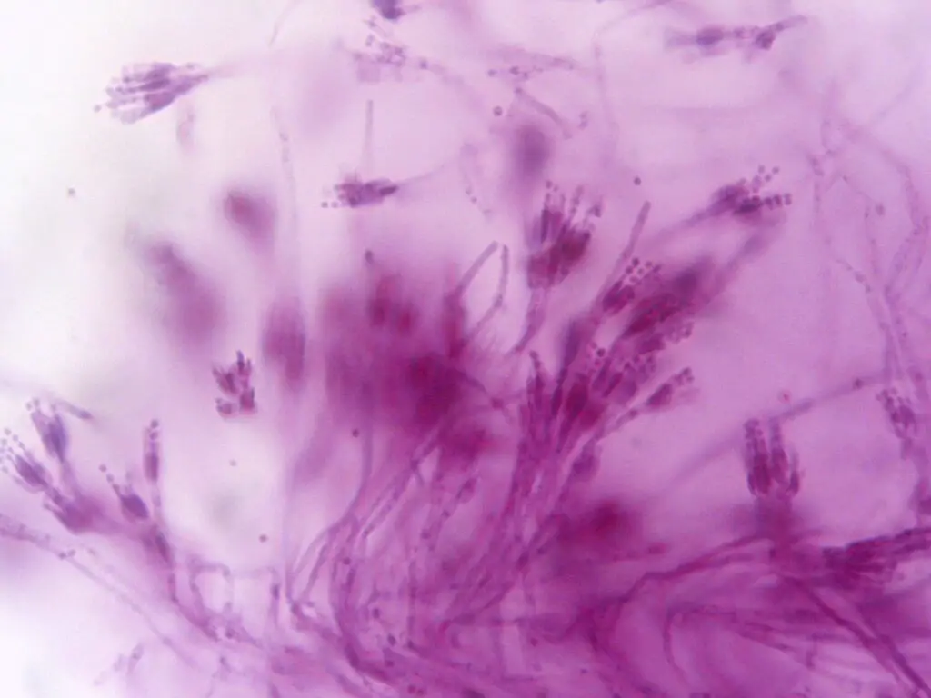
Starting with the objective set at a 40X total magnification, the hematoxylin-stained fungi sample exhibited tortuous masses of thin stalks, known as hyphae. These hyphae formed intriguing patterns, sometimes terminating in complex structures that caught the eye and beckoned for closer inspection.
Advancing to a 100X total magnification, the complex structures appeared clearer and resembled the tentacles of a sea anemone or the intricate patterns of squashed flowers. The delicate intricacy of these formations hinted at the sophistication of Penicillium’s reproductive strategies.
Further elevating the magnification to 400X, the observer could discern individual spherical structures known as conidia. These conidia, essential for the dispersal and reproduction of the fungus, appeared like tiny jewels scattered amidst the hyphal network.
Notably, the “squashed flower-like” structure, the conidiophore, initially posed a challenge to bring into focus. However, with careful adjustments and transitioning to higher magnifications, its 3-dimensional appearance distinguished it from the rest of the 2-dimensional hyphae. This unique conidiophore played a pivotal role in the production and release of conidia, adding to the captivating spectacle of Penicillium’s life cycle.
Each step of the observation journey offered fresh insights into the world of Penicillium, uncovering the intricacies of its structures and reproductive mechanisms. The tortuous hyphal masses, the tentacle-like formations, and the gem-like conidia collectively demonstrated the fungal ingenuity perfected over millennia of evolution.
As researchers and enthusiasts continue their exploration, the observation of Penicillium opens doors to a deeper understanding of the fungus’s ecological roles, its significance in food production, and its vital contributions to the pharmaceutical industry. This small but extraordinary organism exemplifies the astonishing diversity of life on Earth, reminding us of the hidden marvels that await those who dare to venture into the microscopic wonders of the natural world.

FAQ
What does Penicillin look like under a microscope?
Under a microscope, Penicillin appears as tufted masses of thin stalks called hyphae, terminating in complex structures resembling squashed flowers or sea anemone tentacles.
How can I prepare Penicillin samples for microscopy?
Penicillin samples can be obtained from moldy food produce using forceps. The mold is then placed on a clean microscope slide and stained with hematoxylin for better visualization.
What magnification should I use to observe Penicillin in detail?
To observe Penicillin in detail, start with a 40X total magnification objective and gradually increase to 100X and 400X for a closer look at complex structures and individual conidia.
What are the complex structures observed in Penicillin?
The complex structures observed in Penicillin are known as conidiophores. They play a crucial role in the production and release of conidia, essential for the fungus’s reproduction.
What do conidia look like under the microscope?
Conidia appear as individual spherical structures amidst the hyphal network. These small structures are crucial for dispersal and the formation of new fungal colonies.
How do I focus on the 3-dimensional conidiophores?
Focusing on the 3-dimensional conidiophores can be challenging initially. However, adjusting the microscope focus and transitioning to higher magnifications will reveal their distinct appearance.
What are some other key features of Penicillin observed under the microscope?
Apart from hyphae, conidiophores, and conidia, Penicillin may exhibit flask-shaped structures known as phialides, which produce the spores called penicilli.
What is the staining method used for Penicillin microscopy?
Hematoxylin stain is commonly used for Penicillin microscopy as it enhances visibility and brings out the details of fungal structures.
How can I avoid air bubbles in my microscope slide setup?
To avoid air bubbles, gently place the cover slip over the stained sample and carefully expel any present bubbles. A smooth and bubble-free setup ensures clearer observations.
What insights can I gain from observing Penicillin under a microscope?
Observing Penicillin under a microscope provides valuable insights into its morphology, reproductive strategies, and intricate structures. This knowledge is essential for understanding its ecological roles and industrial applications in food production and pharmaceuticals.


