Table of Contents
What is Stomata?
- Stomata are essential structures found on the surface of leaves and young stems of plants. These tiny openings or pores play a crucial role in the plant’s life, as they facilitate the exchange of gases and water vapor between the plant and the atmosphere.
- In dicot leaves, stomata are typically found on the lower surface, while in monocot leaves, they can be present on both surfaces. These stomata appear as small elliptical openings on the leaf’s epidermis and contain chloroplasts, the cell organelles responsible for photosynthesis.
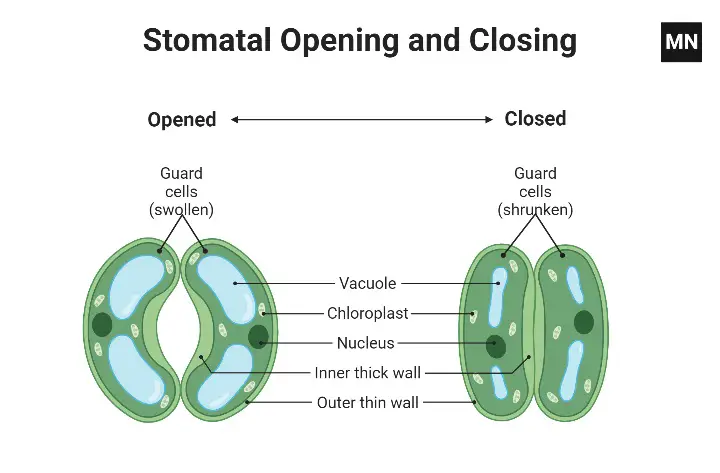
- One of the key features of stomata is their unique arrangement, being surrounded by two kidney-shaped cells called guard cells. These specialized guard cells play a significant role in regulating the opening and closing of the stomatal pores. The guard cells have distinct structural characteristics: a thick inner wall and a thin outer covering.
- When the guard cells become turgid, meaning they are filled with water and have a high internal pressure, they create an opening, causing the stomata to open. In this open state, the plant can exchange gases like carbon dioxide and oxygen with the surrounding atmosphere. This exchange is vital for photosynthesis, where the plant takes in carbon dioxide and releases oxygen.
- On the other hand, when the guard cells lose water and become flaccid, they close the stomatal pores. This closure helps the plant reduce water loss through transpiration, especially in arid conditions or when the plant is under water stress. By controlling the opening and closing of stomata, plants can regulate their water balance and prevent excessive water loss while still obtaining the necessary carbon dioxide for photosynthesis.
- Stomata’s ability to respond to environmental conditions by opening and closing is critical for a plant’s survival and growth. Factors like light intensity, temperature, humidity, and water availability influence the behavior of stomata, ensuring that the plant can adapt and optimize its gas exchange and water regulation according to its immediate surroundings.
- In conclusion, stomata are tiny openings on the surface of leaves and stems that allow plants to exchange gases and water vapor with the atmosphere. The surrounding guard cells play a pivotal role in controlling the opening and closing of these pores, helping the plant maintain a balance between efficient photosynthesis and water conservation. Their ability to respond to environmental cues makes stomata a vital adaptation that enables plants to thrive in various ecological conditions.

Aim
To prepare a temporary mount of a leaf peel in order to show the stomata of a leaf
Principle/Theory
The principle or theory of the stomata under a microscope experiment is based on the understanding that plants, as primary producers, perform essential physiological processes like photosynthesis and respiration. To facilitate these processes, there must be a mechanism for gas exchange between the internal tissues of the plant and the surrounding atmosphere. This gas exchange primarily occurs through specialized structures called stomata, which are tiny openings located on the surface of leaves.
Stomata are vital structures in the plant’s leaves that allow for the exchange of gases such as carbon dioxide and oxygen with the external environment. During photosynthesis, plants take in carbon dioxide through these stomatal openings and release oxygen as a byproduct. In contrast, during respiration, plants require oxygen and release carbon dioxide as a waste product through the same stomatal pores.
The experiment involving stomata under a microscope aims to observe and study these microscopic structures in detail. By carefully preparing a temporary mount of a leaf peel and placing it under a microscope, researchers can examine the stomata’s distribution, shape, and arrangement on the leaf’s epidermis.
The principle of this experiment is grounded in the fact that stomata play a critical role in regulating the plant’s gas exchange and water vapor balance. By studying stomata under a microscope, researchers can make important observations and draw conclusions about various aspects, such as stomatal density, size, and behavior in response to environmental factors.
The experiment can help researchers and students deepen their understanding of plant anatomy and physiology. It allows them to visualize and explore the structural adaptations that enable plants to thrive and survive in different environmental conditions. Additionally, by studying stomata under a microscope, researchers can gain insights into how plants respond to changes in environmental factors like light intensity, humidity, and water availability.
Overall, the principle of the stomata under a microscope experiment is to provide a practical and hands-on approach to learning about the fundamental processes that sustain plant life. By studying stomata, researchers can appreciate the intricate mechanisms by which plants interact with their surroundings and how these interactions influence their growth, development, and overall ecological significance as primary producers.
Material Required
For the experiment to observe stomata under a microscope, the following materials are required:
- A potted plant of Bryophyllum or Tradescantia: Choose a healthy and mature leaf from the potted plant for the experiment. Bryophyllum and Tradescantia are commonly used plants for this purpose due to their easily visible stomata.
- Needles: Fine needles will be used to carefully handle the leaf and create a leaf peel for observation.
- Forceps: Forceps or tweezers are necessary for delicate handling of the leaf and other materials during the experiment.
- Watch glass: A watch glass will be used as a container to hold the safranin stain, which will help in highlighting the stomata during the observation.
- Dropper: A dropper is needed to apply the safranin stain onto the leaf peel.
- Glass slides: Clean glass slides will be used to place the leaf peel for microscopic observation.
- A brush: A soft brush can be used to gently clean the leaf surface and remove any debris before making the leaf peel.
- Coverslips: Coverslips are placed over the leaf peel on the glass slide to protect it and prevent distortion during microscopic observation.
- Blotting paper: Blotting paper is used to remove excess water from the leaf peel before mounting it on the glass slide.
- Safranin: Safranin is a red dye that will be used as a staining agent to make the stomata more visible under the microscope.
- Compound microscope: A compound microscope with appropriate magnification capabilities is necessary to observe the stomata in detail.
- Glycerine: Glycerine is used as a mounting medium to secure the coverslip in place and preserve the leaf peel for longer-term observation.
These materials are essential for conducting the experiment to observe stomata under a microscope. Proper care and precision should be exercised while handling the plant and conducting the steps to ensure accurate and clear observations of the stomata on the leaf’s surface. The safranin staining and mounting in glycerine help enhance the visibility of stomata and enable researchers or students to study their distribution, structure, and behavior in response to different environmental conditions.
Procedure
The procedure for the experiment to observe stomata under a microscope can be summarized as follows:
- Pick a healthy leaf from the potted plant, preferably from the lower surface where stomata are more commonly found.
- Gently fold the leaf to separate a peeled section from the lower surface. Use forceps to handle the leaf carefully during this step. Place the peeled leaf section in a watch glass containing water, and allow it to soak for a short time.
- In the watch glass, add a few drops of safranin stain using a dropper. The safranin stain will help highlight the stomata and make them more visible under the microscope.
- Allow the leaf peel to be stained with safranin for approximately 2-3 minutes.
- Carefully remove the leaf peel from the watch glass, and place it on a clean glass slide.
- Add a small drop of glycerin on top of the leaf peel. Glycerin will act as a mounting medium, helping to preserve the peel and make it suitable for microscopic observation.
- Gently place a clear coverslip over the glycerin and leaf peel, taking care not to trap air bubbles. Use a needle or forceps for this step.
- If there is any excess glycerin or safranin stain, carefully remove it using blotting paper.
- The prepared slide is now ready for observation. Begin by examining the slide under a low-power magnification of the compound microscope to locate the stomata.
- Once stomata are located, switch to a higher magnification (e.g., 100x) to observe them in more detail. Adjust the focus and lighting as needed to get a clear view of the stomata and their structures.
By following this procedure, researchers or students can obtain a temporary mount of a leaf peel with stained stomata. The safranin stain and glycerin mounting medium will aid in enhancing the visibility of stomata, allowing for detailed microscopic observation. This experiment enables learners to study the distribution, shape, and characteristics of stomata, providing valuable insights into the plant’s adaptation and physiological processes related to gas exchange and water regulation.
Observation
The observation of the leaf peel under the microscope reveals several key features related to stomata and their role in regulating gas exchange in plants:
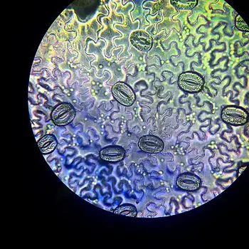
- Visible epidermal cells: The first observation shows the presence of epidermal cells on the surface of the leaf peel. These cells have an irregular outline and appear closely packed with no intercellular spaces.
- Small openings (Stomata): Among the epidermal cells, small openings or pores are scattered throughout the leaf’s surface. These openings are called stomata. Stomata serve as the primary sites for gas exchange between the internal tissues of the plant and the atmosphere.
- Guard cells: Upon closer examination, distinct structures known as guard cells can be observed surrounding the stomatal openings. Guard cells are two kidney-shaped cells that flank the stomata and are responsible for regulating their opening and closing.
- Presence of chloroplasts and nucleus in guard cells: The guard cells show the presence of chloroplasts, the cell organelles responsible for photosynthesis. This is because the guard cells need to perform photosynthesis to produce energy for their function. Additionally, a nucleus is observed within the guard cells, which is essential for controlling cellular activities.
- Thin outer covering and thick inner boundary of guard cells: The guard cells have a specific structural arrangement. They possess a thin outer covering and a thick inner boundary that gives them a concave appearance. This unique configuration allows the guard cells to change shape and control the size of the stomatal pores during the opening and closing process.
- Control of stomatal opening and closing: One of the most important observations is that the guard cells play a crucial role in controlling the opening and closing of the stomata. When the guard cells become turgid (filled with water), they curve and create an opening, allowing gases like carbon dioxide to enter the leaf for photosynthesis. In contrast, when the guard cells lose water and become flaccid, they close the stomatal pores, reducing water loss through transpiration.


Precautions
During the experiment to observe stomata under a microscope, several precautions should be taken to ensure accurate and reliable results:
- Avoid excessive folding of the leaf: When separating the leaf peel, avoid folding the leaf too much, as it may damage the cells or affect the quality of the observation. Snip the peel to an appropriate size to facilitate handling and mounting.
- Proper placement of the peel on the slide: Always place the leaf peel at the center of the glass slide to ensure that it is well-positioned for microscopic observation. Hold the slides from the sides to avoid touching the area where the peel is mounted, preventing any smudging or distortion.
- Avoid overstraining or understraining the peel: While creating the leaf peel, be cautious not to overstrain or understrain it. Overstraining may cause damage to the cells, and understraining might not provide a clear view of the stomata under the microscope.
- Use a brush for handling the peel: To handle the leaf peel, use a soft brush. This gentle approach helps in preventing damage to the cells and maintaining the integrity of the sample.
- Use glycerin for preservation: After staining the leaf peel with safranin, use glycerin as a mounting medium. Glycerin prevents the peel from drying out and helps preserve the sample for prolonged observation.
- Avoid air bubbles when placing coverslip: When placing the coverslip over the mounted peel, take care to avoid trapping air bubbles. Air bubbles can obstruct the view and interfere with the accurate observation of stomata.
- Use blotting paper to remove excess stain: After staining the peel with safranin, use blotting paper to remove any excess stain. This step ensures that only the stomata and relevant structures are clearly visible under the microscope.
FAQ
What is the purpose of observing stomata under a microscope using the temporary mount technique?
The purpose of this technique is to study the microscopic structure and distribution of stomata on the surface of plant leaves. It allows researchers and students to gain insights into the plant’s gas exchange and water regulation mechanisms.
Which plant parts are commonly used for preparing temporary mounts of stomata?
Leaves, especially the lower surface of dicot leaves and both surfaces of monocot leaves, are commonly used for preparing temporary mounts to observe stomata under a microscope.
Why is safranin used during the temporary mount technique?
Safranin is used as a staining agent to highlight the stomata and make them more visible under the microscope. It helps enhance the contrast and makes the stomata stand out from surrounding tissues.
What is the significance of using glycerin in the temporary mount technique?
Glycerin serves as a mounting medium to preserve the leaf peel and maintain its structure over time. It prevents the sample from drying out and allows for longer-term observation.
How can we avoid damaging the leaf cells during the preparation of the leaf peel?
To avoid damaging the leaf cells, it is essential to handle the leaf with care, use fine needles and forceps, and refrain from folding the leaf excessively when creating the peel.
What should be done if air bubbles are trapped under the coverslip during mounting?
Air bubbles can interfere with the observation. To avoid them, gently place the coverslip over the mounted peel, taking care not to trap any air. If air bubbles appear, try repositioning the coverslip or gently tapping it to release the bubbles.
Can any broad flat leaf be used for this experiment?
Yes, any broad flat leaf can be used for this experiment, but it is advisable to test a new leaf type beforehand to ensure it is suitable for observation.
What magnification level should be used to observe stomata under a microscope?
To observe stomata clearly, students will generally need to use at least 100x magnification or higher, depending on the leaf type and the microscope’s capabilities.
How can we properly clean the glass slides and coverslips before mounting?
Before mounting the peel, clean the glass slides and coverslips thoroughly with a suitable cleaning solution, and ensure they are free from dust and debris to avoid any interference during observation.
How should the temporary mounts be stored for further observation or documentation?
After preparing the temporary mounts, store them carefully in a dry and clean environment to avoid any contamination. Seal the edges of the coverslip with nail polish or adhesive to secure the mount and prevent any moisture from entering. Label the slides properly for identification during further study or documentation.
References
- https://sciencelessonsthatrock.com/how-to-view-stomata-under-the-microscope-html/
- https://www.calacademy.org/educators/lesson-plans/stomata-printing-microscope-investigation
- https://www.microscopemaster.com/leaf-structure-under-the-microscope.html
- https://courseware.cutm.ac.in/wp-content/uploads/2020/06/Expt-No-11.pdf
- https://www.gwisd.us/vimages/shared/vnews/stories/4e6fb7eb772cd/Stomata%20Under%20the%20Microscope.pdf
- https://www.microscopeworld.com/p-3384-plant-stomata-under-the-microscope.aspx


