Table of Contents
The development of frogs follows a typical sequence of events observed in sexually reproducing organisms. It begins with the fertilization of an egg or zygote. The zygote then undergoes a series of divisions, dividing and re-dividing to form an embryo. The embryo is the earliest stage of development and remains within the egg or reproductive organs of the mother until hatching or birth.
The study of embryo development is known as embryology, which is a branch of biology dedicated to understanding the processes and stages of embryonic growth. In almost all sexually reproducing organisms, including frogs, the development of the embryo follows a similar sequence of events.
In the case of frogs, the sexes are separate, with females being larger than males. Male frogs possess a nuptial pad at the base of the first finger of their forelimbs and also have a pair of vocal sacs. During mating, known as amplexus, the male grasps the female’s trunk with his forelimbs. Amplexus is a special kind of embrace where the male holds onto the female during reproduction. It is important to note that frogs and toads do not possess penises.
During amplexus, the female frog releases her eggs, typically into water, while the male sheds his sperm over the eggs. This allows for fertilization to occur externally, as the eggs are exposed to the surrounding water where the sperm can reach and fertilize them. This external fertilization is a common reproductive strategy observed in many amphibians, including frogs.
Sperm
- The sperm of a frog is a microcosmic marvel of biological engineering, designed with precision to fulfill its role in the perpetuation of the species. This expository account will elucidate the structure and function of frog spermatozoa, adhering to a detailed and sequential explanation.
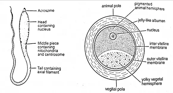
- The mature spermatozoon of a frog is diminutive in size, with an average length of approximately 0.03mm, a testament to its specialized function rather than its stature. The head of the spermatozoon is elongated and robust, harboring the genetic blueprint essential for the inception of new life. At the forefront of the head resides the acrosome, a bead-like structure that houses enzymes. These enzymes are pivotal during fertilization, as they facilitate the breaching of the egg’s protective barriers, allowing the sperm to impart its genetic material.
- Transitioning from the head, the middle piece of the spermatozoon, though not visible in the provided content, is traditionally short and houses mitochondria. These organelles are the veritable power plants of the cell, generating adenosine triphosphate (ATP), the molecular currency of energy that propels the spermatozoon on its journey.
- Then, there is the tail, or flagellum, a filamentous appendage that is markedly longer than the head, often exceeding four times its length. This component is instrumental in the motility of the spermatozoon, enabling it to navigate the aqueous environment of the female reproductive tract. The flagellum’s undulations propel the spermatozoon, ensuring its passage towards the ovum.
- Therefore, the frog’s spermatozoon is a cellular entity exquisitely crafted for its singular purpose. The head, with its genetic cargo and acrosomal enzymes, is designed for penetration and fusion with the egg. The middle piece, though not detailed here, is presumed to supply the energy required for the journey. The tail ensures locomotion, an essential function for the spermatozoon to meet the egg. Each component, though distinct in structure, collaborates seamlessly to achieve fertilization, underscoring the intricate design of these reproductive cells.
Egg
The egg of a frog, at the time of ovulation, measures approximately 2mm in diameter. This vital reproductive cell consists of several distinct components, each serving specific functions in the development of the frog embryo. This expository piece will provide a detailed and sequential explanation of the various components and their roles.
- Accessory Egg Membranes: Surrounding the frog’s egg are two essential structures, apart from the plasma membrane. The first of these is the vitelline membrane, a transparent, non-living membrane developed by the ovum itself. Beyond the vitelline membrane lies the jelly coat or albumen, secreted by the walls of the oviduct. When the egg comes into contact with water, the jelly coat swells due to water absorption. This swelling not only protects the egg but also shields it against injuries and potential infections by bacteria and other microorganisms. Therefore, the accessory egg membranes serve as a protective barrier.
- Polarity and Radial Symmetry: Frog’s eggs exhibit a well-developed polarity and radial symmetry. Within the egg, the cytoplasm is divided into two distinct regions, the cortex, and the endoplasm.a. Egg Cortex (Ectoplasm): The cortex is a jelly-like viscous layer of cytoplasm that adheres closely to the plasma membrane. It houses membrane-bound spherical bodies known as cortical granules, which contain acid mucopolysaccharides. These granules are arranged in a layer near the plasma membrane. Notably, the animal hemisphere of the cortex contains dark-brown pigment granules, imparting a distinctive dark brown color to this region. In contrast, the vegetal pole is relatively whitish and contains fewer pigment granules. The cortex remains stable, unaffected by the movement of cytoplasm or centrifugal forces. Its primary function lies in establishing polarity, bilateral symmetry, and overall organization during the egg’s development. Therefore, it plays a crucial role in shaping the embryo.
- Endoplasm: The inner ooplasm, known as the endoplasm, exhibits a colloidal nature. It contains various cellular organelles, including mitochondria and ribosomes, along with both organic and inorganic substances. A distinctive feature of the endoplasm is the presence of a cup-shaped mass of white yolk platelets known as the vitelline cupola. The germinal vesicle or nucleus is positioned near the animal pole. Yolk granules are present in the endoplasm but vary in size and distribution. In the animal pole, these granules are smaller and less abundant, while the vegetal pole accumulates a higher concentration of yolk granules. Frog’s eggs are described as mesolecithal and moderately telolecithal due to their moderate yolk content, which is unevenly distributed in the cytoplasm, with the vegetal pole containing the highest concentration.
Embryonic Development Stages of Frog
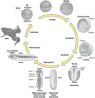
Embryonic development is a complex and intricate process that transforms a single cell, the zygote, into a fully developed organism. In frogs, this process is marked by a series of sequential events that ensure the proper formation and differentiation of various structures and organs. The following is a detailed and sequential explanation of the embryonic development stages of a frog, presented in an expository style.
- Gametogenesis: This is the initial stage where gametes or sex cells are formed. In frogs, the male produces sperm, while the female produces ova. These gametes are specialized cells, carrying half the genetic material required to form a new organism. Therefore, their formation is crucial for the subsequent stages of development.
- Mating: Following gametogenesis, the next step is mating. During this process, the male frog transfers its sperm into the female’s body through a process called copulation. This ensures that the male gametes are in close proximity to the female gametes, setting the stage for fertilization.
- Fertilization: At this juncture, the male and female gametes fuse to form a zygote. This fusion ensures that the zygote has the complete set of genetic material required for the development of a new organism. Besides the genetic combination, fertilization also activates the zygote to begin its developmental journey.
- Cleavage: Post-fertilization, the zygote undergoes a series of rapid cell divisions known as cleavage. These divisions are crucial as they increase the number of cells without increasing the overall size of the embryo.
- Morulation: As a result of cleavage, a solid ball of cells is formed, termed as the morula. This stage is characterized by the compact arrangement of cells, which are almost indistinguishable from one another.
- Blastulation: Then, the morula undergoes further differentiation to form a hollow ball of cells known as the blastula. This cavity, termed the blastocoel, plays a pivotal role in the subsequent stages of development.
- Gastrulation: This stage involves significant movement and rearrangement of cells. The blastula transforms into a structure with three distinct germ layers: the ectoderm, mesoderm, and endoderm. Each of these layers will give rise to specific tissues and organs in the developing frog.
- Organogenesis: As the name suggests, this stage is marked by the development and differentiation of various organs from the primary germ layers. For instance, the ectoderm gives rise to the nervous system and skin, the mesoderm forms muscles, bones, and the circulatory system, while the endoderm develops into the digestive and respiratory systems.
- Morphogenesis: Following organogenesis, there is a focus on the growth and differentiation of form and structure. This ensures that the developing frog has all the necessary anatomical features required for its survival.
- Growth: The final stage of embryonic development is characterized by an increase in size and weight. This is achieved through both cell division and cell enlargement. The developing frog continues to grow until it reaches its adult size.
1. Gametogenesis
Gametogenesis is a fundamental biological process that ensures the continuation of species through sexual reproduction. This process is marked by the formation of specialized reproductive cells, known as gametes, in both male and female organisms. The following is a detailed and sequential explanation of gametogenesis, presented in an expository style.
- Formation of Gametes: Gametogenesis begins with the formation of gametes. These gametes, namely sperms in males and ova in females, are produced by their respective gonads. The process involves a type of cell division called meiosis, which reduces the chromosome number by half, ensuring that the offspring inherit the correct number of chromosomes from both parents. Therefore, meiosis is pivotal for the production of gametes.
- Characteristics of Sperms: The male gametes, or sperms, are distinct in their appearance and function. They are microscopic entities, measuring approximately 0.03mm in length. Structurally, sperms are thread-like, which aids in their motility. This motility is crucial as it allows the sperms to swim towards the female gamete, the ovum, facilitating fertilization.
- Characteristics of Ova: In contrast to sperms, the female gametes, or ova, are significantly larger. They are nearly spherical in shape and, unlike sperms, are non-motile. This means they remain stationary, waiting for the sperm to reach them for fertilization. Besides their size and motility, ova have another distinguishing feature related to their yolk content. Eggs can be classified as mesolecithal or telolecithal based on the distribution of yolk. Mesolecithal eggs have a moderate amount of yolk, while telolecithal eggs possess a large amount of yolk concentrated at one end. This yolk provides nourishment to the developing embryo.
2. Mating
Mating is a critical phase in the reproductive cycle of many organisms, ensuring the transfer of genetic material from one generation to the next. In frogs, this process is marked by specific behaviors and interactions between the male and female. The following is a detailed and sequential explanation of the mating process in frogs, presented in an expository style.
- Initiation of Mating Behavior: The breeding season, typically coinciding with the rainy season, triggers a series of behaviors in frogs that facilitate mating. During this period, male frogs produce distinct croaking sounds. These sounds serve a dual purpose. Firstly, they establish territorial dominance among competing males. Secondly, and more importantly, the croaking acts as a call to attract female frogs. Therefore, the croaking is not just a random sound but a calculated and purposeful act to initiate the mating process.
- Amplexus or Pseudo-Copulation: Once the female frog is attracted to the calling male, the actual process of mating begins. This unique mating embrace is technically termed as “amplexus” or “pseudo-copulation.” Unlike many other organisms, frogs do not have a direct copulatory organ. Instead, the male frog positions himself over the back of the female. Using his forelimbs, he firmly clasps her body, ensuring a close physical connection. This position is crucial for the subsequent steps of the mating process.
- Release of Gametes: In the climax of the amplexus, the female frog releases her ova, often amounting to several hundred, through a specialized opening called the cloaca. Simultaneously, the male frog deposits his spermatic fluid over the released ova. This synchronized release ensures that the sperm meets the ova in the external environment, typically water, facilitating external fertilization. Besides the sheer number of ova released, the emphasis here is on the timing and coordination between the male and female frogs, ensuring a high probability of successful fertilization.
3. Fertilization
Fertilization is a pivotal event in the reproductive sequence, marking the union of male and female gametes to form a new organism. In frogs, this process exhibits several distinctive features and follows a precise sequence of events. The following is a detailed and sequential explanation of fertilization in frogs, presented in an expository style.
- External Fertilization: In frogs, fertilization occurs outside the female’s body, in the aquatic environment. This external mode of fertilization necessitates the simultaneous release of gametes by both sexes to ensure the successful union of sperm and egg.
- Monospermy: The process of fertilization in frogs is characterized by monospermy, meaning that only one sperm is permitted to fuse with the egg. This specificity prevents polyspermy, which can lead to an abnormal number of chromosomes and is detrimental to the embryo’s development.
- Orientation of the Fertilized Egg: After fertilization, the egg exhibits a rotational movement so that the animal hemisphere, which is pigmented, is positioned above the yolk-laden vegetal hemisphere. This orientation is essential for the subsequent developmental processes.
- Jelly Coat Swelling: The jelly coat surrounding the egg swells and increases in thickness upon contact with water. This swelling serves as a protective barrier and aids in preventing polyspermy.
- Completion of Meiosis: The second meiotic division is completed with the release of the second polar body following fertilization. This step is crucial as it ensures the egg has the correct haploid number of chromosomes before it unites with the sperm.
- Sperm Entry: The sperm enters the egg in the animal hemisphere at a specific angle, which is approximately 40 degrees from the center of the animal pole. This precise entry point is critical for the correct alignment and fusion of the male and female genetic material.
- Formation of the Fertilization Membrane: Subsequent to sperm entry, the vitelline membrane elevates and transforms into the fertilization membrane. The creation of the perivitelline space, filled with perivitelline fluid, allows the fertilized egg to rotate freely, which is an inevitable part of normal development.
- Jelly Coat Dynamics: Before the egg’s release into water, the jelly coat remains thin. Upon release, it absorbs water, swells, and its thickness can double, providing additional protection to the egg.
- Second Maturation Division: The second maturation division is promptly completed after fertilization, leading to the release of the second polar body and ensuring the egg’s readiness for the fusion of genetic material.
- Amphimixis: The fusion of the egg and sperm pronuclei to form the zygotic nucleus is termed amphimixis. This fusion results in a zygote with a diploid chromosome number, which is essential for the development of a genetically complete organism.
- Formation of the Grey Crescent: A grey crescent forms on the surface of the egg opposite the point of sperm entry. This crescent indicates the future dorsal side of the embryo and establishes bilateral symmetry, as opposed to the radial symmetry of the unfertilized egg.
- Sperm Penetration Path: The sperm penetrates the egg perpendicular to the cortex and follows a marked path, known as the penetration path, before changing direction towards the egg nucleus, marking the copulation path. These paths are essential for the proper fusion of gametes.
4. Cleavage or segmentation
Cleavage, also known as segmentation, is a fundamental phase in the early development of an embryo. In frogs, this process follows fertilization and is characterized by a series of rapid cell divisions that transform the single-celled zygote into a multicellular entity. The following is a detailed and sequential explanation of cleavage in frogs, presented in an expository style.
- Initiation of Cleavage: Approximately 2-3 hours post-fertilization, the zygote embarks on its journey of division. This series of repeated divisions, termed as cleavage or segmentation, is pivotal for the subsequent developmental stages.
- Nature of Division: The divisions occurring during cleavage are mitotic in nature. Therefore, each resulting cell, or blastomere, retains the diploid chromosome number, ensuring genetic consistency throughout the embryo.
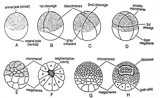
- First Cleavage: The onset of cleavage is marked by a small depression at the animal pole, which progressively extends, enveloping the zygote. This first cleavage is vertical in orientation, resulting in a two-celled stage. Each of these cells is referred to as a blastomere.
- Second Cleavage: Following the initial division, the second cleavage also occurs in a vertical plane. However, it is oriented at a right angle to the first cleavage, culminating in a four-celled stage.
- Third Cleavage: The third cleavage introduces a change in orientation. It is horizontal but occurs above the equatorial line, producing cells of unequal sizes. The upper quartet of smaller, pigmented cells is termed micromeres or epiblast. In contrast, the lower quartet of larger, yolk-laden cells is known as megameres or hypoblast.
- Subsequent Cleavages: The fourth and fifth cleavages are vertical, leading to a 16-celled zygote. These divisions are succeeded by two horizontal cleavages: one towards the animal pole and the other towards the vegetal pole. The culmination of these divisions results in a 32-celled stage.
- Blastomere Identification: The individual cells resulting from cleavage are termed blastomeres. These blastomeres play a crucial role in the formation of various tissues and organs as the embryo develops.
- Significance of the Grey Crescent: Post-fertilization, a grey crescent forms on the zygote’s surface, opposite the sperm entry point. This crescent delineates the future dorsal side of the embryo and establishes its bilateral symmetry. The presence of the grey crescent is vital for the embryo’s proper orientation and subsequent development.
- Sperm’s Path: The sperm’s trajectory within the egg is noteworthy. Initially, it penetrates perpendicular to the cortex, following the penetration path. Upon traversing the cortex, the sperm alters its course, moving towards the egg nucleus along the copulation path. These paths, marked by pigment granules, are essential for the successful fusion of the male and female genetic material.
5. Morulation (formation of morula)
The development of an embryo is a series of intricate and sequential events, each playing a pivotal role in shaping the future organism. Following the cleavage or segmentation phase in frogs, the embryo progresses to the morula stage. The following is a detailed and sequential explanation of the morula stage in frogs, presented in an expository style.
- Formation of the Morula: As a consequence of repeated and irregular cleavage, the embryo transforms into a compact ball of cells. This cellular aggregation is termed the “morula,” drawing its name from its resemblance to a mulberry. Therefore, the term “morula” aptly captures the essence of this developmental stage.
- Composition of the Morula: The morula is not a homogenous entity but exhibits distinct cellular differentiation. The embryo at this stage can be visualized as having two hemispheres, each with its unique set of cells.
- Micromeres: One hemisphere of the morula is densely packed with a large number of small cells that are black in color and devoid of yolk. These cells are referred to as “micromeres.” The presence of micromeres is indicative of the animal pole of the embryo, which is characterized by rapid cell divisions and minimal yolk content. These cells play a crucial role in the formation of the embryo’s ectodermal tissues.
- Megameres: In contrast to the micromeres, the opposite hemisphere of the morula consists of a fewer number of cells that are considerably larger, white in color, and laden with yolk. These cells are termed “megameres.” Representing the vegetal pole of the embryo, the megameres are essential for the formation of endodermal tissues. Besides their role in tissue formation, the yolk within the megameres provides vital nourishment to the developing embryo.
- Significance of the Morula Stage: The morula stage is not merely a transient phase but holds significant importance in embryonic development. The clear distinction between micromeres and megameres sets the foundation for the subsequent differentiation of tissues and organs. Moreover, the compact arrangement of cells in the morula ensures that the embryo remains protected and retains its structural integrity.
6. Blastulation (formation of blastula)
Embryonic development is a meticulously orchestrated sequence of events, with each stage laying the foundation for the next. Following the morula stage in frogs, the embryo transitions to the blastula stage. The following is a detailed and sequential explanation of the blastula stage in frogs, presented in an expository style.
- Formation of the Blastocoel: As the micromeres undergo rapid divisions, a significant developmental event unfolds. These divisions result in the creation of a small, fluid-filled cavity within the embryo. This cavity is termed the “Blastocoel” or “segmentation cavity.” The emergence of the blastocoel is a hallmark of the blastula stage and plays a pivotal role in the embryo’s subsequent development.
- Composition of the Blastula: An embryo at the blastula stage, bearing the blastocoel, is aptly termed a “Blastula.” The blastula exhibits a distinct structural organization. The floor of the blastocoel is primarily composed of yolk-laden megameres, while its roof is densely packed with micromeres. This arrangement ensures the embryo’s stability and lays the groundwork for future tissue differentiation.
- Presumptive Areas: At the blastula stage, the embryo begins to exhibit early signs of tissue differentiation. Through staining techniques, specific regions, known as “presumptive areas,” can be identified. These areas are precursors to the various tissues and organs that will form as the embryo develops.
- Presumptive Ectoderm: The entire animal pole of the blastula represents the presumptive ectoderm. This region is further subdivided into two areas: the presumptive epidermis and the presumptive neural plate. These areas will eventually give rise to the skin and nervous system, respectively.
- Presumptive Notocord: Situated near the vegetal pole is a small area designated as the presumptive notocord. The notocord plays a crucial role in vertebrate development, providing structural support and guiding the formation of the spinal cord.
- Presumptive Mesoderm: Adjacent to the presumptive notocord lies the grey crescent region. This region is identified as the presumptive mesoderm. The mesoderm is essential for the formation of various tissues, including muscles, bones, and the circulatory system.
- Presumptive Endoderm: The remaining vegetal region of the blastula is the presumptive endoderm. This region will eventually differentiate into tissues that form internal organs such as the digestive tract and lungs.
7. Gastrulation (formation of gastrula)
The progression of embryonic development in frogs is marked by a series of intricate stages, each laying the groundwork for the subsequent phase. Following the blastula stage, the embryo transitions into the gastrula stage. This stage is characterized by the formation of a two-layered embryo through a process known as gastrulation. The following is a detailed and sequential explanation of the gastrula stage in frogs, presented in an expository style.
- Definition of Gastrula: The gastrula is an embryonic stage that results from the migration and rearrangement of the blastula’s cells. Gastrulation transforms the embryo from a single-layered structure (monoblastic) to a two-layered entity (diploblastic), culminating in the formation of three primary germ layers.
- Gastrulation Process: Gastrulation encompasses several critical changes, including cell differentiation and the establishment of the primary germ layers. This process unfolds in the following steps:
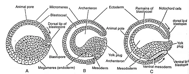
- Epiboly: At the animal pole, micromeres divide rapidly and repeatedly, eventually encasing the megameres, except in the yolk plug region. This spreading or overgrowth of micromere cells is termed “Epiboly.”
- Emboly or Intucking (Invagination): A small groove appears near the grey crescent region due to the invagination of megameres. As this invagination deepens, cells migrate inward, leading to the “Yolk plug stage.” The blastopore, which narrows over time, exerts pressure on the underlying yolk-laden megameres, causing some megamere cells to protrude as a yolk plug. As invagination continues, the archenteron enlarges, reducing and eventually obliterating the blastocoel. The groove that forms is the beginning of the archenteron, with its anterior opening termed the “blastopore.” The blastopore is bordered by an anterior margin called the dorsal lip and a backward-projecting lateral lip.
- Involution: The enlargement of the archenteron and the formation of the yolk plug trigger a rapid migration of presumptive areas within the embryo. This movement is termed “involution.” Additionally, gastrulation shifts the embryo’s center of gravity. While the blastula stage sees the embryo floating with its animal pole upward, the formation of the archenteron causes the embryo to rotate, positioning the blastopore near the vegetal pole.
- Formation of Three Germ Layers: Gastrulation culminates in the formation of three primary germ layers: ectoderm, mesoderm, and endoderm. These layers are pivotal as they give rise to all the organs and structures of the body. Specifically: – The blastopore becomes the presumptive gut. – The archenteron’s roof develops into the chordamesoderm. – The archenteron’s floor differentiates into the endoderm.
Fate of Germ Layers
In the intricate journey of embryonic development, the formation of the three primary germ layers – ectoderm, mesoderm, and endoderm – is a pivotal event. These layers, established during the gastrula stage, play a foundational role in determining the fate of various tissues and organs in the developing organism. Presented in an expository style, here is a detailed and sequential explanation of the fate of these germ layers:
1. Ectoderm: The outermost germ layer, the ectoderm, is responsible for giving rise to a diverse array of structures and tissues. Derived from the primary ectoderm layer, the following are its contributions:
- Epidermis: The outer layer of the skin.
- Cutaneous Glands: Glands present in the skin.
- Eye Components: This includes the lens, cornea, retina, and conjunctiva.
- Central Nervous System: Comprising the brain and spinal cord.
- Glandular Structures: The pineal gland and pituitary gland originate from the ectoderm.
- Dental Structures: The enamel of the teeth is ectodermal in origin.
2. Mesoderm: Positioned between the ectoderm and endoderm, the mesoderm is instrumental in forming several vital structures. Derived from the primary mesoderm layer, its contributions include:
- Notochord: A rod-like structure that provides support during early development.
- Body Cavities: The pericardium (surrounding the heart) and peritoneum (lining the abdominal cavity).
- Musculature: All muscles in the body.
- Skeletal System: Bones and cartilage.
- Connective Tissues: This encompasses blood, lymph, adipose tissue, and the dermis of the skin.
- Visceral Organs: Organs within the body cavities.
3. Endoderm: The innermost germ layer, the endoderm, is the precursor to several internal structures. Derived from the primary endoderm layer, its contributions are:
- Digestive Tract: The epithelium or lining of the digestive tract.
- Respiratory Tracts: The linings of the trachea, bronchi, and lungs.
- Eustachian Tubes: Tubes connecting the middle ear to the nasopharynx.
- Glandular Structures: Gastric and intestinal glands, liver, and pancreas.
- Ducts: Bile and pancreatic ducts that transport digestive enzymes.
- Urinary System: The lining of the urinary bladder.
8. Organogenesis
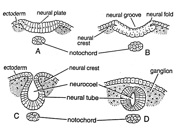
Organogenesis is a pivotal phase in embryonic development that follows the gastrulation process. This intricate phase is characterized by the formation and differentiation of various embryonic tissues derived from the three primary germinal layers: ectoderm, mesoderm, and endoderm. These layers, established post-gastrulation in triploblastic metazoans, lay the groundwork for the subsequent development of the embryo.
1. Initiation of Organogenesis:
Approximately thirty hours post-fertilization, gastrulation culminates, resulting in the formation of the three germinal layers. The subsequent developmental phase, organogenesis, is instrumental in leading to the emergence of an active, free-swimming larval stage known as the tadpole larva.
2. Neurulation (Formation of Neural Tube):
- The ectoderm cells located on the mid-dorsal region undergo thickening, leading to the formation of the neural plate.
- Adjacent to the neural plate are the neural folds, which expand and eventually fuse at the mid-dorsal region, giving rise to the neural canal or tube.
- Initially, the neural tube possesses an anterior opening known as the neuropore.
- The neural tube remains temporarily connected to the archenteron via the neurenteric canal.
- Ultimately, the neural tube transforms into a closed canal, with its anterior segment developing into the brain and the posterior segment forming the spinal cord. At this juncture, the embryo is termed a neurula.
3. Formation of Notochord and Mesoderm:
- The chordamesoderm, situated at the mid-dorsal region, evolves into a rod-like structure known as the notochord.
- The remaining chordamesoderm differentiates into the mesoderm.
- Adjacent to the notochord, the mesoderm can be categorized into three segments: epimere (dorsally located), mesomere (middle mesoderm), and hypomere (ventral mesoderm).
4. Formation of Endoderm:
- Cells constituting the floor of the archenteron proliferate and extend dorsally, encapsulating the archenteron to form the endoderm.
- The embryo, now elongated, distinctly exhibits the three primary germinal layers: the outer ectoderm, the inner endoderm, and the intervening mesoderm.
5. Fate of the Three Germinal Layers:
- Ectoderm: This layer differentiates into the epidermis, cutaneous glands, central nervous system, components of the eye, and olfactory and auditory organs.
- Endoderm: It gives rise to the epithelial lining of the digestive system, respiratory system components, urinary bladder lining, and specific glands like the thymus and thyroid.
- Mesoderm: This layer forms the dermis, skeletal system components, blood vascular system, excretory and reproductive organs, and certain eye structures.
9. Morphogenesis
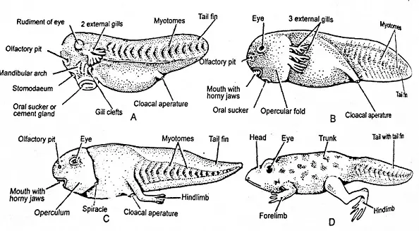
Morphogenesis is the biological process that causes an organism to develop its shape. It is one of the three fundamental aspects of developmental biology along with the control of cell growth and cellular differentiation.
1. Transition to Pre-tadpole Stage:
- Approximately four days after fertilization, the embryo, now approximately 4mm in length, remains encased within the egg membrane.
- The embryonic structure is distinguishable into three regions: the head, trunk, and tail.
- The head features bilateral elevations indicating the future tympanum, while the ventral side of the anterior end exhibits a U-shaped sucker, the cement gland, formed by mucous gland cells.
- A small depression between the sucker and nasal pit marks the stomodaeum, and a similar depression at the posterior end, the proctodaeum, is evident.
- The tail extends posteriorly from the proctodaeum, and internally, the embryo comprises the central nervous system, notochord, a closed alimentary canal, liver, heart, and the rudiment of the urinary bladder.
- The development of these organs signals the readiness of the embryo to hatch.
2. Hatching Process:
- After approximately two weeks post-fertilization, the embryo, now about 6mm in length, emerges from the egg membrane in a process known as hatching, resulting in the tadpole larva.
3. Development of the Tadpole Larva:
- The tadpole, a small, blackish, fish-like creature, initially clings to aquatic plants via its ventral sucker.
- Respiration is facilitated by the development of two, and subsequently three pairs of branched external gills, complemented by cutaneous respiration.
- Nutritional needs during the first week post-hatching are met by yolk reserves within the archenteron cells.
- The mouth, bounded by two horny jaws, forms seven days after hatching, allowing the tadpole to consume aquatic vegetation.
- The stomodaeum and proctodaeum connect to form a complete alimentary canal, initially short and broad, later elongating and coiling.
- External gills give way to four pairs of internal gills, concealed by an operculum.
- The mesonephric kidney, characteristic of the adult, begins to form.
- The lateral line system, crucial for aquatic navigation, is well-developed.
- Limb development ensues, with hind limbs appearing before the more slowly developing forelimbs, which remain hidden by the operculum.
- Lungs develop and, upon maturation, the tadpole utilizes both pulmonary and gill respiration.
Throughout morphogenesis, the embryo undergoes significant structural changes, transitioning from a simple layered structure to a complex organism with distinct organs and systems. This process is meticulously regulated by genetic and molecular cues that ensure the proper development of the organism’s body plan.
10. Metamorphosis and growth
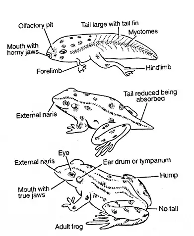
Metamorphosis is a fascinating biological process that describes the drastic transformation of an organism from one developmental stage to another, often resulting in significant changes in form and function. In the context of amphibians, such as frogs, metamorphosis is the transition from the aquatic tadpole stage to the terrestrial adult frog stage.
1. The Tadpole Larva: The tadpole larva is markedly different from the adult frog in both form and nature. This aquatic stage is characterized by:
- A fish-like appearance.
- The presence of external gills for respiration.
- A herbivorous diet, initially sustained by yolk reserves from the archenteron cells.
- A sectorial mouth bound by two horny jaws.
- A long, coiled intestine suited for a herbivorous diet.
2. Initiation of Metamorphosis: Approximately two to three weeks after the tadpole begins breathing through its lungs, it undergoes a series of dramatic changes, marking the onset of metamorphosis. These changes are driven by the secretion of the hormone thyroxine from the newly developed thyroid gland.
3. Key Metamorphic Changes:
- Feeding Habits: The tadpole ceases feeding. As the tail begins to recede, its tissues provide nutrition to the transforming larva.
- Mouth and Digestive System: The sectorial mouth widens, and a large sticky tongue develops. The once long and coiled intestine, suited for a herbivorous diet, shortens in preparation for a carnivorous diet.
- Respiratory System: External gills disappear, and the lungs become the primary respiratory organs.
- Skeletal System: The cartilaginous skeleton transitions to a bony endoskeleton.
- Sensory Organs: The eyes enlarge, and the middle ear and tympanum develop.
- Limbs: Forelimbs emerge from beneath the operculum, and hind limbs elongate.
- Skin: The skin undergoes vascularization, pigmentation, and glandular development.
4. Completion of Metamorphosis: Upon the completion of these changes, the young frog, now with a vestigial tail, leaves its aquatic environment to inhabit damp terrestrial areas. It adopts a carnivorous diet, feeding primarily on insects, and continues to grow, eventually leading an amphibious life.
In summary, metamorphosis in frogs is a complex and meticulously regulated process that ensures the successful transition from an aquatic tadpole to a terrestrial adult frog. This transformation is vital for the survival and propagation of the species, allowing it to exploit different ecological niches at various stages of its life.
Neurulation, Notogenesis, and Formation of coelom
1. Neurulation: The Genesis of the Neural Tube
Neurulation refers to the intricate process that leads to the formation of the neural tube, which eventually gives rise to the central nervous system, encompassing the brain and spinal cord. This process commences post-gastrulation, with the prospective neural plate positioning itself along the mid-dorsal region. As the process unfolds:
- Neural plate edges elevate to form neural folds.
- These folds grow in prominence and converge at the median line, culminating in the formation of the neural tube. This tube encases the neural canal.
- The closure of this tube initiates anteriorly, progressing both forwards and backwards.
- While the anterior end of the neural tube remains temporarily open via the neuropore, the posterior end establishes a brief connection with the archenteron through the neurenteric canal.
- The culmination of this process results in a sealed tubular neural tube, which subsequently differentiates into the brain and spinal cord.
2. Notogenesis: The Birth of the Notochord
Notogenesis pertains to the formation of the notochord, a pivotal structure during embryonic development. As the process unfolds:
- Meso-endodermal cells, situated in the mid-dorsal region atop the archenteron, segregate from the mesoderm layer.
- These cells solidify, forming a cylindrical, rod-like structure known as the notochord.
- Subsequently, a protective sheath, the notochordal sheath, envelops the notochord.
- In mature stages, the notochord undergoes transformation, being replaced by the vertebral column.
3. Formation of Coelom: Crafting the Body Cavity
The coelom, a mesodermal-derived body cavity, plays a crucial role in housing various organs. The genesis of the coelom involves:
- The mesodermal layer bifurcating into two distinct layers: the outer somatic (or parietal) layer and the inner visceral (or splanchnic) layer.
- A cavity, termed the splanchnocoel, materializes between these layers, extending downwards and externally beneath the gut.
- The outer somatic layer amalgamates with the ectoderm, giving rise to the body wall, known as the somatopleure.
- Conversely, the inner visceral layer merges with the endoderm, forming the gut wall or splanchnopleure.
- The splanchnocoel evolves to form the coelom, creating a cavity nestled between the gut wall and the body wall. This type of coelom is termed a Schizocoel coelom.
In essence, neurulation, notogenesis, and the formation of the coelom are pivotal processes during embryonic development, each contributing to the intricate architecture and functionality of the mature organism.
FAQ
What is pre-embryonic development in frogs?
Pre-embryonic development refers to the initial stage after fertilization when the zygote undergoes cleavage and forms a multicellular embryo.
What are the major events during embryonic development in frogs?
During embryonic development, the fertilized egg undergoes various stages, including gastrulation, neurulation, and organogenesis. The body plan and major organs of the frog start to form during this stage.
What happens during gastrulation in frog embryonic development?
Gastrulation is the process in which the embryo transforms from a hollow ball of cells into a three-layered structure known as the gastrula. This process establishes the basic body plan of the frog.
What is neurulation in frog embryonic development?
Neurulation is the process by which the neural tube forms from the ectoderm. The neural tube will eventually give rise to the central nervous system of the frog.
What is organogenesis in frog embryonic development?
Organogenesis is the stage in which the major organs and organ systems develop. During this stage, the heart, liver, central nervous system, digestive system, and other organs begin to take shape.
What are the key stages in post-embryonic development of a frog?
Post-embryonic development in frogs includes the pre-tadpole stage, hatching, and the tadpole larval stage. These stages involve further growth, metamorphosis, and the transition to adult frog form.
What are some notable features of the pre-tadpole stage in frog development?
The pre-tadpole stage is characterized by the growth of the embryo within the egg membrane. The body differentiates into head, trunk, and tail regions, and important organs and structures begin to develop.
What happens during hatching in frog development?
Hatching is the process in which the embryo breaks free from the egg membrane and emerges into the external environment. This marks the transition from the embryonic stage to the larval stage known as tadpoles.
What are the main characteristics of tadpole larval stage in frog development?
Tadpoles are aquatic and possess external gills for respiration. They undergo feeding, growth, and development while living in water. Their body shape and structures are adapted for an aquatic lifestyle.
What is metamorphosis and its significance in frog development?
Metamorphosis is the process through which tadpoles undergo remarkable changes to transform into adult frogs. This includes the reabsorption of the tail, development of limbs, changes in organs, and adaptation for a terrestrial lifestyle. Metamorphosis is a crucial step in the frog’s life cycle, enabling it to transition from an aquatic larva to a fully formed adult frog.
References
- https://www.wardsci.com/store/product/8875044/frog-development-slide-set
- http://www.svc.ac.in/SVC_MAIN/Departments/Zoology/Museum/MuseumData/5.%20DEVELOPMENTAL%20BIOLOGY/1_FROG%20EMBRYOLOGY%20SLIDES/5.1%20FROG%20EMBRYOLOGY%20SLIDES.pdf
- http://bcs.whfreeman.com/webpub/biology/sadavalife9e/animated%20tutorials/life9e_3301_script.html
- https://onlinesciencenotes.com/sequential-events-and-stages-in-the-development-of-frog-pre-embryonic-embryonic-and-post-embryonic-development/
- https://old.amu.ac.in/emp/studym/100003759.pdf
- https://www.notesonzoology.com/frog/development-of-frog-with-diagram-vertebrates-chordata-zoology/8626
- https://alrasheedcol.edu.iq/modules/lect/lect/2925-blatolation.pdf
- https://www.yourarticlelibrary.com/science/development-of-frog-stages-in-gastrulating/23140
